MEDICAL BIOLOGY, HUMAN ANATOMY, PHYSIOLOGY AND PATHOLOGY - Y.I. Fedoniuk 2010
ANATOMY, PHYSIOLOGY, PATHOLOGY
CHAPTER 2. THE LOCOMOTOR SYSTEM (THE MECHANISM OF MOVEMENT)
MUSCLES AND FASCIAE OF BODY REGIONS
MUSCLES AND FASCIAE OF THE TRUNK
2. MUSCLES AND FASCIAE OF THE CHEST
The chest Muscles are divided into two groups: 1) muscles acting on the JOINTS OF THE Pectoral Girdle; 2) intrinsic chest muscles. The first group includes:
1. The pectoralis major Muscle originates from the anterior surface of the clavicle, the Sternum, and the cartilages of Ribs II–VII. It inserts into the crest of the greater tubercle of the humerus (Fig. 2.83). Function: adduces the arm to the trunk and rotates it medially. The clavicular part of the muscle flexes the arm (lifts it forward). When the upper limb is fixed, it elevates the ribs, thus participating in the act of inspiration.
Class="center">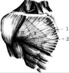
Fig. 2.83. Pectoralis major muscle (1) and the contours of the pectoralis minor muscle (2), which form the anterior wall of the axillary fossa; PROJECTION OF THE clavipectoral triangle (I) and the pectoral triangle (II)
2. The pectoralis minor muscle originates from ribs II–V and inserts into the coracoid process of the scapula (Fig. 2.84).
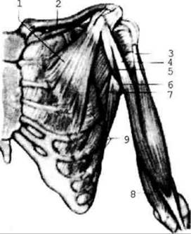
Fig. 2.84. Muscles of the chest and arm
Function: pulls the scapula anteriorly and inferiorly. When the scapula is fixed, it elevates the ribs.
3. The subclavius muscle extends from rib I to the clavicle.
Function: pulls the clavicle inferiorly and medially.
4. The serratus anterior muscle originates from the upper nine ribs and inserts into the inferior angle and medial border of the scapula.
Function: pulls the scapula anteriorly, rotating its inferior angle laterally.
All these muscles, when the pectoral girdle and upper limb are fixed, participate in Respiration. This explains the forced posture of patients with Bronchial Asthma. They sit gripping the back of a bed or chair tightly. In this position, the contraction of the chest muscles enhances breathing and relieves dyspnea.
The second group includes the external and internal intercostal muscles located in the intercostal spaces. The former elevate the ribs (inspiration),
whereas the latter depress them (expiration).
Fasciae of the chest
1. The pectoral fascia has superficial and deep layers.
2. The endothoracic fascia lines the thoracic cavity from the inside and continues onto the Diaphragm.
The diaphragm (diaphragma) is a thin, flat muscle that separates the thoracic cavity from the Abdominal cavity. Its muscle bundles originate from the sternum, ribs, and lumbar vertebrae (Fig. 2.85). Accordingly, the sternal, costal, and lumbar PARTS OF THE diaphragm are distinguished. The muscle fibers of these parts converge toward the center of the diaphragm and transition into an aponeurosis, forming the central tendon, which contains the opening for the INFERIOR VENA CAVA.
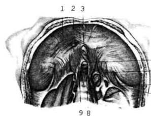
Fig. 2.85. The diaphragm; inferior view
The lumbar part is the strongest. It originates with medial and lateral crura. The medial crura bound the openings through which the Esophagus and aorta pass.
Between the lumbar and costal parts, as well as between the COSTAL AND STERNAL parts, there are areas lacking muscle fibers. These triangular areas are known as the lumbocostal and sternocostal triangles. They are paired, and diaphragmatic hernias may form within their regions; therefore, these triangles are referred to as the weak spots of the diaphragm.
The diaphragm is the primary respiratory muscle. Upon contraction, it descends, increasing the volume of the thoracic cavity to cause inspiration. Upon relaxation, the diaphragm rises into a dome shape, the Lungs recoil, and expiration occurs.
Last update: 08/08/2026
Editorial and Educational Adaptation: This material has been compiled based on the primary/original source text. The project team performed an editorial review, corrected technical inaccuracies, structured sections, and adapted the content for an educational format.
What was processed:
- elimination of formatting defects (OCR errors, structural breaks, corrupted characters);
- editorial organization of content;
- standardization of terminology in accordance with academic sources;
- verification of factual statements against the original source text.
All mentions of the author, publication year, and origin of the primary text have been preserved in accordance with the source.