HUMAN ANATOMY ATLAS - G.L. Bilich - 2014
Articular System
JOINTS OF THE RIBS AND STERNUM. THORAX
The Thorax (thorax) forms the skeletal framework of the walls of the thoracic cavity (cavitas thoracis), which houses Internal Organs (Heart, Lungs, Trachea, Esophagus, major Blood Vessels, etc.). The thorax is formed by interconnected thoracic vertebrae, Ribs, and the Sternum. The ribs articulate with the vertebrae via the costovertebral joints and with the sternum (Table 28, see Fig. 36).
The joint of the HEAD of the rib (articulatio capitis costae) is formed by the articular surfaces of the rib head and the costal demifacets of the II—X thoracic vertebrae (Figs. 140, 141). The Joint Capsule attaches along the margins of the articular surfaces. Each joint of the heads of the II—X ribs contains an intra-articular ligament of the head of the rib (ligamentum capitis costae intraarticulare), which originates on the crest of the head of the rib and attaches to the intervertebral disc separating the articular demifacets of the adjacent superior and inferior vertebrae. The JOINTS OF THE I, XI, and XII ribs lack intra-articular ligaments, as they are formed by the heads of these ribs and complete facets on the bodies of the I, XI, and XII thoracic vertebrae. Externally, the capsule of each joint of the head of the rib is reinforced by the radiate ligament of the head of the rib (ligamentum capitis costae radiatum), which originates on the anterior surface of the rib head and attaches to the intervertebral disc and the bodies of adjacent vertebrae. This ligament includes the superior and inferior anterior ligaments of the head of the rib — Barkow's ligaments (Barkow, Hans Karl Leopold, 1798–1873, German anatomist).
The costotransverse joint (articulatio costotransversaria) is formed by the articular surface of the tubercle of the rib and the costal facet of the transverse process of the I—X ribs (see Fig. 140). The capsules of these joints are reinforced by three ligaments. These are the costotransverse ligament (ligamentum costotransversarium) — Cruveilhier's costotransverse ligament (Cruveilhier, Jean, 1791–1874, French anatomist, pathologist, and clinician); the superior costotransverse ligament (ligamentum costotransversarium superius), connecting the neck of the rib to the adjacent superior transverse process; and the lateral costotransverse ligament (ligamentum costotransversarium laterale), connecting the tubercle of the rib to the tip of the transverse process.
The joint of the head of the rib and the costotransverse joint together form a combined uniaxial joint. The axis of Rotation of the rib passes through both joints. Movement in these joints results in the elevation and depression of the ribs.
The sternocostal joints (articulationes sternocostales) are formed by the anterior ends of the cartilages of the II—VII ribs and the costal notches of the sternum (Figs. 142, 143). The Cartilage of the I rib fuses with the sternum, forming a synchondrosis. The capsules of the sternocostal joints are continuations of the perichondrium of the costal cartilages, transitioning into the periosteum of the sternum. The radiate sternocostal ligaments (ligamenta sternocostalia radiata) reinforce the joint capsule on the anterior and posterior surfaces of the joints. Anteriorly, the radiate sternocostal ligaments fuse with the periosteum of the sternum, forming a dense sternal membrane. The joint of the II rib features an intra-articular sternocostal ligament (ligamentum sternocostale intraarticulare) — Quain's ligament (Quain, John, 1795–1851, English physician and anatomist). The inferior end of the body of the sternum connects to the cartilages of the VI and VII ribs via flat costoxiphoid ligaments (ligamenta costoxiphoideae) — Lannelongue's ligaments, from which fibers extend to the Pericardium (Lannelongue, Odilon Marc, 1840–1911, French surgeon and pathologist).
Class="center">Table 28. Connections of the Ribs with THE Vertebral Column and Sternum
Joint Name |
Articular Surfaces |
Joint Ligaments |
Type of Movement Axis |
Function and Muscles Responsible |
Joint of the head of the rib |
Articular surface of the head of the rib, costal demifacets of the thoracic vertebrae (except I, XI, and XII ribs; the heads of these ribs articulate with the costal facets of I, XI, and XII vertebrae) |
Intra-articular ligament of the head of the rib, radiate ligament of the head of the rib |
Combined, rotational, uniaxial (along the neck of the rib) |
Rib elevation, muscles: external intercostals, levatores costarum, serratus posterior superior, scalenes. Rib depression, muscles: internal intercostals, subcostals, transversus thoracis, serratus posterior inferior |
Costotransverse joint |
Articular surface of the tubercle of the rib, costal facet of the transverse process, except for XI and XII ribs, which lack this joint |
Costotransverse ligament |
||
Sternocostal joint |
Anterior ends of the costal cartilages, II–VII costal notches of the sternum |
Radiate sternocostal ligaments |

Fig. 140. Ligaments of the costovertebral joints, superior view:
1 — Anulus fibrosus; 2 —Nucleus pulposus; 3 = 1 + 2 — Intervertebral disc; 4 — Radiate ligament of head of rib; 5 — Costotransverse liga- ment: 6 — Superior costotransverse ligament; 7 — Lateral costotransverse ligament; 8 — Superior articular facet; 9 — Yertebral arch; 10 — Spinous process; 11 — Costotransverse joint; 12 — Articular facet; 13 — Tubercle; 14 —Neck of rib; 15 — Head ofrib; 16 — Joint of hcad ofrib; 17 — Vertebral foramen; 18 — Thoracic vertebra [VIII], vertebral bodv
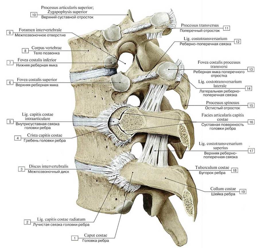
Fig. 141. Ligaments of the costovertebral joints, lateral view, left side (V–VIII thoracic vertebrae):
1— Head of rib; 2 — Radiate ligament of head ofrib: 3 — Intervcrtebral disc; 4 — Crest of head of rib; 5 — Intra-articular ligantem of head of rib: 6 — Superior costal facct; 7 — Inferior costal facct; 8 — Vcrtcbral body; 9 — Intervcrtebral foramen; 10 — Superior articular process; 11 — Transverse process; 12 — Costotransverse ligament; 13 — Transverse costal facet; 14 — Lateral costotransverse ligament; 15 — Spinous process; 16 — Articular facet; 17—Superior costotransverse ligament; 18 — Tubercle ofrib; 19 — Neck ofrib
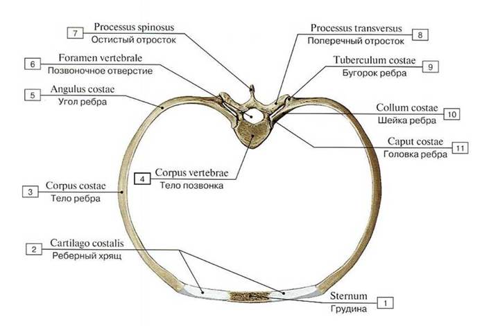
Fig. 142. Connections of the ribs with the Vertebral Column and sternum:
1 — Sternum; 2 — Costal cartilage; 3 — Body of rib; Sliaft of rib; 4 — Vertebral body; 5 — Angle of rib; 6 — Vertebral foramen; 7 — Spinous process; 8— Transverse process: 9 —Tubercle; 10 — Neck of rib: 11 — Hlead of rib
The cartilages of the VIII–X ribs do not connect directly to the sternum. They fuse with one another and with the cartilage of the superior (VII) rib, forming the costal margin (arcus costalis). Occasionally, interchondral joints (articulationes interchondrales) are present between the cartilaginous ends of the lower ribs, enclosed by joint capsules formed by the perichondrium.
The anterior ends of all ribs are connected to one another by the external intercostal membrane (membrana intercostalis externa), whose fibers run downward and forward. Between the posterior ends of the ribs, the internal intercostal membrane (membrana intercostalis interna) is stretched, with fibers running upward and backward. Thanks to these joints and interchondral connections, the thorax possesses a high degree of mobility.
During inhalation and exhalation, the posterior ends of the ribs rotate within the costovertebral joints, while the RIBS AND STERNUM simultaneously elevate or depress. During inhalation, the anterior ends of the ribs and the sternum rise, and the intercostal spaces widen; during exhalation, the anterior ends of the ribs and the sternum descend.
The costovertebral joints receive their blood supply from the posterior intercostal Arteries. Venous blood drains into the vertebral venous plexus and subsequently into the intercostal Veins. Lymph drains into the intercostal Lymph Nodes located near the spine. Innervation is supplied by the posterior Branches of the thoracic Spinal Nerves. The sternocostal joints are vascularized by branches of the internal thoracic artery, with venous drainage into the corresponding veins. Lymph drains into the parasternal and deep cervical lymph nodes. Innervation is provided by the Anterior branches of the intercostal nerves.
Thorax. The human thorax is shaped roughly like an irregular barrel, expanded transversely and flattened in the anteroposterior direction. Depending on body type, three shapes of the thorax are distinguished. In individuals with a brachyorphic body type, the thorax is cone-shaped, with the lower part significantly wider than the upper; the subcostal angle is obtuse, the ribs have a slight downward inclination, and the anteroposterior and transverse diameters are nearly equal. In individuals with a dolichomorphic body type, the thorax is flat and flattened anteroposteriorly, with a pronounced downward inclination of the ribs and an acute subcostal angle. In individuals with a mesomorphic body type, the thorax is cylindrical in shape.

Fig. 143. Costosternal joints, anterior view:
1 — Xiphoid process; 2 — Costa cartilage; 3— Sternum; 4 — Clavicular notch; 5 — First rib [I]; 6— Radiate sternocostal ligamenls; 7— Sternocostal joints; 8 — Costoxiphoid ligaments
The thoracic cage has four walls and two apertures (see Fig. 36). The anterior wall is formed by the sternum and costal cartilages, the lateral walls by the ribs, and the posterior wall by the thoracic vertebrae and the posterior ends of the ribs (Fig. 144, see Fig. 38). The ribs are separated from one another by intercostal spaces (spatium intercostale), which contain intercostal muscles, ligaments, blood vessels, and nerves. The superior thoracic aperture (apertura thoracis superior) is bounded by the first thoracic vertebra [I], the first pair of ribs, and the superior margin of the sternum. The trachea, esophagus, major blood vessels, Lymphatic vessels, and nerves pass through this aperture. The plane of the superior thoracic aperture is tilted forward and downward, as its anterior margin is lowered in accordance with the course of the ribs. The jugular notch of the sternum lies at the level of the intervertebral disc between the second [II] and third [III] thoracic vertebrae. The inferior thoracic aperture (apertura thoracis inferior), bounded by the twelfth thoracic vertebra [XII], the lower ribs, costal cartilages, and the xiphoid process of the sternum, is closed by the Diaphragm, through whose openings pass the aorta, esophagus, INFERIOR VENA CAVA, lymphatic vessels, and nerves.
The right and left costal arches laterally bound the infrasternal angle (angulus infrasternalis), which opens inferiorly and has its apex at the level of the ninth thoracic vertebra, formed by the xiphoid process. The thoracic cage is somewhat shorter anteriorly than posteriorly, and the sternum is shorter than the thoracic region of the vertebral column. Dorsally, the bodies of the thoracic vertebrae project into the thoracic cavity, dividing it into right and diminutive left halves that house the lungs. On either side of the thoracic spine lie vertically oriented deep depressions known as the pulmonary sulci (sulci pulmones), which accommodate the posterior borders of the lungs. Table 29 presents the average dimensions of the adult male thoracic cage.
Movements of the thoracic cage are driven by inspiration and expiration. Because the anterior ends of the ribs are attached to the sternum, both the ribs and the sternum move during Respiration. Elevation of the anterior ends of the ribs and the sternum during inspiration leads to an increase in the transverse and sagittal (anteroposterior) dimensions of the thoracic cage and a widening of the intercostal spaces. This results in an increased volume of the thoracic cavity. Conversely, during expiration, the anterior ends of the ribs and the sternum descend, the anteroposterior dimension of the thoracic cage significantly decreases, the intercostal spaces narrow, and the volume of the thoracic cavity diminishes. Fig. 145 illustrates the movements of the upper and lower ribs.

Fig. 144. Thoracic cage, position of the scapula and clavicle (right side), lateral view:
1 — Rib [XII]; 2 — Lateral border; 3 — Scapula, posterior surface; 4 — Glenoid cavity; 5 — Cora-coid process; 6 — Acromion; 7 — Acromiocla-vicular joint; 8 — Vertebra prominens [CVII]; 9 — First rib [I]; 10 — Clavicle; 11 — Stemo- clavicular joint; 12 — Manubrium of sternum; 13 — Body of sternum; 14 —Costal cartilagc

Fig. 145. Comparative characteristics of upper and lower rib movements:
1 — Lower rib; 2 — Neck of rib; 3 — Upper rib
Table 29. Dimensions of the Thoracic Cage (during quiet breathing, adult male measurements)
Parameter |
Measurement, cm |
Length of posterior wall (along the midline) |
27-30 |
Length of anterior wall (along the midline) |
16-19 |
Length of lateral wall (along the midaxillary line) |
28-30 |
Dimensions of superior aperture: transverse anteroposterior |
10-12 5-6 |
Dimensions of inferior aperture: transverse anteroposterior (at the level of the xiphoid process) |
19-20 16-20 |
Maximum circumference (at the level of the eighth rib) |
80-84 |
Depression of the ribs occurs not only through the action of specialized rib-depressing muscles (see "Muscles of the Chest"), but also due to the elasticity of the costal cartilages and the weight of the thoracic cage. The shape and dimensions of the thoracic cage are subject to significant age-related and individual variations.
Last update: 08/08/2026
Editorial and Educational Adaptation: This material has been compiled based on the primary/original source text. The project team performed an editorial review, corrected technical inaccuracies, structured sections, and adapted the content for an educational format.
What was processed:
- elimination of formatting defects (OCR errors, structural breaks, corrupted characters);
- editorial organization of content;
- standardization of terminology in accordance with academic sources;
- verification of factual statements against the original source text.
All mentions of the author, publication year, and origin of the primary text have been preserved in accordance with the source.