ATLAS OF HUMAN ANATOMY - G.L. Bilich - 2014
Skeletal System and Joints
JOINTS OF THE TRUNK BONES. THE VERTEBRAL COLUMN AS A WHOLE
THE Vertebral Column, or spine (columna vertebralis), is formed by vertebrae stacked one upon another (Fig. 125, see also Figs. 31, 33), which are interconnected by various TYPES OF JOINTS: intervertebral discs, symphyses, synovial joints, and ligaments (Table 26). The spine contains 122 joints, 365 ligaments, and 26 cartilaginous joints. The spine performs a supportive function, serves as the flexible axis of the trunk, contributes to The formation of the posterior walls of the thoracic and abdominal cavities and the pelvis, and houses and protects the Spinal Cord.
The vertebral foramina, aligned vertically, form the vertebral canal (canalis vertebralis), the cross-sectional area of which in adults ranges from 2.2 to 3.2 cm2. The canal is narrowest in the thoracic region, where it is rounded in shape, and widest in the lumbar region, where its cross-section approaches a triangular shape. The vertebral notches of adjacent upper and lower vertebrae form symmetrical intervertebral foramina that enclose spinal ganglia, passing corresponding Spinal Nerves and Blood Vessels. The vertebral canal contains the spinal cord enclosed within its Meninges, with the anterior and posterior roots of the spinal nerves emerging from it, along with venous plexuses and adipose tissue. Muscles Attached to the vertebrae contract to alter THE POSITION OF the vertebral column as a whole or its individual segments, with individual vertebrae acting as bony levers.
Class="center">
Fig. 125. Cervical region of the vertebral column, anterior view, frontal section at the level of the vertebral bodies:
1 — Groove for spinal nerve; 2 — Nucleus pulposus; 3 — Vertebral body; 4 — Intervertebral disc; 5 — Anterior tubercle; 6 — Uncus of body; Uncinate process; 7 — Posterior tubercle; 8 — Anulus fibrosus, outer zone; 9 — Anulus fibrosus
JOINTS OF THE vertebral bodies. Intervertebral symphyses (synchondroses columnae vertebralis, symphyses intervertebrales). Between the vertebral bodies lie the intervertebral discs (disci intervertebrales), ranging in thickness from 3–4 mm in the thoracic region to 5–6 mm in the cervical region and 10–12 mm in the lumbar region. The first disc is located between the bodies of the II and III cervical vertebrae, and the last between the bodies of the V lumbar and I sacral vertebrae. Each disc resembles a biconvex lens, in the center of which lies the gelatinous nucleus pulposus (a remnant of the notochord), surrounded by the anulus fibrosus composed of fibrocartilage (Figs. 126, 127, 128). A horizontal cleft is frequently present within The Nucleus pulposus, leading such joints to be classified as hemiarthroses or intervertebral symphyses. Because the diameters of the intervertebral discs exceed those of the vertebral bodies, the discs project beyond the margins of the adjacent vertebrae they connect.
The anulus fibrosus, which firmly fuses with the vertebral bodies, consists of ordered lamellae formed predominantly of type I and II Collagen arranged parallel to The surface of the vertebral bodies. Thick collagen fibers (about 70 nm in thickness) from adjacent layers intersect at a 60° angle and penetrate the hyaline Cartilage and the periosteum of the vertebrae. Along with collagen, the ground substance of the anulus fibrosus contains Elastin, Proteoglycans, and hyaluronic acid. Sparse chondrocytes of the anulus fibrosus lie between bundles of collagen fibers in isogenous groups, enclosed within narrow lacunae bounded by a well-defined wall formed of collagen microfibrils. Ellipsoid chondrocytes, 15–20 µm in diameter, feature a spherical nucleus with partially condensed Chromatin, a well-developed granular Endoplasmic reticulum and Golgi apparatus, a moderate number of Cell/35.html">Mitochondria, and numerous granules (proteoglycan aggregates).
Table 26. CHARACTERISTICS OF THE Joints of the Trunk
Joint Name |
Articular Surfaces |
Articular Capsule |
Ligaments |
Joint Type |
Axes of Motion |
and Muscles Involved |
Atlanto- occipital (paired) |
Right and left occipital condyles, superior articular surfaces of the atlas |
Wide |
The anterior atlanto-occipital membrane extends between the basilar part of the Occipital bone and the upper margin of the anterior arch of the atlas. The posterior atlanto-occipital membrane is thin but wider than the anterior; it spans between the posterior circumference of the foramen magnum and the upper margin of the posterior arch of the atlas |
Ellipsoid combined |
Movement in both articulations occurs simultaneously around two axes: frontal and sagittal |
Flexion (tilting) of the HEAD forward (up to 20°); muscles: longus capitis, rectus capitis anterior and lateralis. Extension (up to 30°); muscles: trapezius, sternocleidomastoid, splenius capitis, longissimus capitis, semispinalis capitis, rectus capitis posterior Major and minor, obliquus capitis superior. Lateral flexion of the head (15–20°) with simultaneous contraction of the extensor and flexor muscles on the corresponding side |
Median atlanto- axial (anterior and posterior) |
Facet for the dens on the anterior arch of the atlas, anterior and posterior articular surfaces of the dens of the axis, and the articular facet on the posterior surface of the transverse ligament of the atlas, which spans behind the dens of the axis between the inner surfaces of the lateral masses of the atlas |
The anterior and posterior dens articulations have separate joint capsules |
The unpaired, thin apical ligament of the dens extends between the posterior margin of the anterior circumference of the foramen magnum and the apex of the dens. Two strong alar ligaments, which limit excessive Rotation of the head to the right and left in the median atlantoaxial joint, originate from the lateral surfaces of the dens, course superolaterally, and attach to the inner surfaces of the occipital condyles. |
Uniaxial pivot (cylindrical) |
Movement in both joints occurs simultaneously around the longitudinal (vertical) axis |
Rotational movements (turning) of the atlas together with the cranium around the dens (30–40°) in each direction; muscles: splenius capitis, longissimus capitis, obliquus capitis inferior, sternocleidomastoid |
Lateral atlanto- axial (paired) |
Inferior articular facets of the atlas, superior articular surfaces of the axis |
The right and left joints have separate joint capsules |
The cruciform ligament of the atlas is formed by the transverse ligament of the atlas and fibrous longitudinal bundles passing superiorly and inferiorly from it. The superior bundle terminates on the anterior circumference of the foramen magnum of the occipital bone, and the inferior bundle on the posterior surface of the body of the axis |
Plane, multiaxial, combined |
Slightly movable |
Gliding movement with minor Displacement of the articular surfaces relative to each other occurs simultaneously with rotation in the median atlantoaxial joint |
Zygapo- physeal (paired) |
Articular processes of adjacent upper and lower vertebrae |
Each joint has a separate capsule |
Plane multiaxial combined |
Slightly movable |
Movements of the vertebral column occur simultaneously in the zygapophyseal joints and intervertebral discs. Lateral bending of the spine to the right and left from the starting vertical position involves simultaneous contraction of the trunk flexors and extensors, as well as the quadratus lumborum of the corresponding side (approx. 55°). Rotation (torsion) of the spine around the vertical axis (standing — 90°, sitting — 54°); muscles: transversospinales, external abdominal oblique, scalene (corresponding side), internal abdominal oblique, splenius capitis and cervicis (opposite side). Extension, muscles: erector spinae, trapezius, splenius capitis and cervicis, quadratus lumborum. Flexion, muscles: rectus abdominis, external and internal abdominal obliques, scalenes, longus colli, sternocleidomastoid, iliopsoas |
|
Lumbosacral |
Inferior articular processes of the V lumbar vertebra, superior articular processes of the sacrum |
Plane multiaxial combined |
Slightly movable |
Extension: erector spinae. Flexion: rectus abdominis, external and internal abdominal obliques, iliopsoas |
The avascular nucleus pulposus is formed of cartilage tissue poor in Cells. The number of collagen fibers within it (type II collagen) increases from the center toward the periphery. The center of the nucleus contains a small number of randomly oriented fibrous elements. At the periphery, these concentrically arranged collagen fibers blend directly into the tissue of the anulus fibrosus. Due to an Abundance of unaggregated proteoglycans, the nucleus pulposus is rich in Water, which accounts for its gelatinous consistency. The cells located at the periphery of the nucleus pulposus are typical chondrocytes, as described above. The center of the nucleus contains two cell types: one consists of small, branched cells with sparse Organelles, clear Cytoplasm, and a small nucleus containing mainly decondensed chromatin; the other consists of large, rounded cells with a prominent nucleus rich in peripherally distributed condensed chromatin, a well-developed granular endoplasmic reticulum, a Golgi apparatus, and numerous Ribosomes and polyribosomes. These latter cells synthesize Proteins and proteoglycans. Nutrition of the nucleus pulposus occurs via diffusion.

Fig. 126. Structural Organization of an intervertebral disc (A — isolated intervertebral disc,
B — intervertebral disc resting on the superior surface of a vertebral body, C — anatomical position of the intervertebral disc,
D — disc with nucleus pulposus removed, E — outer zone of the anulus fibrosus (inner zone removed),
F — hyaline cartilage endplate):
1 — Anulus fibrosus; 2 — Nucleus pulposus; 3 — Superior vertebral notch; 4 — Vertebral foramen; 5 — Spinous process; 6 — Superior articular process; 7 — Costal process; 8 — Inferior articular process; 9 — Vertebral body; 10 — Anular epiphysis; Marginal ridge; 11 — Vertebral arch

Fig. 127. Intervertebral disc (photographs)
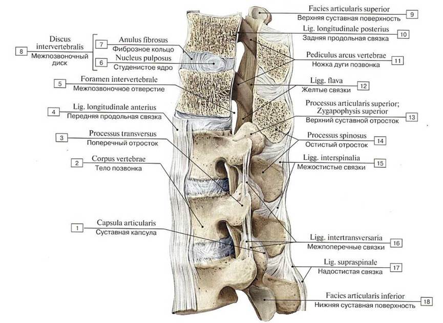
Fig. 128. Intervertebral connections (TXI — LIII), lateral view, left side.
The upper two vertebrae are sectioned in the sagittal plane:
1 — Joint Capsule; Articular capsule; 2 — Vertebral body; 3 — Transverse process; 4 — Anterior longitudinal ligament; 5 — Intervertebral foramen; 6 — Nucleus pulposus; 7 — Anulus fibrosus; 8= 6 + 7 — Intervertebral disc; 9 — Superior articular facet; 10 — Posterior longitudinal ligament; 11 — Pedicle; 12 — Ligamenta flava; 13 — Superior articular process; 14 — Spinous process; 15 — Interspinous ligaments; 16 — Intertransverse ligaments; 17 — Supraspinous ligament; 18 — Inferior articular facet
The Structure of the intervertebral disc is ideally adapted to perform mobility and cushioning functions. Because the discs are elastic, the vertebrae they connect possess a certain degree of mobility.
The connected vertebral bodies are reinforced by strong ligaments. The anterior and posterior longitudinal ligaments, formed by dense regular Connective Tissue, reinforce the joints of the vertebral bodies anteriorly and posteriorly (Figs. 129, 130, Table 27). The anterior longitudinal ligament (ligamentum longitudinale anterius) runs along the anterior surface of the vertebral bodies, firmly fusing with the intervertebral discs all the way from the pharyngeal tubercle of the occipital bone and the anterior tubercle of the anterior arch of the atlas down to the second and third transverse lines of the pelvic surface of the sacrum. Between the atlas and the occipital bone, the anterior longitudinal ligament thickens to form the anterior atlanto-occipital membrane (membrana atlantooccipitalis anterior), which attaches superiorly to the anterior margin of the foramen magnum and inferiorly to the anterior arch of the atlas. From the latter, a strong atlanto-axial membrane extends to the body of the second cervical vertebra. The posterior longitudinal ligament (ligamentum longitudinale posterius) runs along the posterior surface of the vertebral bodies within the vertebral canal. From the inferior margin of the clivus of the occipital bone, it extends across the Articulations of the First and Second cervical vertebrae and further down to the first coccygeal vertebra. The ligaments fuse with the cartilage of the intervertebral discs, but are only loosely connected to the vertebral bodies. At the level of the median atlanto-axial joint, the posterior longitudinal ligament fuses with the bundles of the cruciform ligament of the atlas, which lies anterior to it, and continues superiorly into the tectorial membrane.

Fig. 129. Ligaments of the vertebral column (A — anterior longitudinal ligament, anterior view; B — posterior longitudinal ligament, posterior view, with the vertebral arches, except for the lowest one, sawn through; C — intertransverse and yellow ligaments, anterior view):
1 — Intervcrtebral disc; 2 — Costal process; 3 — Vcrlcbral body; 4 — Anterior longitudinal ligament; 5 — Posterior longitudinai ligament; 6 — Nutrient foramen; 7 — Vertebral arch, pedicle; 8 — Intervcrtebral foramen; 9 — Transverse process; 10 — Inlertransverse ligaments; 11 —Superior articular process; 12 — Vertebral arch, lamina; 13 — Ligamenta flava; 14 — Inferior articular facet; 15 — Spinous process
Table 27. Syndesmoses of the vertebral column
Ligaments |
Connected structures |
Interspinous ligaments |
Spinous processes |
Ligamenta flava |
Vertebral arches |
Intertransverse ligaments (absent in the cervical region) |
Transverse processes |
Supraspinous ligament |
Tips of the spinous processes |
Nuchal ligament |
External occipital crest and spinous processes of the cervical vertebrae |
Anterior longitudinal ligament |
Runs along the anterior surface of the vertebral bodies, firmly fusing with the intervertebral discs |
Posterior longitudinal ligament |
Runs along the posterior surface of the vertebral bodies within the vertebral canal |

Fig. 130. Ligaments of the vertebral column, posterior view, vertebral arches sawn through:
1 — Supraspinous ligament; 2 — Inferior articular process; 3 — Intervertebral foramen; 4 —Transverse process; 5 — Interverlebral disc; 6 — Posterior longitudinal ligament; 7 — Pediclc; 8 — Superior articular facet; Superior articular process; 9 — Intertransverse ligaments; 10 — Zygapophysial joints
Joints of the vertebral arches. The vertebral arches are interconnected by strong ligamenta flava, which occupy the spaces between the vertebral arches (Fig. 131; see also Figs. 128, 129, 130). These ligaments are formed by yellowish elastic connective tissue. Consisting of parallel bundles of elastic fibers interspersed with reticular and collagen microfibrils and a small number of fibrocytes, the ligamenta flava counteract excessive forward flexion of the vertebral column. Their elastic resistance opposes gravity, which tends to tilt the torso anteriorly, and also assists in the extension of the spine.
Joints of the vertebral processes. The articular processes of adjacent vertebrae are connected by flat, multiaxial, slightly movable zygapophysial joints (articulationes zygapophysiales). The facing articular surfaces of corresponding processes, including those of the fifth lumbar and first sacral vertebrae, are covered with articular cartilage (Fig. 131). The articular capsule is attached along the periphery of the articular cartilage and reinforced by thin bundles of connective tissue fibers. These joints allow for flexion and extension of the spine, lateral bending to the right and left, and rotation around the vertical axis.
The spinous processes of the vertebrae are connected by interspinous and supraspinous ligaments. The interspinous ligaments (ligamenta interspinalia), formed by dense regular connective tissue, are very thin in the cervical region of the Vertebral Column and significantly thicker in the lumbar region. The supraspinous ligament (ligamentum supraspinale) is a long fibrous cord attached to the tips of the spinous processes of all vertebrae. The nuchal ligament (ligamentum nuchae) is a strong, triangular, connective-tissue upper portion of the supraspinous ligament, stretched between the external occipital crest and the spinous processes of the cervical vertebrae. In mammals, the nuchal ligament plays an important role in supporting the head; in humans, due to bipedalism, this function is negligible. The transverse processes are connected by intertransverse ligaments (ligamenta intertransversalia), which stretch between the tips of the transverse processes of adjacent vertebrae. These ligaments are absent in the cervical region of the spine.
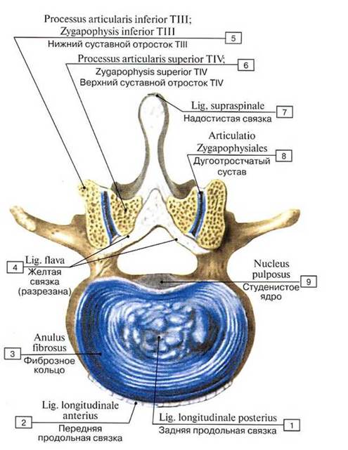
Fig. 131. Intervertebral disc and zygapophysial (intervertebral) joints. Horizontal section between the twelfth thoracic and first lumbar vertebrae, superior view:
1 — Posterior longitudinal ligament; 2 — Anterior longitudinal liga- inent; 3 — Anulus fibrosus; 4 — Ligament flava; 5 — Inferior articular process TIII; 6 — Superior articular process TIV; 7 — Supraspi-notis ligament; 8— Zygapophysial joint; 9— Nucleus pulposus
Joints of the sacrum with the coccyx. The apex of the sacrum is connected to the first coccygeal vertebra via an intervertebral disc and a series of ligaments. The intervertebral disc typically contains a cleft that obliterates in individuals over the age of 50. This joint is reinforced by the paired lateral sacrococcygeal ligament (ligamentum sacrococcygeum laterale), which originates at the inferior margin of the lateral sacral crest and attaches to the vestige of the transverse process of the first coccygeal vertebra. In terms of origin and Location, this ligament is the analogue of the intertransverse ligaments of the vertebral column. The ventral sacrococcygeal ligament (ligamentum sacrococcygeum ventrale), located on the anterior surface of the sacrococcygeal junction, is a continuation of the anterior longitudinal ligament. The superficial posterior sacrococcygeal ligament (ligamentum sacrococcygeum posterius superficiale; ligamentum sacrococcygeum dorsale superficiale), which originates from the margins of the sacral hiatus and attaches to the posterior surface of the coccyx, corresponds to the supraspinous and yellow ligaments. It almost completely closes the opening of the sacral hiatus. The deep posterior sacrococcygeal ligament (ligamentum sacrococcygeum posterius profundum; ligamentum sacrococcygeum dorsale profundum), located on the posterior surfaces of the bodies of the first coccygeal and fifth sacral vertebrae, is analogous to the posterior longitudinal ligament. The sacral and coccygeal cornua are interconnected by syndesmoses. Mobility in the sacrococcygeal joints is well-expressed at a young age, when a slit-like cavity is present, as well as in women (especially during Pregnancy), allowing the coccyx to deflect posteriorly during childbirth.
Connections between the vertebral column and the Skull. The vertebral column connects to the skull via the atlanto-occipital, median, and lateral atlanto-axial joints, which are reinforced by ligaments (Figs. 132, 133, 134, 135, 136, 137).
The paired, combined atlanto-occipital joint (articulatio atlantooccipitalis) is formed by the articular surfaces of the occipital condyles and the superior articular fossae of the atlas. Each joint is surrounded by a broad articular capsule. Both capsules are reinforced by the anterior and posterior atlanto-occipital membranes. The anterior atlanto-occipital membrane (membrana atlantooccipitalis anterior) is stretched between the basilar part of the occipital bone and the upper margin of the anterior arch of the atlas. The posterior atlanto-occipital membrane (membrana atlantooccipitalis posterior) is thin yet broader than the anterior one, stretching between the posterior semicircle of the foramen magnum and the upper margin of the posterior arch of the atlas. The vertebral artery passes through this membrane into the vertebral canal, heading toward the cranial cavity to supply blood to the Brain. Each joint is ellipsoidal (condyloid); movements occur around the frontal and sagittal axes: flexion up to 20°, extension up to 30°, and lateral head tilting up to 15–20°.
The median atlanto-axial joint (articulatio atlantoaxialis mediana) comprises two independent joints formed by the anterior and posterior articular surfaces of the dens of the second cervical vertebra (see Fig. 134). The Formation of the anterior joint involves a facet located on the posterior aspect of the arch of the atlas. The posterior joint is formed by the posterior surface of the dens and a facet on the anterior surface of the transverse ligament of the atlas (ligamentum transversum atlantis). This ligament is stretched posterior to the dens of the axis between the inner surfaces of the lateral masses of the atlas. The anterior and posterior articulations of the dens possess their own joint cavities and enclosing articular capsules. The unpaired, thin apical ligament of the dens (ligamentum apicis dentis) is stretched between the posterior margin of the anterior circumference of the foramen magnum and the apex of the dens. Two strong alar ligaments (ligamenta alaria) restrict excessive right and left head rotation within the median atlanto-axial joint. Each ligament originates on the lateral surface of the dens, runs obliquely upward and outward, and attaches to the inner surface of the corresponding occipital condyle. Within the cylindrical, uniaxial median atlanto-axial joint, the atlas rotates around the dens by 30–40° in each direction around the longitudinal (vertical) axis. The paired, combined, multiaxial lateral atlanto-axial joint (articulatio atlantoaxialis lateralis) is formed by the inferior articular fossae of the atlas and the superior articular surfaces of the axis (see Figs. 135, 136). The right and left joints possess separate articular capsules. The joints are reinforced by the cruciform ligament of the atlas (ligamentum cruciforme atlantis), which consists of the transverse ligament of the atlas and fibrous longitudinal bundles extending upward and downward from it. The superior bundle lies posterior to the apical ligament of the dens and terminates on the anterior semicircle of the foramen magnum. The inferior bundle extends downward to attach to the posterior surface of the body of the axis. The joint has limited mobility, permitting gliding movements with slight displacement of the articular surfaces relative to each other.
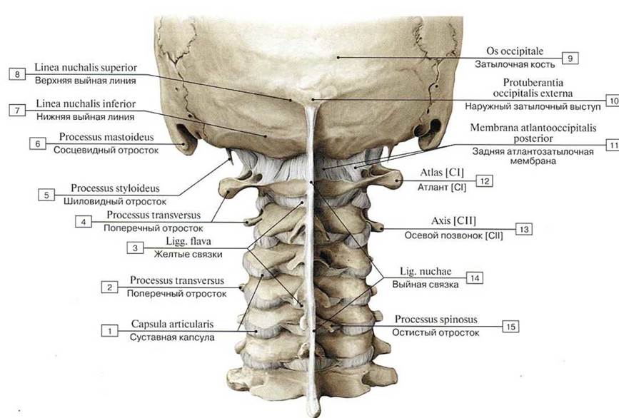
Fig. 132. Atlanto-occipital joint and connection of the cervical vertebrae, posterior view:
1 — Joint capsule; Articular capsule; 2 — Transverse process; 3 — Ligamenta Rava; 4 — Transverse process; 5 — Styloid process; 6 —- Mastoid process; 7— Inferior nuchal line; 8 —Superior nuchal line; 9 — Occipital bone; 10 — External occipital protuberance; 11 — Posterioratlanto-occipital membrane; 12 — Atlas [CI]; 13 — Axis [CII]; 14— Ligamentum nuchae; Nuchal ligament; 15 — Spinous process
Posteriorly, from the side of the vertebral canal, the median and lateral atlanto-axial joints and their ligaments are covered by a strong, broad fibrous sheet—the tectorial membrane (membrana tectoria)—which at the level of the body of the axis continues into the posterior longitudinal ligament, while superiorly it arches over the anterior margin of the foramen magnum and terminates on the inner surface of the basilar part of the occipital bone (in the region of the clivus).
Movements in the right and left lateral atlanto-axial joints occur in conjunction with movements in the median atlanto-axial joint. Simultaneously with head rotation in the median atlanto-axial joint, only a gliding movement occurs, with the articular surfaces shifting relative to one another. During rotation, the dens of the axis is held in position by strong ligaments: the apical ligament of the dens (ligamentum apicis dentis), the alar ligaments (ligamenta alaria), and the cruciform ligament of the atlas (ligamentum cruciforme atlantis).
The joints of the vertebral column are supplied with blood in the cervical region by Branches of the vertebral artery. In the thoracic region, the spine receives branches from the posterior intercostal Arteries, in the lumbar region — from the lumbar arteries, and in the sacral region — from the lateral sacral branches. Venous blood from the spine drains into the vertebral venous plexus, and from there into Veins corresponding to the arteries. The venous outflow is directed, respectively, into the occipital, auricular, deep cervical, intercostal, lumbar, and sacral veins. The Innervation of the joints and ligaments of the vertebral column is provided by the posterior rami of the corresponding spinal nerves.
-

Fig. 133. Connection between The Skull and the vertebral column (A — anterior view, B — posterior view):
1 — Axis [СII]; 2 — Lateral atlanto-axial joint; 3 — Atlas [CI]; 4 — Atlanto-occipital joint; 5 — Occipital bone, lateral part; 6 — Occipital bone, basi lar part; 7 — Anterior atlanto-occipital membrane; 8 — Joint capsule; Articular capsule; 9 — Anterior longitudinal ligament; 10 — Intervertebral disc; 11 — Cervical vertebrae [CIII], vertcbral body; 12 — Axis [CII] vertebral arch; 13 — Atlas [CI], lateral mass; 14 — Occipital bone; 15 — Posterior atlanto-occipital membrane; 16 — Lateral atlanto-occipital ligament; 17 — Lateral atlanto-axial joint;
Joint capsule; Articular capsule; 18 — Posterior tuberclc
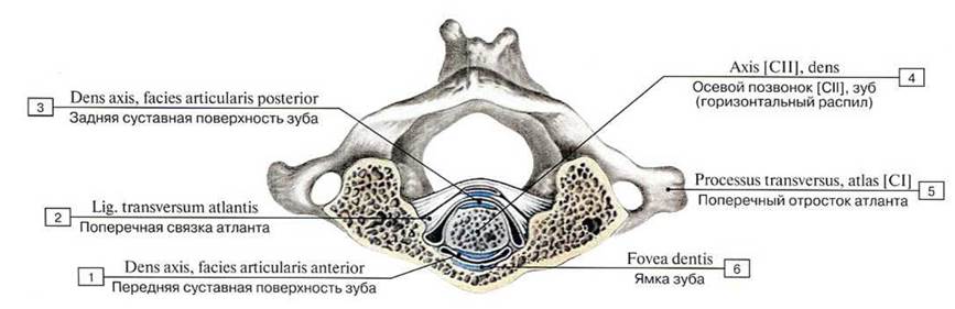
Fig. 134. Median atlanto-axial joint, superior view:
1 — Dens, anterior articular facet; 2 — Transverse ligament of atlas; 3 — Dens, posterior articular facet; 4 — Axis [CII], dens; 5 — Transverse
process atlas [CI]; 6 — Fovea for dens
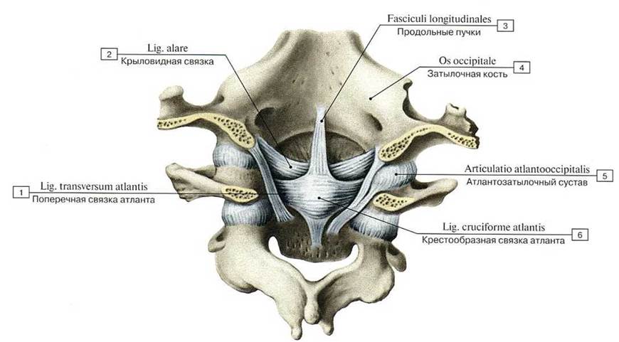
Fig. 135. Ligaments and joints of the cervical vertebrae and occipital bone, posterior view, from the side of the vertebral canal:
1 — Transverse ligameni of atlas; 2 — Alar ligament; 3 — Longitudinal fasciculus; 4 — Occipital bone; 5 — Atlanto-occipital joint; 6 — Caiciate
ligament of atlas
Curves of the vertebral column. The human spine features curves known as physiological curves (see Figs. 18, 30). The anterior convexities of the vertebral column are called lordoses, the posterior convexities — kyphoses, and the lateral convexities to the right and left — scolioses. The cervical lordosis transitions into the thoracic Kyphosis, which is followed by the lumbar lordosis, and then the sacrococcygeal kyphosis. Thoracic kyphosis and lumbar lordosis are more pronounced in women than in men. While physiological lordoses and kyphoses are permanent structures, aortic Scoliosis — observed in 30% of cases at the level of the III–V thoracic vertebrae as a slight rightward convexity — is associated with the position of the Thoracic Aorta at this level. The functional role of these curves is substantial. Thanks to them, shocks and jolts transmitted to the spine during various movements and falls are dampened before reaching the skull, and most importantly, the brain. In the horizontal position of the body, the spinal curves straighten out slightly; in the upright position, they become more pronounced, and with increased load, they increase proportionally to the weight. In the morning after a night's Sleep, the spinal curves are slightly less pronounced and its height is greater. By evening, the curvature increases, while the length of the vertebral column decreases. Human posture affects The Nature and prominence of spinal curves. A dropped head and slouching increase thoracic kyphosis while reducing cervical and lumbar lordoses. In old age, thoracic kyphosis increases (senile hump).
The vertebral column of the human embryo and fetus forms an arch with its convexity facing posteriorly. The spine of a newborn infant lacks curves; they develop gradually in connection with BODY POSITION AND Muscle pull. Cervical lordosis appears when the child begins to hold up their head (around 3 months), thoracic kyphosis when the child begins to sit (around 6 months), and lumbar lordosis when the child begins to stand (9–12 months). At the same time, the body's center of gravity shifts posteriorly. The definitive Development of the curves is completed by 6–7 years of age.

Fig. 136. Atlanto-occipital and atlanto-axial joints, posterior view (A — posterior atlanto-occipital membrane,
B — atlanto-occipital joint, vertebral arches sawed through):
1 — Zygapophysial joints; Joint capsule; Articular capsule; 2 — Posterior atlanto-occipital mcmbrane; 3 — Styloid process; 4 — Mastoid process; 5 — Lateral atlanto-occipital Iigament; 6 — Superior nuchal line; 7— External occipital protuberancc; 8— Ligamentum nuchae; Nuchal ligament; 9 — Foramen magnum; 10 — Occipital bone; 11 — Occipital condyle; 12 —Tectorial membrane; 13 — Atlas [CI]; 14 — Axis [CII]; 15 —Transverse process; 16 —Ligamenta flava; 17 — Spinous process; 18 — Temporal bone; 19 — External occipital crest; 20-Lateral atlanto- occipital joint; 21 — Posterior arch; 22 — Vertebral arch; 23 — Posterior longitudinal ligament
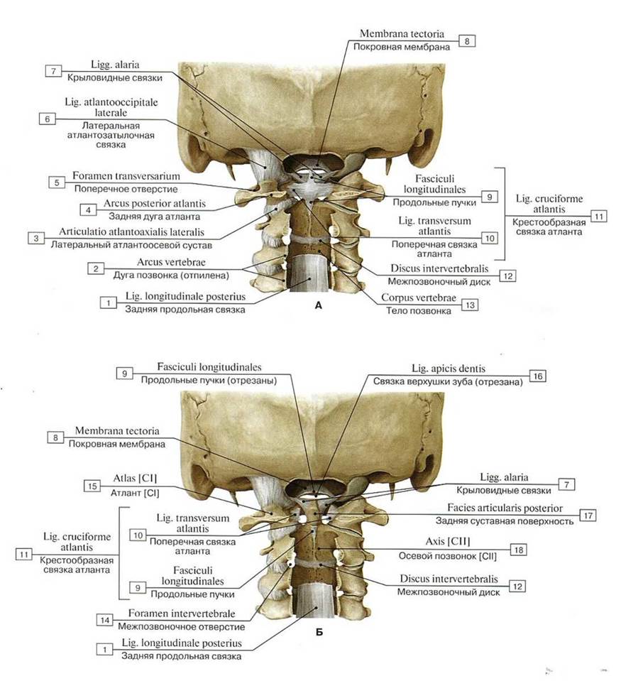
Fig. 137. Atlanto-occipital and atlanto-axial joints, posterior view (A — atlanto-occipital and median atlanto-axial joints, tectorial membrane removed, vertebral arches sawed through, B — atlanto-occipital and median atlanto-axial joints, tectorial membrane and cruciate ligament removed, vertebral arches sawed through):
1 — Posterior longitudinal ligament; 2 — Vertebral arch; 3 — Lateral atlanto-axial joint; 4 — Posterior arch; 5 — Foramen transversarium; 6 — Lateral atlanto-occipital ligament; 7— Alar ligaments; 8 — Tectorial membranc; 9 — Longitudinal fasciculus; 10 — Transverse ligament ofatlas; 11 = 9 + 10— Cruciate ligament ofatlas; 12 — Intervertebral disc; 13 —Vertebralbody; 14 — Intervertebral foramen; 15-Atlas [Cl]; 16 — Apical ligament of dens; 17 — Posterior articular facct; 18 — Axis [СII]
The vertebral column on radiographs. On anteroposterior radiographs, constrictions (the "waist") are visible in the area of the vertebral bodies. The superior and inferior margins of the vertebral bodies have a rounded-corner angular shape. Sacral foramina are visible against the Background of the sacrum, and spaces mark the location of intervertebral discs. The pedicles of the vertebral arches appear as ovals superimposed on the vertebral bodies. The vertebral arches are also superimposed on the images of the vertebral bodies. Spinous processes, lying in the sagittal plane, look like a teardrop against the background of the vertebral bodies. The images of the inferior articular processes are superimposed on the contours of the superior processes. The Head and Neck of the corresponding rib are superimposed on the transverse processes of the thoracic vertebrae. On lateral radiographs, the arches of the first cervical vertebra, the dens of the axis, and the contours of the atlanto-occipital and atlanto-axial joints can be distinguished.
In other sections of the vertebral column, the vertebral arches, spinous and articular processes, articular clefts (spaces), and intervertebral foramina are clearly defined (Fig. 138, see Figs. 30, 31, 32, 33).
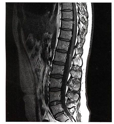
Fig. 138. Sagittal slice of the lumbosacral spine, T2-weighted magnetic Resonance image (MRI):
1 — lumbar vertebra [LV]; 2 — intervertebral disc; 3 — epidural adipose tissue; 4 — anterior longitudinal ligament; 5 — ligamentum flavum; 6 — spinous process; 7 — NERVES OF THE cauda equina; 8 — thoracic vertebra [TXII]; 9 — vertebral veins; 10 — posterior longitudinal ligament (according to S.K. Ternovoy)
Movements of the vertebral column. The human vertebral column exhibits a high degree of mobility (Fig. 139). This is facilitated by resilient, thick intervertebral discs, the STRUCTURE OF THE vertebrae, their articular processes, ligamentous apparatus, and muscles. Individual movements of small amplitude between adjacent vertebrae summate, allowing the spinal column to perform extensive movements that occur around three axes:
1) around the transverse (frontal) axis, flexion (bending forward) and extension (bending backward) of the spine take place. The range of these movements reaches 170–245°. During trunk flexion, the vertebral bodies tilt forward, and the spinous processes move apart. The anterior longitudinal ligament of the spine relaxes, while the posterior longitudinal, ligamenta flava, interspinous, and supraspinous ligaments become taut, resisting this movement. During spinal extension, all ligaments except the anterior longitudinal relax; the anterior longitudinal ligament tightens, limiting extension. The thickness of the intervertebral discs decreases on the side of spinal inclination during flexion and extension, and increases on the opposite side;
2) around the sagittal axis, lateral flexion to the right and left occurs, with a total range of motion of about 165°. These movements take place primarily in the lumbar region. In this case, the ligamenta flava, intertransverse ligaments, and the capsules of the zygapophyseal joints located on the opposite side become taut, which restricts the movement;
3) around the longitudinal (vertical) axis, rotational movements (rotation) occur, with a total range of about 120°. During rotation, the nucleus pulposus of the intervertebral discs acts akin to a joint head, while the annulus fibrosus of the intervertebral discs and the ligamenta flava become taut, limiting this movement;
4) circumduction around the longitudinal (vertical) axis. The fulcrum is located at the level of the Lumbosacral junction; the upper end of the vertebral column moves freely in space, describing a circle, while the entire vertebral column traces a cone.
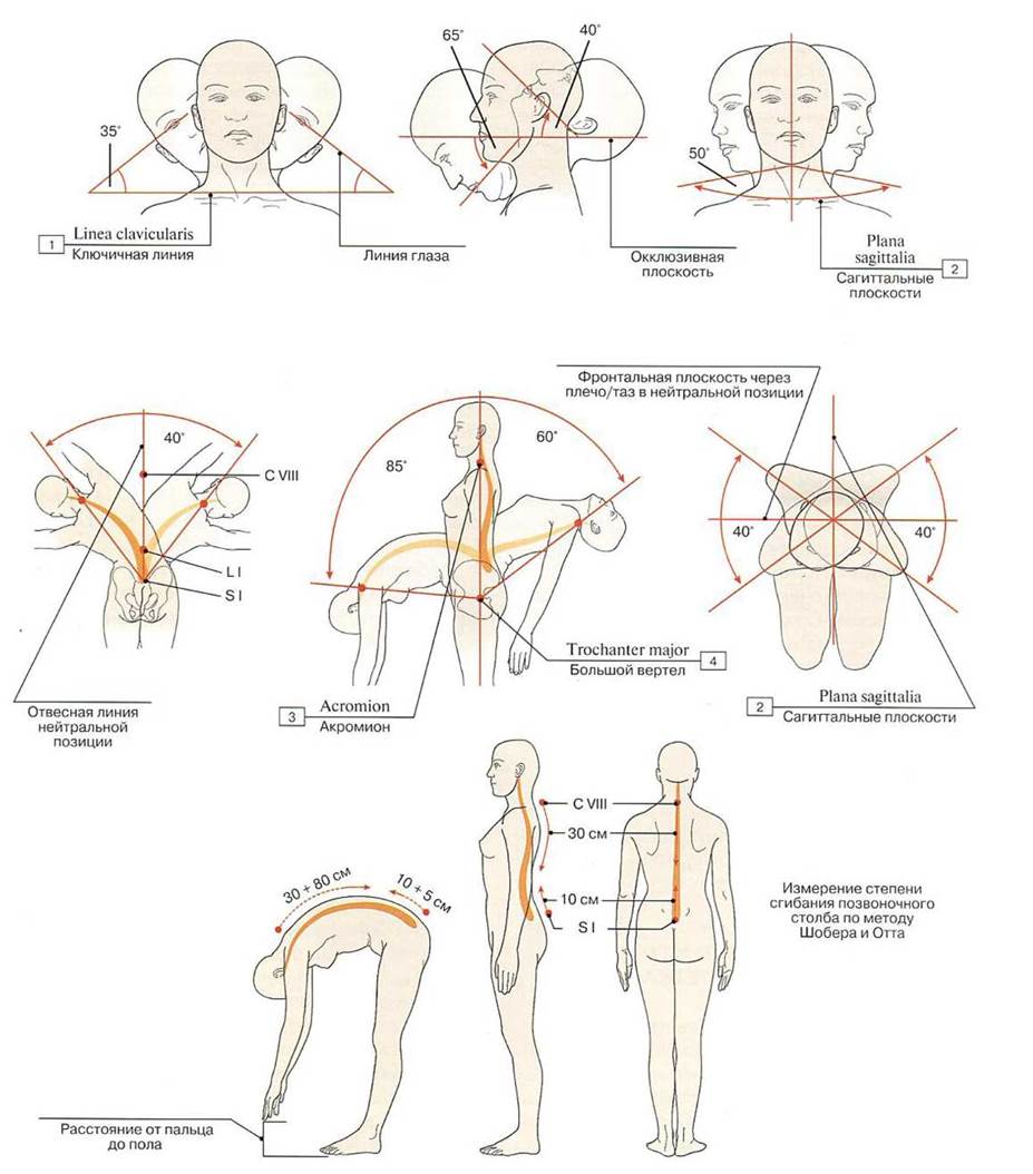
Fig. 139. Range of motion of the vertebral column:
1 — Clavicular line; 2 — Sagittal planes; 3 — Acromion; 4 — Greater trochanter
The range and direction of movements in each region of the vertebral column (cervical, thoracic, lumbar) vary. The range of motion is greatest in the cervical and lumbar regions. In the cervical spine, the range of motion is 70–75° for flexion, 95–105° for extension, and 80–85° for rotation. In the thoracic region, mobility is restricted by the Ribs AND Sternum, the thinness of the intervertebral discs, and the spinous processes, which are directed obliquely downward. Flexion, extension, and lateral bending here are limited: flexion up to 35°, extension up to 50°, and rotation up to 20°. In the lumbar region, thick intervertebral discs promote greater mobility (flexion up to 60°, extension up to 45–50°). The structure of the articular processes of the lumbar vertebrae hinders rotation and lateral movements.
Throughout all spinal regions, maximum mobility is attained by the end of adolescence. The greatest range of motion is observed in the cervical region, and the least in the lower thoracic region. After 50–60 years of age, the mobility of the vertebral column declines. Spinal mobility depends primarily on the structure of the intervertebral discs. With age, the number and thickness of collagen fiber bundles within the annuli fibrosus increase, their architecture is disrupted, the bundles become deformed, and many collagen fibers undergo destruction and hyalinization. Concurrently, elastic fibers also change: they thicken, become tortuous, and fragment. Furthermore, starting from the age of five to six years, chondrocytes appear in the nucleus pulposus and collagen fibers begin to form, gradually increasing in number.
Last update: 08/08/2026
Editorial and Educational Adaptation: This material has been compiled based on the primary/original source text. The project team performed an editorial review, corrected technical inaccuracies, structured sections, and adapted the content for an educational format.
What was processed:
- elimination of formatting defects (OCR errors, structural breaks, corrupted characters);
- editorial organization of content;
- standardization of terminology in accordance with academic sources;
- verification of factual statements against the original source text.
All mentions of the author, publication year, and origin of the primary text have been preserved in accordance with the source.