MEDICAL BIOLOGY, HUMAN ANATOMY, PHYSIOLOGY AND PATHOLOGY - Ya.I. Fedoniuk 2010
ANATOMY, PHYSIOLOGY, PATHOLOGY
CHAPTER 2. MUSCULOSKELETAL SYSTEM (THE MECHANISM OF MOVEMENT)
THE TRUNK SKELETON
1. VERTEBRAL COLUMN
1.2. TYPES OF JOINTS BETWEEN VERTEBRAE
All types of joints are present between the vertebrae, namely (Fig. 2.37): syndesmoses, such as the anterior and posterior longitudinal ligaments, as well as the ligamentum flavum between the vertebral arches (synelastosis); synchondroses, represented by the intervertebral discs between the vertebral bodies; synostoses, which are the sacral vertebrae fused into a single sacral bone; and diarthroses (synovial joints), formed by the Articulation of the superior articular processes of the inferior vertebra with the inferior articular processes of the superior vertebra. Structurally, these joints are combined, flat in shape, and functionally multiaxial, yet exhibiting limited mobility.
Class="center">
FIG. 2.37. Intervertebral connections (lumbar region, part of the vertebral canal opened):
The bodies of two adjacent vertebrae are connected by means of intervertebral discs. Each disc has the shape of a biconvex lens, the peripheral part of which is formed by fibrocartilage; its fibers constitute the anulus fibrosus, while the central part consists of an elastic substance known as The Nucleus pulposus (Fig. 2.38). The bodies of adjacent vertebrae are connected to each other by the anulus fibrosus. The nucleus pulposus, located inside the anulus fibrosus and compressed by the adjacent vertebral bodies, acts as a Shock absorber.

Fig. 2.38. Intervertebral Cartilage
Along THE Vertebral Column, the vertebral bodies and intervertebral discs are connected by the anterior and posterior longitudinal ligaments. Short ligaments connect the transverse processes (intertransverse ligaments) and spinous processes (interspinous ligaments). Running along the tips of the spinous processes is the long supraspinous ligament, which in the cervical region transitions into the nuchal ligament (which is well developed in ungulates) (Fig. 2.39).

Fig. 2.39. Ligaments of the thoracic vertebral column (posterior ends of the Ribs preserved); posterior view.
Between the first cervical vertebra (atlas) and the Occipital bone, the right and left atlanto-occipital joints are formed; these are structurally combined, ellipsoidal in shape, and functionally Biaxial joints (Fig. 2.40). Movements possible in these joints occur around the frontal axis (flexion and extension of the HEAD) and the sagittal axis (abduction and adduction). Rotational Movements of the head to the right and left take place in the cylindrical atlanto-axial joint. In this movement, the atlas rotates around the dens of the second cervical vertebra along with the cranium.
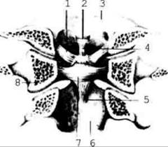
Fig. 2.40. Ligaments of the median atlanto-axial joint; posterior view (frontal section at the level of the lateral masses of the atlas), semi-schematic
Last update: 08/08/2026
Editorial and Educational Adaptation: This material has been compiled based on the primary/original source text. The project team performed an editorial review, corrected technical inaccuracies, structured sections, and adapted the content for an educational format.
What was processed:
- elimination of formatting defects (OCR errors, structural breaks, corrupted characters);
- editorial organization of content;
- standardization of terminology in accordance with academic sources;
- verification of factual statements against the original source text.
All mentions of the author, publication year, and origin of the primary text have been preserved in accordance with the source.