MEDICAL BIOLOGY, ANATOMY, HUMAN PHYSIOLOGY AND PATHOLOGY - Ya.I. Fedoniuk 2010
ANATOMY, PHYSIOLOGY, PATHOLOGY
CHAPTER 2. THE MUSCULOSKELETAL SYSTEM (THE MECHANISM OF MOVEMENT)
THE AXIAL SKELETON
1. THE VERTEBRAL COLUMN
1.1. Structure OF THE VERTEBRAE
All vertebrae, with the exception of the first (I) and second (II) cervical vertebrae, share a common structure: each vertebra consists of an anteriorly positioned body from which an arch extends (Fig. 2.32). The body and the arch enclose the vertebral foramen. When stacked upon one another, these foramina form the vertebral canal, which houses the Spinal Cord. The canal communicates with the cranial cavity via the foramen magnum in the Occipital bone. The arch of each vertebra is attached to the body by pedicles, which feature superior and inferior notches. The latter form the intervertebral foramina through which the Spinal Nerves emerge. Processes project from the arch of each vertebra: superior and inferior articular processes (paired) extending upward and downward, a spinous process (unpaired) projecting posteriorly, and transverse processes (paired) extending laterally.
Class="center">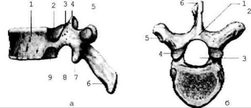
Fig. 2.32. Thoracic vertebra
Cervical vertebrae (vertebrae cervicales). A characteristic feature of the cervical vertebrae is the presence of a foramen in the transverse processes, while the spinous process is bifid (except for CVII) (Fig. 2.33). The first cervical vertebra is called the atlas (atlant); it lacks a body and a spinous process, consisting only of two arches (anterior and posterior) and lateral masses that bear articular facets (superior and inferior) for articulation with the occipital bone and the second cervical vertebra (Fig. 2.34). The second cervical vertebra (axis) does possess a body, which features the odontoid process, or dens (projecting upward), for articulation with the anterior arch of the atlas (Fig. 2.35).
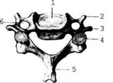
Fig. 2.33. Cervical vertebra
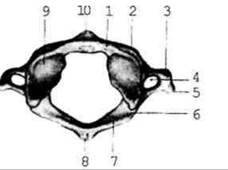
Fig. 2.34. First cervical vertebra - atlas
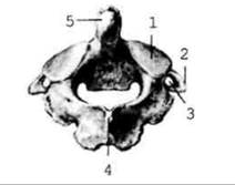
Fig. 2.35. Second cervical vertebra
The seventh cervical vertebra (vertebra prominens) features a long, non-bifid spinous process, which serves as an anatomical landmark for counting vertebrae during lumbar puncture to obtain CEREBROSPINAL FLUID, as well as for subarachnoid anesthesia.
Thoracic vertebrae (vertebrae thoracicae). A characteristic feature of the thoracic vertebrae is the presence of articular facets on the lateral surfaces of the body and the transverse processes for articulation with the HEAD and tubercle of a rib.
Lumbar vertebrae (vertebrae lumbales). The lumbar vertebrae have massive bodies because they bear significant weight loads and feature additional processes.
Sacral vertebrae (vertebrae sacrales) are classified as false vertebrae because they fuse into a single sacral bone. It presents: an anterior (pelvic) surface, a posterior (dorsal) surface, a base (directed superiorly), and an apex (directed inferiorly) (Fig. 2.36). The posterior surface bears five crests: a median crest, and paired medial and lateral crests. These are formed by the fusion of the spinous, transverse, and articular processes. On the pelvic surface, the upper margin of the body of the first sacral vertebra and the lower margin of the body of the fifth lumbar vertebra form a prominent projection known as the sacral promontory, which is used by obstetricians to measure pelvic dimensions. A canal runs through the interior of the sacrum, formed by the fusion of the vertebral foramina of the sacral segments. The lateral PARTS OF THE sacrum feature an auricular surface for articulation with the hip bone.
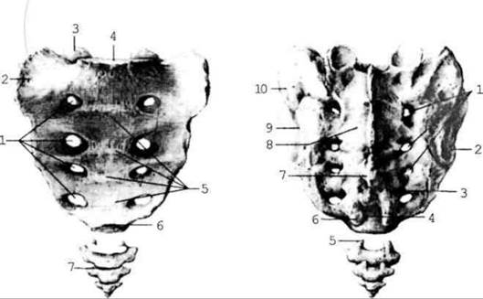
Fig. 2.36. Sacrum and coccyx
Coccygeal vertebrae (vertebrae coccygeae): the human coccygeal region corresponds to the tail of vertebrate animals. The coccygeal vertebrae also fuse into a single bone—the coccyx. Occasionally, children are born with a tail-like appendage.
Last update: 08/08/2026
Editorial and Educational Adaptation: This material has been compiled based on the primary/original source text. The project team performed an editorial review, corrected technical inaccuracies, structured sections, and adapted the content for an educational format.
What was processed:
- elimination of formatting defects (OCR errors, structural breaks, corrupted characters);
- editorial organization of content;
- standardization of terminology in accordance with academic sources;
- verification of factual statements against the original source text.
All mentions of the author, publication year, and origin of the primary text have been preserved in accordance with the source.