HUMAN ANATOMY ATLAS - G.L. Bilich - 2014
THE LOCOMOTOR SYSTEM
THE SKELETAL SYSTEM
RIBS AND STERNUM
The bones of The thoracic cage consist of the Sternum and 12 pairs of Ribs, which articulate posteriorly with THE Vertebral Column (Figs. 36, 37, 38).
The sternum (sternum) is a flat bone situated in the frontal plane, comprising three parts. Its upper part is the manubrium of the sternum, the middle part is the body, and the lower part is the xiphoid process. In adults, these three parts fuse into a single bone. The manubrium of the sternum (manubrium sterni) is broad and thick, featuring the jugular notch (incisura jugularis) on its upper margin. Flanking this notch are the clavicular notches (incisurae claviculares) for articulation with the clavicles. On the right and left margins of the manubrium, inferior to the clavicular notch, lie depressions for the Cartilage of the 1st rib. Further inferiorly is a hemifacet that, upon uniting with the corresponding hemifacet on the body of the sternum, forms a complete costal notch for articulation with the cartilage of the 2nd rib. At the junction of the manubrium and the sternal body, a slight anteriorly facing sternal angle (angulus sterni) is formed; it corresponds to the level of the second ribs and serves as a key landmark during clinical examinations of the thoracic cavity Organs. The body of the sternum (corpus sterni) is wider in its middle and lower sections than superiorly. The anterior surface of the body exhibits transverse lines (the sites of fusion of the bone segments), while its margins bear costal notches (incisurae costales) for articulation with the cartilages of the true ribs. The costal notch for the VII rib is located at the junction between the sternal body and the xiphoid process. The xiphoid process (processus xiphoideus) varies in shape; it is occasionally bifurcated inferiorly or possesses an aperture formed during The Development of the process from two ossification centers (see Fig. 37).
The ribs (costae) are curved bony plates that transition anteriorly into cartilaginous parts (Fig. 39). The osseous portion of a rib—the rib bone (os costale)—is longer, whereas the anterior cartilaginous portion—the costal cartilage (cartilago costalis)—is short. The upper seven pairs of ribs (I–VII) connect via their cartilaginous parts directly to the sternum; these are termed true ribs (costae verae). The cartilages of the VIII, IX, and X rib pairs do not attach to the sternum but rather to the cartilage of the rib immediately superior to them, and are therefore designated as false ribs (costae spuriae). The XI and XII ribs feature short cartilaginous parts that terminate within the musculature of the anterior abdominal wall. Exhibiting greater mobility than the others, these ribs are referred to as floating ribs (costae fluctuantes).
At the posterior extremity of each rib lies the HEAD (caput costae), which articulates with the costal facets on the body of one or two adjacent thoracic vertebrae. Because ribs II through X articulate via their heads with two neighboring vertebrae, their heads feature a crest of the head of the rib (crista capitis costae) that divides the head into two articular facets. A ligament attaches to this crest, securing the rib head to the corresponding vertebra. However, ribs I, XI, and XII lack these crests, as their heads articulate exclusively with a complete facet on the body of their corresponding vertebra. The head of the rib tapers into a narrower segment—the neck of the rib (collum costae). At the boundary between the neck and the shaft lies the tubercle (tuberculum costae). On the tubercle of each of the 10 upper ribs is the articular facet of the rib tubercle (facies articularis tuberculi costae) for articulation with the transverse costal facet of the corresponding vertebra. Just superior to this articular facet lies the attachment site for the costotransverse ligament; ribs XI and XII lack an articular facet for the transverse process, and their tubercles are poorly developed or entirely absent. Extending beyond the tubercle is the wider and longest anterior portion of the rib bone—the body of the rib (corpus costae)—which is slightly twisted around its own longitudinal axis and bends sharply forward a short distance from the tubercle. This region is designated as the angle of the rib (angulus costae). The body of the rib is flat, presenting external and internal surfaces, as well as superior and inferior margins. The internal surface is smooth, and along the entire length of the inferior margin runs the costal groove (sulcus costae), which lodges the intercostal Vessels and nerves. The anterior, thickened end of the rib body features a depression for articulation with the costal cartilage. Unlike the others, the first rib presents superior and inferior surfaces, as well as medial and lateral margins. Its superior surface bears the scalene tubercle (tuberculum musculi scaleni anterioris) for the attachment of the anterior scalene Muscle. Posterior to this tubercle runs the groove for the Subclavian Artery (sulcus arteriae subclaviae), while anterior to it lies the groove for the subclavian vein (sulcus venae subclaviae). The angle of the 1st rib coincides with its tubercle.
Class="center">
Fig. 36. Skeleton of the thoracic cage (A — anterior view, B — posterior view):
1 — Superior thoracic aperture; Thoracic inlet; 2 — Jugular notch; Suprasternal notch; 3 — Clavicular notch; 4 — Manubrium of sternum; 5 — Sternal angle; 6 — Body of sternum; 7 — Xiphoid process; 8 — Sternum; 9 — Costal cartilage; 10 — Costal margin; Costal arch; 11 — Inferior thoracic aperture; Thoracic outlet; 12 — Spinous process; 13 — Tubercle; 14 — Angle of rib; 15 — Rib [XII]

Fig. 37. Sternum (A — anterior view, B — lateral view, right):
1 — Jugular notch; Suprasternal notch; 2 — Clavicular notch; 3 — Manubrium of sternum; 4 — Sternal angle; 5 — Body of sternum; 6 — Xiphoid process; 7 — Sternum; 8 — Costal notch [I]; 9 — Costal notches [II–VII]

Fig. 38. Chest X-ray, posteroanterior projection:
1 — clavicle; 2 — scapula; 3 — anterior part of rib; 4 — posterior part of rib; 5 — ROOT of the right lung; 6 — root of the left lung; 7 — cardiac silhouette; 8 — aortic arch; 9 — bifurcation of the Trachea; 10 — trachea; 11 — right costodiaphragmatic recess; 12 — costomediastinal recess; 13 — left dome of the Diaphragm; 14 — right dome of the diaphragm; 15 — gastric air bubble (after A.Yu. Vasiliev)
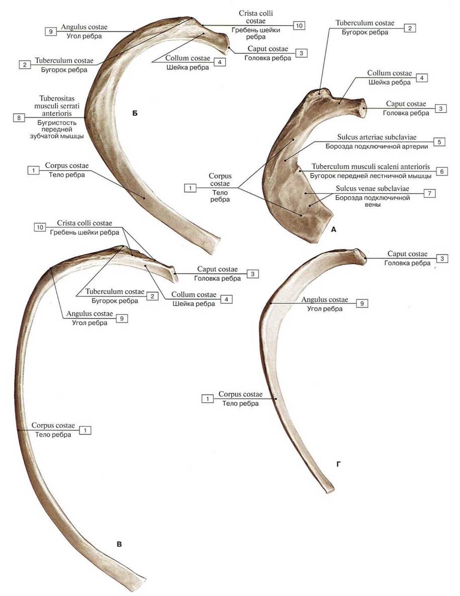
Fig. 39. Ribs (A — first [I] rib, right, superior view; B — second [II] rib, right, superior view; C — fifth rib [V], right; D — eleventh rib [XI], right):
1 — Body of rib; Shaft of rib; 2 — Tubercle; 3 — Head of rib; 4 — Neck of rib; 5 — Groove for subclavian artery; 6 — Scalene tubercle; 7 — Groove for subclavian vein; 8 — Tuberosity for serratus anterior; 9 — Angle of rib; 10 — Crest of neck of rib
Last update: 08/08/2026
Editorial and Educational Adaptation: This material has been compiled based on the primary/original source text. The project team performed an editorial review, corrected technical inaccuracies, structured sections, and adapted the content for an educational format.
What was processed:
- elimination of formatting defects (OCR errors, structural breaks, corrupted characters);
- editorial organization of content;
- standardization of terminology in accordance with academic sources;
- verification of factual statements against the original source text.
All mentions of the author, publication year, and origin of the primary text have been preserved in accordance with the source.