ATLAS OF HUMAN ANATOMY - G.L. Bilich - 2014
Articular System
JOINTS OF THE UPPER LIMB
The BONES OF THE Pectoral Girdle are connected to each other and to the Sternum through various TYPES OF JOINTS, which provide high mobility for the shoulder, forearm, and hand (Table 30, Fig. 146).
Five joints in the shoulder region ensure a wide range of motion for the upper limb at the shoulder joint. There are three true shoulder joints and two functional ones.
✵ True joints:
1. Sternoclavicular joint;
2. Acromioclavicular joint;
3. Shoulder joint (glenohumeral joint).
✵ Functional joints:
4. Subacromial space, located adjacent to synovial bursae (subacromial bursa and subdeltoid bursa). It allows for gliding between the acromion and the rotator cuff (the muscular cuff of the shoulder joint, consisting of the supraspinatus, infraspinatus, subscapularis, and teres minor Muscles, which press the HEAD of the humerus against the glenoid cavity of the scapula).
5. Scapulothoracic joint — Connective Tissue located between the subscapularis and serratus anterior muscles, facilitating the gliding of the scapula along the chest wall.
In addition to true and functional joints, two more ligaments contribute to the mobility of the upper limb: the costoclavicular ligament (between the clavicle and the first rib) and the coracoclavicular ligament (between the clavicle and the coracoid process). However, the high mobility of the shoulder is achieved at the expense of its stability. Since the shoulder joint has a loose capsule and weak reinforcing ligaments, the stabilization function falls upon the tendons of the rotator muscles. Evolutionary Changes in the upper limb, transforming it from a support organ into a manipulating organ, have made the pathology of periarticular soft Tissues a highly relevant issue. In a large percentage of shoulder injuries, periarticular soft tissues are involved in the pathological process.
The sternoclavicular joint (articulatio sternoclavicularis) is a flat, complex, multiaxial joint formed by the articular surface of the sternal end of the clavicle and the clavicular notch of the manubrium of the sternum (Fig. 147-A). A thin capsule attaches along the margins of the articular surfaces. The articular disc (discus articularis), which fuses with the capsule, divides the joint cavity into two compartments. The joint is reinforced by several ligaments. The anterior and posterior sternoclavicular ligaments (ligamenta sternoclavicularia anterius et posterius) blend into the periosteum of the sternum. The interclavicular ligament (ligamentum interclaviculare) connects the sternal ends of the right and left clavicles. The short, broad costoclavicular ligament (ligamentum costoclaviculare) connects the Inferior surface of the sternal end of the clavicle to the Cartilage and bony part of the I rib.
The acromioclavicular joint (articulatio acromioclavicularis) is a flat, multiaxial joint formed by the articular surface of the acromion and the clavicular articular surface of the clavicle (Fig. 147-B). In 1/3 of cases, an articular disc—the cartilage of Vater-Baudhin (Josias Weitbrecht [Vaitbrekht] (1702–1747), a native anatomist and student of D. Bernoulli)—is located between these articular surfaces. A thin Joint Capsule attaches along the margins of the articular surfaces. The joint is reinforced by the acromioclavicular ligament (ligamentum acromioclavicular), which connects the tip of the acromion and the acromial end of the clavicle, and the robust coracoclavicular ligament (ligamentum coracoclaviculare)—Caldani's ligament (Leopoldo Marco Antonio Caldani (1725–1813), an Italian anatomist and physician). The coracoclavicular ligament consists of two bundles of fibers originating at the Base of the coracoid process of the scapula and attaching to the conoid tubercle and trapezoid line on the inferior surface of the acromial end of the clavicle. More medially lies the trapezoid ligament (ligamentum trapezoideum), and lateral to it is the conoid ligament (ligamentum conoideum).
Class="center">Table 30. JOINTS OF THE Upper Limb
Joint Name |
Articular Surfaces |
Articular Ligaments |
Type of Movement Axis |
Function and Muscles Acting on the Joint |
|
Sternoclavicular |
Articular surface of the sternal end of the clavicle, clavicular notch of the manubrium of the sternum (articular disc present) |
Anterior and posterior sternoclavicular ligaments, costoclavicular ligament |
Flat complex Multiaxial (sagittal, longitudinal, frontal axes) |
Elevation of the scapula and clavicle; muscles: levator scapulae, rhomboid Major and minor, sternocleidomastoid, trapezius (upper fibers). Depression of the scapula and clavicle; muscles: trapezius (lower fibers), serratus anterior (lower fibers), pectoralis minor, subclavius. Forward and lateral movement; muscles: serratus anterior, pectoralis minor |
|
Acromioclavicular |
Articular surface of the acromion, acromial articular surface of the clavicle |
Acromioclavicular ligament, coracoclavicular ligament |
Flat multiaxial (sagittal, longitudinal, frontal axes), slightly movable |
Backward and medial movement of the scapula (toward the spine); muscles: trapezius, rhomboid major and minor. Rotation of the scapula around the sagittal axis: turning the inferior angle outward; muscles: serratus anterior (lower digitations), trapezius (upper fibers). Turning the scapula with the inferior angle medially (toward the spine); muscles: rhomboid, pectoralis minor |
|
Shoulder (Glenohumeral) |
Head of the humerus, flattened glenoid cavity of the scapula (features a glenoid labrum) |
Coracohumeral ligament |
Ball-and-socket multiaxial (sagittal, longitudinal, frontal axes) |
Flexion of the arm; muscles: deltoid (anterior fibers), pectoralis major (clavicular part), coracobrachialis, biceps brachii. Extension of the arm (range: flexion-extension — 120°); muscles: deltoid (posterior fibers), latissimus dorsi, teres major, long head of the triceps brachii, infraspinatus. Abduction of the arm; muscles: deltoid (acromial part), supraspinatus, biceps brachii (long head), deltoid (clavicular part and fibers originating from the scapular spine when abducted beyond 60°). Adduction of the arm (abduction-adduction — 100°); muscles: pectoralis major, coracobrachialis, latissimus dorsi, infraspinatus, teres major and minor, deltoid (clavicular part and fibers from the scapular spine when the arm is abducted less than 60°). Medial (internal) rotation; muscles: deltoid (clavicular part), pectoralis major, subscapularis, latissimus dorsi, teres major. Lateral (external) rotation (rotation around the longitudinal axis — 135°); muscles: deltoid (posterior fibers), infraspinatus, teres minor. All the aforementioned muscles participate in circular movements |
|
Elbov (consists of three joints) |
Ulnar and radial collateral ligaments, anular ligament |
Compound biaxial (frontal and longitudinal axes) |
Flexion and extension of the forearm (170°). During flexion, the forearm deviates slightly medially, and the hand rests on the chest. At maximum extension, the olecranon abuts against the olecranon fossa of the humerus, bringing the arm and forearm almost into a straight line. Flexion; muscles: biceps brachii, brachialis, brachioradialis, flexor carpi radialis, palmaris longus, extensor carpi radialis longus, extensor carpi radialis brevis. Extension; muscles: triceps brachii, anconeus |
||
Humeroulnar |
Trochlea of the humerus, trochlear notch of the ulna |
Hinged (ginglymus / trochlear) uniaxial |
Flexion and extension of the forearm |
||
Humeroradial |
Capitulum of the humerus, articular facet of the head of the radius |
Multiaxial ball-and-socket (longitudinal, frontal axes) |
Rotation of the radius around the longitudinal axis — pronation, supination, flexion |
||
Proximal radioulnar |
Articular circumference of the radius, radial notch of the ulna |
Anular ligament |
Trochoid (pivot) uniaxial (longitudinal axis of the forearm) |
Rotation of the radius around the ulna — pronation and supination. In this process, the proximal epiphysis of the radius rotates in place because its head is held by the anular ligament of the radius and the radial notch, while the distal epiphysis glides along the articular circumference of the ulna, describing an arc around the stationary head of the ulna. The maximum range of motion of the forearm in the radioulnar joints (supination and pronation) reaches 180° |
|
Distal radioulnar |
Articular circumference of the ulna, ulnar notch of the radius |
PALMAR AND DORSAL intercarpal ligaments, interosseous membrane |
Trochoid (rotational) uniaxial |
Supination; muscles: supinator, biceps brachii, brachioradialis, extensor indicis, extensor pollicis longus and brevis, abductor pollicis longus (with a semi-flexed elbow joint), biceps and brachioradialis. Pronation; muscles: pronator teres and quadratus, flexor carpi radialis, palmaris longus, extensor carpi radialis longus (Displacement of the radius around the ulna is about 150°) |
|
Radiocarpal |
Carpal articular surface of the radius, proximal surface of the 1st row of Carpal Bones (an articular DISC is present on the medial side) |
Radial collateral ligament of the wrist, ulnar collateral ligament, palmar radiocarpal ligament, dorsal radiocarpal ligament |
Ellipsoid, complex, compound biaxial (sagittal, frontal) |
Flexion of the hand (about 80°); muscles: flexor carpi radialis and ulnaris, palmaris longus, flexor digitorum superficialis and profundus (especially with extended fingers). Extension of the hand (about 70°); muscles: extensor carpi ulnaris, extensor carpi radialis longus and brevis, extensors of the fingers (especially with flexed fingers). Adduction (ulnar deviation) of the hand (about 40°); muscles: flexor carpi ulnaris and extensor carpi ulnaris. Abduction (radial deviation) of the hand (about 15°); muscles: flexor carpi radialis and extensor carpi radialis longus and brevis. Circular movement of the hand is possible—performed sequentially by the muscles that flex and extend the hand (about 150°) |
|
Midcarpal |
Bones of the First and Second rows of the carpus (except the pisiform) |
Radiate carpal ligament, palmar intercarpal ligaments, dorsal intercarpal ligaments, interosseous intercarpal interarticular ligaments |
Hinged compound uniaxial (frontal axis) |
Functionally connected with the radiocarpal joint. Participates in flexion and extension of the hand |
|
Intercarpal |
Opposing surfaces of the carpal bones |
Pisohamate and pisometacarpal |
Flat, slightly movable |
||
Carpometacarpal II–V |
Distal articular surfaces of the second row of carpal bones, articular surfaces of the bases of the II–V Metacarpal bones |
Dorsal and palmar carpometacarpal ligaments, pisometacarpal ligament |
Gliding by 5–10° |
||
Carpometacarpal of the thumb |
Articular surfaces of the trapezium and the base of the I metacarpal bone |
Dorsal and palmar carpometacarpal ligaments of the thumb |
Saddle biaxial (frontal, sagittal axis) |
Muscles perform movements in this joint, as well as in the metacarpophalangeal and interphalangeal joints of the thumb. Flexion; muscles: flexor pollicis longus and brevis. Extension; muscles: extensor pollicis longus and brevis. Abduction; muscles: abductor pollicis longus and brevis. Adduction; Muscle: adductor pollicis. Opposition; muscle: opponens pollicis |
|
Intermetacarpal |
Adjacent surfaces of the bases of the II–V metacarpal bones |
Dorsal and palmar metacarpal ligaments, interosseous metacarpal ligaments |
Multiaxial flat, slightly movable |
The range of motion simultaneously in the radiocarpal and midcarpal joints during flexion is 75–80°, during extension—about 45°, during abduction—15–20°, during adduction—30–40°. Circular motion in these joints is the result of compounding sequential movements around the sagittal and frontal axes, with the fingertips describing a circle |
|
Metacarpophalangeal |
Articular surfaces of the heads of the metacarpal bones and the bases of the proximal Phalanges |
Collateral, palmar, and deep transverse metacarpal ligaments |
Approaching ellipsoid, biaxial (frontal, sagittal axes) |
Flexion and extension of the finger (about 90°), abduction and adduction of the finger (45°–50°); circular movements are possible. Flexion; muscles: flexor digitorum superficialis and profundus, flexor digiti minimi brevis, lumbricals. Extension; muscles: extensor digitorum, extensors of the II and V fingers (intrinsic). Abduction; muscles: dorsal interosseous, abductor digiti minimi |
|
Interphalangeal |
Articular surfaces of the heads and bases of adjacent phalanges |
Collateral and palmar ligaments |
Hinged uniaxial (frontal axis) |
Flexion and extension of the phalanges (about 90°). Adduction; palmar interosseous muscles |
|

Fig. 146. Articulation of the right shoulder and pectoral girdle bones:
1 — Humerus, 2 — Coracoid process; 3 — Head of humerus; 4 — Acromion; 5 — Clavicle; 6 — First rib [I]; 7 — Manubrium of sternum; 8 — Scapula, costal surface; 9 — Ribs

Fig. 147. Selected joints of the upper limb (A — sternoclavicular joints, anterior view, capsule opened on the left; B — acromioclavicular and shoulder joints, right, anterior view):
1 — Intra-articular sternocostal ligament; 2 — Costal cartilage; 3 — Clavicle; 4 — Anterior sternoclavicular ligament; 5 — Interclavicular ligament; 6 — Articular disc; 7 — Coracoclavicular ligament; 8 — First rib [I]; 9 — Manubrium of sternum; 10 — Sternocostal joint; 11 — Humerus; 12 — Intertubercular sulcus; Bicipital groove: 13 — Lesser tubercle; 14 — Greater tubercle; 15 — Head of humerus; 16 — Coracoid process; 17 — Coraco-acromial ligament; 18 — Acromion; 19 — Acromioclavicular ligament; 20 — Acromial end; 21 — Trapezoid ligament; 22 — Conoid ligament; 23 = 20 + 21 + 22 — Costoclavicular ligament; 24 — Sternal end; 25 — Superior angle; 26 — Superior transverse scapular ligament; 27 — Suprascapular notch; 28 — Glenoid cavity; 29 — Scapula, costal surface; 30 — Medial border
Various PARTS OF THE scapula are connected to each other by extracapsular and intrinsic ligaments of the scapula. The coracoacromial ligament (ligamentum coracoacromiale) is a triangular band stretched between the tip of the acromion and the coracoid process of the scapula, forming an arch over the shoulder joint. It protects the shoulder joint from above and limits the upward movement of the humerus during arm abduction. The superior transverse scapular ligament (ligamentum transversum scapulae superius) bridges the scapular notch, converting it into a foramen. The inferior transverse scapular ligament (ligamentum transversum scapulae inferius) connects the base of the acromion to the posterior margin of the glenoid cavity of the scapula. This ligament is located on the posterior surface of the scapula.
The acromioclavicular and sternoclavicular joints play a crucial role in executing Movements of the pectoral girdle. Abduction of the arm up to the horizontal line occurs at the shoulder joint. The arm is raised higher due to the elevation of the clavicle as it moves around the sagittal axis in the sternoclavicular joint and the rotation of the scapula in the acromioclavicular joint. When the clavicle moves around the anteroposterior axis, the pectoral girdle shifts upward or downward. When the clavicle moves around the vertical axis, the pectoral girdle shifts forward or backward. The range of motion of the scapula around the anteroposterior axis reaches 30–35°, and abduction reaches 15–20°.
The bones of the Free part of the upper limb are connected to each other by joints, as well as to the scapula. The free part of the upper limb is joined to the pectoral girdle (shoulder girdle) via the shoulder joint.
The shoulder joint (articulatio humeri, articulatio glenohumeralis) is a multiaxial ball-and-socket joint formed by the flattened glenoid cavity of the scapula and the head of the humerus (Fig. 148, cf. Fig. 146), which is nearly three times the surface area of the glenoid cavity.
The glenoid cavity is deepened by the fibrocartilaginous glenoid labrum (labrum glenoidale). A thin, loose articular capsule, particularly capacious in its inferior part, attaches to the outer surface of the glenoid labrum and partially along the margin of the glenoid cavity of the scapula. The wider portion of the capsule attaches to the anatomical neck of the humerus, bridging across the upper part of the intertubercular sulcus. The single coracohumeral ligament (ligamentum coracohumerale), which reinforces the superior part of the capsule, originates from the lateral margin and base of the coracoid process of the scapula and, passing anterolaterally and inferiorly, inserts onto the upper part of the anatomical neck of the humerus (Figs. 149, 150).
The synovial membrane of the shoulder joint capsule forms the intertubercular synovial sheath (Vagina synovialis intertubercularis), which surrounds the tendon of the long head of the biceps brachii muscle as it traverses the joint superficial to the humeral head. The subtendinous bursa of the subscapularis muscle (bursa subtendinea musculi subscapularis), also formed by the synovial membrane, lies at the base of the coracoid process of the scapula, beneath the tendon of the subscapularis muscle. Near the shoulder joint lie the subdeltoid and other synovial bursae that do not communicate with the joint cavity.
Dislocations of the shoulder joint are common, accounting for approximately 45% of all dislocations. Typically, the humeral head is displaced anteriorly, or anteriorly and inferiorly, in response to forceful external rotation of the elevated arm. While major trauma is required for the initial dislocation of the humeral head from the glenoid fossa, recurrent dislocations may be triggered by trivial forces, such as an awkward turning movement of the shoulder during Sleep (recurrent dislocation).
A fall onto the shoulder or a strain frequently causes displacement of the acromioclavicular joint and injury to the coracoclavicular ligaments. Due to ligamentous damage, the lateral end of the clavicle begins to move independently of the scapula, displacing superiorly. Although the clavicle may be depressed downward (accompanied by significant pain), it springs back upward when pressure is released (the "piano key sign"). Depending on the severity of the ligament injury, Three types of acromioclavicular injuries are distinguished:
1) the acromioclavicular and coracoclavicular ligaments are stretched but intact;
2) the acromioclavicular ligament is ruptured, resulting in subluxation of the joint;
3) both ligaments are ruptured, resulting in complete dislocation of the acromioclavicular joint.
Foramen of Weitbrecht — an aperture in the capsule of the shoulder joint through which the joint cavity communicates with the bursa located beneath the subscapularis muscle (Josiah Weitbrecht [1702–1747], Russian anatomist).

Fig. 148. Right shoulder joint (A — anterior view, B — posterior view):
1 — Neck of scapula; 2 — Intertubercular sulcus; Bicipital groove; 3 — Joint capsule; Articular capsule; Glenohumeral ligaments; 4 — Intertubercular tendon sheath; Bicipital groove; 5 — Coracohumeral ligament; 6 — Coracoid process; 7 — Coraco-acromial ligament; 8 — Acromion; 9 = 6+7+8 — Fornix of humerus; 10 — Acromioclavicular ligament; 11 — Clavicle; 12 — Coracoclavicular ligament; 13 — Suprascapular notch; 14 — Scapula, costal surface; 15 — Lateral margin; 16 — Infraspinous fossa; 17 — Spine of scapula; 18 — Superior transverse scapular ligament; 19 — Greater tubercle; 20 — Joint capsule; Articular capsule; 21 — Humerus; 22 — Axillary recess
Reynier ligaments (syn.: glenohumeral ligaments, ligamenta glenohumeralia) — thickened bands of the shoulder joint capsule connecting the anatomical neck of the humerus superiorly, centrally, and inferiorly to the articular surface (Jean-Baptiste Reynier, 19th-century French orthopedic surgeon).
Flood ligament (syn. superior glenohumeral ligament) — a thickened region of the fibrous layer of the shoulder joint capsule connecting the anatomical neck of the humerus to the glenoid labrum (Valentine Flood [1800–1847], Irish physician and anatomist).

Fig. 149. Capsule and ligaments of the right shoulder joint, anterior view:
1 — Humerus; 2 — Biceps brachii, long head; Tendon; 3 — Subscapularis; Tendon; 4 — Coracohumeral ligament; 5 — Acromion; 6 — Coracoacromial ligament; 7 — Coracoid process; 8 — Scapula; 9 — Joint capsule; Articular capsule

Fig. 150. Right shoulder joint, anterior view (frontal section):
1 — Biceps brachii, long head; Tendon; 2 — Head of humerus; 3 — Joint capsule; Articular capsule; 4 — Acromion; 5 — Superior transverse scapular ligament; 6 — Scapula; 7 — Articular cavity
Struthers ligament — bands of fibrous fibers extending from the medial border of the humerus to its medial epicondyle, forming an osteofascial canal for the brachial artery (John Struthers [1823–1899], Scottish anatomist).
A capsular ligament of the shoulder joint that occurs infrequently is Gantzer's accessory ligament; it originates from the cartilage of the second rib and inserts into the shoulder joint capsule (Carl Friedrich Ludwig Gantzer, German anatomist).
Brodie transverse ligament — a duplication of the coracohumeral and coracoacromial ligaments; frequently absent (Charles Gordon Brodie [1860–1933], English surgeon).
Movements of the scapula (Fig. 151 — A, B, C).
The sternoclavicular and acromioclavicular joints are mechanically linked in such a way that all clavicular movements are accompanied by scapular movements. The scapula moves by gliding along the chest wall within the functional scapulothoracic joint. Both movement and stabilization are achieved through muscular action. The following types of scapular movements are distinguished.
A. Elevation and depression (during elevation and depression of the shoulder girdle): displacement of the shoulder in the superior-inferior direction. B. Protraction and retraction (during retraction and protraction of the shoulder girdle): horizontal Introduction/27.html">Translation of the scapula in the posterior-medial to anterior-lateral direction. C. Lateral rotation of the inferior angle of the scapula (during abduction or elevation of the arm): rotation of the scapula around the anteroposterior axis passing through the center of the scapula. When the range of rotation is about 60°, the inferior angle of the scapula moves laterally by about 10 cm, while the superior angle moves 2–3 cm in the inferior-medial direction.
Movements in the sternoclavicular joint (Fig. 151-G, D).
G. Elevation and depression of the shoulder around the parasagittal axis. D. Adduction and abduction of the shoulder around the longitudinal (vertical) axis.
Range of motion of the clavicle (Fig. 151-E).
Right clavicle, lateral view. Viewed laterally, the range of motion of the clavicle in the sternoclavicular joint shows that the clavicle moves roughly within a conical shell whose apex points toward the sternum, with the diameter of the slightly oval base measuring 10–13 cm. During elevation of the shoulder girdle, the clavicle rotates around its own axis, and the degree of shoulder elevation is significantly increased due to the S-shaped curvature of the clavicle. The range of clavicular rotation is 45°. The sternoclavicular joint Functions as a ball-and-socket joint.

Fig. 151. Movements of the bones of the pectoral girdle:
1 — Clavicle; 2— Sternoclavicular joint

Fig. 152. Shoulder movements:
1 — Lateral rotation; External rotation: 2 — Medial rotation; Internal rotation
Movements in the shoulder joint (Fig. 152-A-E).
As a typical ball-and-socket joint, the shoulder joint features three mutually perpendicular axes of movement with three degrees of freedom and six primary directions of motion. All movements in the shoulder joint can be divided into vertical, horizontal, and rotational. During vertical movements, the arm is elevated in various directions from the neutral position (adducted to the trunk). During horizontal movements, the arm, abducted to 90°, moves forward and backward. Rotational movements can be performed by the arm in any position. The maximum range of various movements can be achieved only through accompanying movements in the shoulder girdle.
A. Forward and backward displacement (flexion and extension) around the horizontal axis. B. Forward and backward movement of the arm abducted to 90° (horizontal movement). C. Abduction and adduction occur around the sagittal axis; these movements are often referred to as elevation movements. At 80°–90° of abduction, automatic external rotation takes place, which prevents the greater tubercle from impinging against the coracoacromial arch. When the arm is abducted in internal rotation, the range of abduction is reduced to 60°. G–E. Internal and external rotation of the arm occurs around the longitudinal axis of the humerus. When the forearm is flexed at the elbow during these movements, it can serve as an indicator. With the arm hanging freely, the maximum range of internal rotation is limited by the trunk. Placing the arm behind the back is equivalent to 95° of internal rotation. When the arm is abducted to 90°, the range of external rotation increases, while the maximum range of internal rotation slightly decreases (E).
Scapulohumeral rhythm (Fig. 152-Ж).
The arm and scapula move in a 2:1 ratio during abduction. This means that when the arm is abducted, for example, to 90°, two-thirds of the movement (60°) occurs in the shoulder joint, while the remaining third of the range of motion (30°) is performed by the shoulder girdle. This "scapulohumeral rhythm" depends on the freedom of scapular movement during abduction. In shoulder joint pathology, this rhythm may be altered, causing the scapula to begin rotating much earlier, such as in ankylosis or arthrodesis of the shoulder joint—where joint immobilization results from disease or surgery—after which movements in the shoulder joint allow the arm to be abducted by 40°–60°, preserving 1/3 of the normal range of flexion/extension.
Connections of the bones of the forearm. The forearm bones articulate with the humerus via the elbow joint and with each other via two joints (the proximal and distal radioulnar joints), as well as through the fibrous interosseous membrane of the forearm stretched between the interosseous borders of the radius and ulna. Within the interosseous membrane, inferior to the proximal radioulnar joint, runs a thick band of Collagen fibers known as the oblique cord.
The elbow joint (articulatio cubiti) is formed by the articulation of the humerus with the ulna and radius. It is a compound joint consisting of three articulations: the humeroulnar, humeroradial, and proximal radioulnar joints (Fig. 153, 154).
The hinge uniaxial humeroulnar joint (articulatio humeroulnaris) is formed by the trochlea of the humerus and the trochlear notch of the ulna (see Fig. 153-B, 154). The ball-and-socket humeroradial joint (articulatio humeroradialis) is formed by the capitulum of the humerus and the articular facet of the radial head. The pivot uniaxial proximal radioulnar joint (articulatio radioulnaris proximalis) is formed by the articular circumference of the radius and the radial notch of the ulna.
The articular capsule, common to all three joints, is loose, slack, thicker laterally than anteriorly and posteriorly, and particularly thin at the level of the olecranon fossa. In the posterior part of the elbow joint capsule, There is a thickening known as Bardinet's ligament, which the author referred to as the humeroulnar ligament (Bardinet, Barthélemy Alphonse, 1809–1874, a French anatomist).
The capsule attaches to the humerus superior to the coronoid and olecranon fossae, leaving them within the joint cavity. The articular capsule is reinforced by ligaments. The ulnar collateral ligament (ligamentum collaterale ulnare) originates from the base of the medial epicondyle of the humerus, expands inferiorly, and attaches to the medial margin of the trochlear notch of the ulna. Some fibers of the ulnar collateral ligament pass from the olecranon to the coronoid process, forming Cooper's ligament (ligamentum olecrano-humerale) (Cooper, Astley Paston, 1768–1841, an English surgeon and anatomist). The radial collateral ligament (ligamentum collaterale radiale) is thick and strong, originating on the lateral epicondyle of the humerus. Descending toward the head of the radius, it divides into two bundles. The anterior bundle runs forward and attaches to the anterolateral margin of the trochlear notch of the ulna. The posterior bundle passes posterior to the neck of the radius, loops around it, and blends into the annular ligament of the radius. The annular ligament of the radius (ligamentum anulare radii) encircles the neck of the radius and attaches to the anterior and posterior margins of the radial notch of the ulna, holding the radius against the lateral surface of the ulna. The quadrate ligament (ligamentum quadratum), also known as Denuce's ligament (Dénucé, Jean Louis Paul, 1824–1889, a French surgeon), connects the distal margin of the radial notch of the ulna to the neck of the radius. Fibrous bundles run obliquely downward and outward from the ulnar tuberosity to the radial tuberosity, forming Weitbrecht's cord (syn.: oblique cord, chorda obliqua).

Fig. 153. Humeroulnar joint, right (A — capsule and ligaments of the elbow joint, B — vertical section):
1 — Radius; 2 — Biceps brachii: Tendon; 3 — Ulnar collateral ligament; 4 — Anular ligament of radius; 5 — Radial collatcral ligament; 6 — Joint capsulc: Articular capsule: 7 — Humerus; 8 — Oblique cord: 9 —- Ulna; 10 — Articular cartilage; 11 — SynoviaI membrane; Synovial laver: 12— Coronoid fossa; 13— Fibrous layer; Fibrous membrane; Joint capsule; Articular capsule; 14- Olecranon fossa; 15 — Olecranon; 16 — Trochlea; 17— Articular cartilage
There are several synovial bursae located around the joint. These include the subcutaneous olecranon bursa (bursa subcutanea olecrani), situated between the olecranon of the ulna and the Skin; the subtendinous bursa of the triceps brachii muscle (bursa subtendinea musculi tricipitis brachii), located between the tendon of the triceps muscle and the olecranon; and the bicipitoradial bursa (bursa bicipitoradialis), positioned at the insertion of the distal tendon of the biceps brachii into the radial tuberosity.
Movements in the elbow joint occur around the frontal and longitudinal axes. Flexion and extension of the forearm around the frontal axis have a range of up to 170º. During flexion, the forearm deviates slightly medially, and the hand rests against the chest. At maximum extension, the olecranon abuts against the olecranon fossa of the humerus, bringing the arm and forearm into an almost straight line. Flexion and extension of the forearm take place in the humeroulnar and humeroradial joints. Rotation of the radius around its longitudinal axis (pronation and supination) occurs in the humeroradial joint as well as the proximal and distal radioulnar joints (Fig. 155).
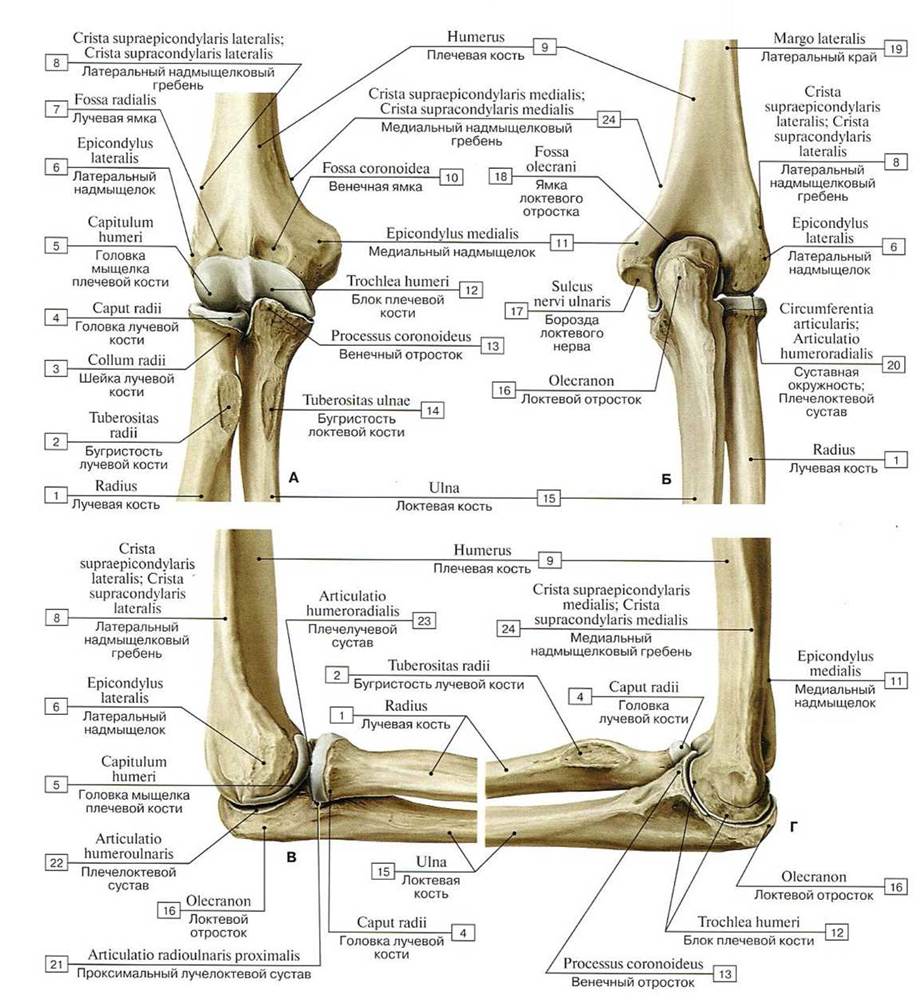
Fig. 154. Relationships between the humerus and the bones of the right forearm (A — anterior view, B — posterior view, C — lateral view, D — medial view):
1 — Radius; 2 — Radial luberosity; 3 — Neck of radius; 4 — Head of radius; 5 —Capitulum; 6 — Lateral epicondyle; 7 — Radial fossa; 8 — Lateral supraepicondvlar ridge; Lateral supracondylar ridge; 9 — Humerus; 10 —Coronoid fossa; 11 — Media!epicondyle; 12—- Trochlea; 13 — Coronoid process; 14 — Tuberosity of ulna; 15 — Ulna; 16 —Olecranon; 17 — Groove for ulnar nerve; 18 — Olecranon fossa; 19— Lateral margin; 20 — Articular circumference; Humero-ulnar joint; 21 — Proximal radio-ulnar joint; 22 — Humeroulnar joint; 23 — Humeroradial joint; 24 — Medial supraepicondylar ridge; Medial supracondylar ridge

Fig. 155. Movements of the elbow joint and hand (A — elbow joint with the forearm extended, B — range of motion in the elbow joint, C — range of pronation and supination of the elbow joint, D — range of motion of the right hand, E — displacement of the radius and ulna during pronation):
1 — Head of radius; 2— Ulnarstyloid process
In the region of the elbow joint, There are two clinically important topographic landmarks: Hueter's line and Hueter's triangle. Hueter's line (syn.: Tillaux's line, ulnar line) connects the lateral epicondyle of the humerus to the apex of the olecranon of the ulna. When joint integrity is compromised, this line becomes broken (Paul J. Tillaux, 1834–1904, French surgeon). Hueter's triangle, or the ulnar triangle (trigonum ulnaris), is formed during flexion of the elbow joint by three points of Hueter's line and is isosceles. In fractures around the elbow joint, the shape of the triangle changes.
The distal radioulnar joint (articulatio radioulnaris distalis) is a cylindrical uniaxial joint formed by the articular circumference of the ulna and the ulnar notch of the radius (Fig. 156). The loose articular capsule attaches along the margins of the articular surfaces. A proximally directed pouch of the joint capsule extending between the distal parts of the forearm bones is known as the sacciform recess (recessus sacciformis). A triangular fibrocartilaginous articular disc (discus articularis) lies between the ulnar notch of the radius and the ulnar styloid process. It separates the distal radioulnar joint from the radiocarpal joint and acts as a specialized socket for the head of the ulna. The loose articular capsule of the distal radioulnar joint attaches along the margins of the articular surfaces and the articular disc.
Movements in the proximal and distal radioulnar joints occur simultaneously as a combined joint (see Fig. 155). During rotation of the radius around the ulna, the proximal epiphysis of the radius rotates in place, as its head is held by the annular ligament of the radius against the ulnar notch. Meanwhile, the distal epiphysis of the radius slides along the articular circumference of the ulna, describing an arc around the stationary head of the ulna. Because the radius articulates with the hand, the hand rotates along with the radius during its rotation. The maximum range of forearm motion inward (pronation) and outward (supination) in the proximal and distal radioulnar joints reaches 180°.
Wrist joints. The bones of the hand articulate with the forearm bones to form the radiocarpal joint, and with one another via intercarpal joints (see Figs. 95, 96).
The radiocarpal joint (articulatio radiocarpea) is a complex, ellipsoid, biaxial joint. It is formed by the carpal articular surface of the radius and the intra-articular disc, as well as the proximal surfaces of the first row of carpal bones (scaphoid, lunate, and triquetrum). The triangular fibrocartilaginous intra-articular disc (discus articularis), which attaches to the ulnar styloid process and laterally to the distal margin of the ulnar notch of the radius, separates the radiocarpal joint from the distal radioulnar joint.
The articular capsule, which is thin (especially posteriorly), attaches along the margins of the articular surfaces of the articulating bones. The capsule of the radiocarpal joint is reinforced by ligaments. The radial collateral ligament of the wrist (ligamentum collaterale carpi radiale), also known as Arnold's ligament (Friedrich Arnold, 1803–1890, German anatomist), extends between the radial styloid process and the scaphoid bone. The ulnar collateral ligament of the wrist (ligamentum collaterale carpi ulnare) connects the ulnar styloid process to the triquetrum and pisiform bones. The palmar radiocarpal ligament (ligamentum radiocarpale palmare) originates on the anterior margin of the articular surface of the radius and attaches via separate bundles to the first row of carpal bones and the capitate bone. The dorsal radiocarpal ligament (ligamentum radiocarpeum dorsale) originates on the radius and attaches to the first row of carpal bones (Figs. 157, 158).
Movements in the radiocarpal joint occur around the sagittal and frontal axes: flexion and extension of the hand — approximately 70°, adduction of the hand — approximately 40°, and abduction of the hand — approximately 15° (Fig. 159).
The radiocarpal joint receives its Blood supply from the articular network, which is formed by contributions from the radial, ulnar, anterior interosseous, and posterior interosseous Arteries. Venous blood drains into Veins OF THE same name. Lymph drains into the cubital Lymph Nodes. Innervation is supplied by Branches of the ulnar, median, and radial nerves.
The midcarpal joint (articulatio mediocarpalis) is formed by the first and second rows of carpal bones, excluding the pisiform; its joint space is S-shaped. The joint effectively possesses two heads: one is formed by the scaphoid, which articulates with the trapezium and trapezoid bones; the other is formed by the capitate and hamate bones, which articulate with the triquetrum, lunate, and scaphoid bones. The joint capsule is relatively loose and very thin posteriorly, attaching along the margins of the articular surfaces. Functionally linked to the radiocarpal joint, the hinge-like uniaxial midcarpal joint participates in flexion and extension of the hand (see Fig. 156).

Fig. 156. Frontal section of the right hand:
1 — Interphalangeal joints of hand; 2 — Metacarpophalangeal joints; 3 — Dorsal interossei; 4 — Abductor digiti minimi; 5 — Dorsal inetacarpal ligaments; 6 — Carpometacarpal joints; 7— Hamate; 8— Capitate; 9— Pisiforrn; 10 — Triquetrum; 11 - Ulnar collateral ligament; 12 — Articular disc; 13 — Dislal radio-ulnar joint; 14 — Ulna; 15 — lnterosseous membrane of forearm; 16 — Lunate; 17 — Radius; 18— Wrist joint; 19 — Scaphoid; 20— Midcarpal joint; 21 — Radial collateral ligament of wrist joint; 22 — Trapezium; 23 — Carpometacarpal joint ofthumb; 24 — Opponens pollicis; 25 — Trapezoid; 26 — Metacarpals [1—V]; 27—Collateral ligaments; 28— Proximal phalanx;
29— Middle phalanx; 30 — Dislal phalanx

Fig. 157. Radiocarpal joint and joints of the right hand, anterior view:
1 — Palmar ligaments; 2 — Distal phalanx; 3 — Proximal phalanx; 4 —- Metacarpals [1-V]; 5 — Palmar carpometacarpal ligaments; 6 — Tubercle; 7 — Radial collateral ligament of wrist joint; 8 — Radial styloid process; 9 — Palmar radiocarpal ligament; 10 — Palmar radioulnar ligament; 11 — Radius; 12 — Ulna; 13 — Distal radio-ulnar joint; 14 — Ulnar styloid process; 15 — Palmar ulnocarpal ligament; 16 — Flexor carpi ulnaris; Tendon; 17 — Pisiform; 18 — Radiate carpal ligament; 19 — Hook of hamate; 20 — Palmar meiacarpal ligaments; 21 — Metacarpophalangeal joints, collateral ligaments; 22 — Deep transverse metacarpal ligament; 23 — Middle phalanx; 24 — Proximal interphalangeal joint; Joint capsule; Articular capsule; 25 — Distal interphalangeae joint; Joint capsule; Articular capsule

Fig. 158. Right wrist joint and joints of the hand, posterior view:
1 — Metacarpal, collateral ligaments; 2 — Metacarpophalangeal joints, collateral ligaments; 3— Metacarpals [1—V]; 4— Dorsal metacarpal ligaments; 5 — Hamate; 6 — Triquetrum; 7 — Ulnar collateral ligament of wrist joint; 8 — Dorsal radiocarpal ligament: 9 — Ulnar styloid process; 10 —Dorsal radio-ulnar ligament; 11 — Ulna; 12 — Radius; 13 — Dorsal tubercle; 14— Radial styloid process; 15 — Radial collateral ligament of wrist joint; 16 — Dorsal intercarpal ligaments; 17— Dorsal carpometacarpal ligaments; 18 — Proximal phalanx; 19— Distal phalanx; 20— Middle phalanx

Fig. 159. Range of motion in the wrist joint and finger joints
Range of motion in the finger joints (Fig. 159).
The proximal and distal interphalangeal joints are hinge joints allowing a single type of movement (flexion-extension). The metacarpophalangeal joints of the second through fifth digits are spheroid (condyloid) in shape. Theoretically, three types of movement can occur in these joints, but rotation is restricted by the collateral ligaments; consequently, only Two Types of movement are performed: flexion-extension and abduction-adduction. The following specific movements are distinguished in the finger joints:
A) flexion at the distal interphalangeal joint,
Б) flexion at the proximal interphalangeal joint,
B) flexion at the metacarpophalangeal joint,
Г) extension at the distal interphalangeal joint,
Д) extension at the metacarpophalangeal joint,
Е) abduction and adduction at the metacarpophalangeal joints (affecting the fingers by abducting and adducting them around the dorso-palmar axis passing through the heads of the metacarpal bones).
Abduction and adduction movements are described relative to the middle finger: movements away from the middle finger are classified as abduction, while movements toward the middle finger are classified as adduction.
Movements in the radiocarpal and midcarpal joints (see Fig. 159).
Starting from the neutral position (0’), flexion and extension occur around the transverse axis (Ж), whereas radial and ulnar deviation take place around the dorso-palmar axis (3). The transverse axis passes through the lunate bone of the wrist joint and through the capitate bone of the midcarpal joint. The dorso-palmar axis passes through the capitate bone. Thus, flexion and extension can occur in both the radiocarpal and midcarpal joints, whereas radial and ulnar deviation can occur only in the radiocarpal joint.
II. Functional position of the hand (see Fig. 159).
For postoperative immobilization of the upper extremity when a splint is applied, the desired position for the hand and fingers must be carefully considered. Otherwise, the ligaments may shorten, and the hand will no longer be able to assume its normal relaxed posture.
The intercarpal joints (articulationes intercarpales, articulationes carpi) are plane joints with limited mobility, formed by the apposing articular surfaces of the carpal bones (see Fig. 156). The joint cavities of the midcarpal and intercarpal joints communicate with one another. The thin articular capsule of these joints attaches along the margins of the articular surfaces. The capsule is reinforced by short palmar and dorsal intercarpal ligaments (ligamenta intercarpalia palmaria et dorsalia), as well as the more superficial radiate ligament of the carpus (ligamentum carpi radiatum) — also known as Mayer's ligament (Mayer, F., 1870–1935, American histologist and anatomist). The fibers of the radiate ligament on the palmar side diverge radially from the capitate bone to the adjacent bones. The intercarpal joints also feature intra-articular interosseous intercarpal ligaments (ligamenta intercarpalia interossea intraarticularia). The pisiform bone forms an independent joint with the triquetrum, which is reinforced by the pisohamate ligament (Barken's ligament) and the pisometacarpal ligament (ligamentum pisohamatum et ligamentum pisometacarpale). The pisometacarpal ligament attaches to the bases of the IV–V metacarpals. Both of these ligaments are continuations of the tendon of the flexor carpi ulnaris, which attaches to the pisiform bone, with some of its fibers continuing into these ligaments (see Figs. 157, 158).
The total range of motion in the radiocarpal and intercarpal joints combined is approximately 85° for flexion and extension, and 25–30° for abduction and adduction. Circumduction in these joints results from the combination of sequential movements around the sagittal and frontal axes, during which the fingertips trace a circle.
The carpometacarpal joints of the II–V fingers (articulationes carpometacarpales II–V) are plane, slightly movable joints formed by the distal articular surfaces of the second row of carpal bones and the articular surfaces at the bases of the II–V metacarpals. The common joint space forms a transverse jagged line that communicates with the cavities of the intercarpal and midcarpal joints. The thin articular capsule, common to all four carpometacarpal joints, is tightly stretched and reinforced by strong palmar and dorsal carpometacarpal ligaments (ligamenta carpometacarpalia palmaria et dorsalia) (see Figs. 156, 157, 158).
The carpometacarpal joint of the thumb (articulatio carpometacarpalis pollicis) is a saddle joint, anatomically isolated from the other carpometacarpal joints, formed by the articular surfaces of the trapezium and the base of the first metacarpal bone (Figs. 160, 161, 162, see Figs. 156, 157, 158); its joint capsule is loose. The joint is highly mobile and possesses two axes of rotation: frontal and sagittal. Flexion and extension occur around the frontal axis; during flexion, the thumb tilts toward the palm and opposes the other digits (Fig. 163, 164, see Fig. 159). Adduction (bringing the thumb closer to the index [II] finger) occurs around the sagittal axis. Combining movements around the frontal and sagittal axes allows for circumduction in the carpometacarpal joint of the thumb.
The intermetacarpal joints (articulationes intermetacarpeae) are formed by the contiguous surfaces of the bases of the II–V metacarpals. Their capsules are shared with those of the carpometacarpal joints. The joints are reinforced by palmar and dorsal metacarpal ligaments (ligamenta metacarpalia palmaria et dorsalia), which run transversely and connect the bases of adjacent metacarpal bones.
Movements of the hand relative to the forearm involve the radiocarpal, midcarpal, carpometacarpal, and intercarpal joints. Clinicians often refer to all these functionally unified joints collectively as the wrist joint. The overall range of motion of the hand is the sum of movements across all these joints.
The metacarpophalangeal joints (articulationes metacarpophalangeae) are ellipsoidal Biaxial joints formed by the rounded articular surfaces of the metacarpal heads and the oval-shaped bases of the proximal phalanges. The loose articular capsules attach along the margins of the articular surfaces. The joints are reinforced by collateral ligaments (ligamenta collateralia lateralia) stretching between the bases of the primary phalanges and the metacarpal heads, as well as palmar ligaments (ligamenta palmaria). The metacarpophalangeal joints of digits II–V are additionally reinforced by deep transverse metacarpal ligaments (ligamenta metacarpalia transversa profunda) spanning transversely between the metacarpal heads. Movements around the frontal and sagittal axes are possible in the metacarpophalangeal joints. Flexion and extension around the frontal axis range up to 90°. Abduction and adduction around the sagittal axis measure 45–50°. Circumduction is also possible in the metacarpophalangeal joints (see Figs. 159, 164).

Fig. 160. Articular surfaces of the carpometacarpal joint of the thumb:
1 — Head ofmetacarpal; 2 — Shaft of metacarpal; Body of metacarpal [I]; 3 — Base of metacarpal; 4 — Trapezium; 5 — Tubercle

Fig. 161. Axes of movement in the carpometacarpal joint of the thumb of the right hand (highlighted in red — A, B):
1 — Distal phalanx; 2— Middlc phalanx; 3 — Proximal phalanx; 4— Head of metacarpal; 5— Shaft of metacarpal; Body of metacarpal; 6 — Base of metacarpal; 7 — Trapezoid; 8 —Capitate; 9 — Lunate; 10 — Scaphoid; 11 — Ulnar styloid process; 12 — Ulna; 13 — Radius: 14 — Radial styloid process; 15 — Trapezium; 16 — Metacarpal [I]; 17 — Proximal phalanx [I]; 18 — Distal phalanx [I]; 19 — Base of phalanx; 20 — Shaft of phalanx; Body of phalanx; 21 — Head of phalanx; 22 — Tuberosity of distal phalanx

Fig. 162. Displacement of bones in the carpometacarpal joint of the thumb during opposition:
1 — Trapezium; 2 — Metacarpal [I]

Fig. 163. Thumb of the hand (A — movements of the thumb in the carpometacarpal joint, right hand, palmar view; B — position of the thumb relative to the other fingers in the neutral position (0°), right hand, distal view):
1—Trapezium; 2 — Triquetrum; 3 — Hamate; 4 — Trapezoid; 5 — Capitate; 6 — Hook of hamate; 7 — Pisiform; 8 — Metacarpal [l]
A.
I — normal (neutral) position (0°)
II — axes of movement in the carpometacarpal joint of the first digit
III — adduction
IV — abduction
V — flexion
VI — extension
VII — opposition
VIII — axis of thumb opposition. While the first metacarpal bone rotates, its contact area with the articular surface of the trapezium significantly decreases.
B.
Due to the arched concavity of the carpal bones, the scaphoid and trapezium exhibit a noticeable radiopalmar orientation. As a result, the metacarpal bone does not lie on the same axis as the other fingers, but is rotated by approximately 60° toward the palm.
Four types of grip are distinguished:
A) pinch grip,
B) power grip,
B) key grip,
Г) hook grip.
When performing a Clinical examination of hand function, special attention should be paid to impairments in fine motor skills and gross strength. This allows for an assessment of various grips. The pinch grip between the thumb and index finger is of paramount importance for hand function. Therefore, the loss of the thumb or index finger impacts vocational abilities far more severely than the absence of any other fingers.

Fig. 164. Types of finger grips of the right hand
The interphalangeal joints (articulationes interphalangeae manus) are formed by the bases and heads of adjacent phalanges. These are typical hinge joints, whose joint capsules are reinforced by lateral collateral ligaments (ligamenta collateralia lateralia). The PALMAR ASPECT OF these capsules is strengthened by palmar ligaments (ligamenta palmaria). Movements around the frontal axis (flexion and extension) in these joints have a range of approximately 90°.
Thus, the carpal bones are firmly bound together by numerous ligaments (see Figs. 157, 158), with the dorsal ligaments being weaker than the palmar ones. The bones located in the distal row of the carpus (trapezium, trapezoid, capitate, hamate), interconnected with each other and with the II–V metacarpal bones via joints, form a rigid and highly durable foundation of the hand (V.N. Tonkov).
Of particular importance is the saddle-shaped biaxial carpometacarpal joint of the thumb. The opposition of the thumb to the other fingers plays a crucial role in manual labor.
Radiological Anatomy of the upper extremity joints. When the upper limb is held close to the trunk, the anteroposterior projection reveals the head of the humerus, the glenoid cavity of the scapula, and an arcuate X-ray joint space. In the lower part of the joint, the articular head overlaps the glenoid cavity of the scapula (Figs. 165, 166).

Fig. 165. Radiograph of the humerus, anteroposterior projection:
1 — glenoid cavity of the scapula; 2 — head of the humerus; 3 — anatomical neck of the humerus; 4 — greater tubercle; 5 — lesser tubercle; 6 — surgical neck of the humerus; 7 — shaft of the humerus; 8 — deltoid tuberosity of the humerus; 9 — olecranon fossa of the humerus; 10 — lateral epicondyle of the humerus; 11 — medial epicondyle of the humerus; 12 — trochlea of the humerus; 13 — capitulum of the humerus; 14 — radius; 15 — ulna (adapted from A.Yu. Vasilyev)

Fig. 166. Frontal slice of the right shoulder joint, T1-weighted image (Magnetic Resonance imaging):
1 — coracobrachialis muscle; 2 — deltoid muscle; 3 — surgical neck of the humerus; 4 — greater tubercle of the humerus; 5 — head of the humerus; 6 — glenoid labrum; 7 — scapula; 8 — subscapularis muscle; 9 — supraspinatus muscle; 10 — trapezius muscle; 11 — lung (adapted from S.K. Ternovoy)
In the anteroposterior PROJECTION OF THE elbow joint, the articular surface of the humerus appears as a curved, zigzag line. The 2–3 mm wide joint space of the elbow is superimposed by the image of the articular process of the ulna (Fig. 167-A). The joint space of the proximal radioulnar joint is also visible. In the lateral projection, when the forearm forms a right angle with the humerus at the elbow joint, the joint space is visible between the capitulum of the humerus and the trochlear notch of the ulna, as well as the head of the radius (Fig. 167-B).
Radiographs of the hand clearly show the articulating bones and the intervening X-ray joint spaces. In the radiocarpal joint, the joint space is narrow near the radius and wide near the head of the ulna due to the radiolucency of the articular disc. The shadow of the pisiform bone is superimposed on the triquetrum. The joint spaces of the carpometacarpal joints are slightly curved, while those of the metacarpophalangeal joints are convex distally. In the interphalangeal joints, which are hinge-like in shape, the X-ray joint space precisely follows the contours of the articulating phalangeal surfaces.

Fig. 167-A. Radiograph of the elbow joint in the posteroanterior projection:
1 — olecranon fossa of the humerus; 2 — medial epicondyle of the humerus; 3 — lateral epicondyle of the humerus; 4 — olecranon of the ulna; 5 — medial ridge of the humeral trochlea (not in NA); 6 — capitulum of the humerus (not in NA); 7 — humeroulnar joint; 8 — humeroradial joint; 9 — proximal radioulnar joint; 10 — head of the radius; 11 — radial tuberosity; 12 — ulna; 13 — radius (adapted from A.Yu. Vasilyev)

Fig. 167-B. Radiograph of the elbow joint in the lateral projection:
1 — humerus; 2 — ulna; 3 — radius; 4 — coronoid fossa of the humerus; 5 — olecranon fossa of the humerus; 6 — superimposition of the capitulum and trochlea of the humerus; 7 — olecranon of the ulna; 8 — coronoid process of the ulna; 9 — humeroulnar joint; 10 — radial tuberosity; 11 — humeroradial joint; 12 — head of the radius; 13 — neck of the radius (adapted from A.Yu. Vasilyev)
Last update: 08/08/2026
Editorial and Educational Adaptation: This material has been compiled based on the primary/original source text. The project team performed an editorial review, corrected technical inaccuracies, structured sections, and adapted the content for an educational format.
What was processed:
- elimination of formatting defects (OCR errors, structural breaks, corrupted characters);
- editorial organization of content;
- standardization of terminology in accordance with academic sources;
- verification of factual statements against the original source text.
All mentions of the author, publication year, and origin of the primary text have been preserved in accordance with the source.