Human Anatomy - Kotsan I. Ya. 2009
Brain
Projection of the cranial nerve nuclei onto the rhomboid fossa
The grey matter in the region of the Rhomboid fossa forms distinct clusters, or nuclei, separated from one another by White matter (Fig. 249). To understand the Topography of the grey matter in the rhomboid fossa, recall that the neural tube in the region of the Medulla Oblongata and Pons opened along its posterior (dorsal) surface and unfolded such that its posterior parts shifted to become the lateral portions of the rhomboid fossa. Consequently, the sensory nuclei of the rhomboid Brain, which correspond to the posterior horns of the Spinal Cord, occupy a lateral position in the rhomboid fossa. The motor nuclei, corresponding to the anterior horns of the spinal cord, are located medially. As for the autonomic nuclei, which lie in the lateral horns of the spinal cord, their intermediate position between the posterior and anterior horns meant that during the unfolding of the neural tube they ended up positioned between the motor and sensory nuclei in the rhomboid fossa. As a result, unlike in the spinal cord, the nuclei of the grey matter in the rhomboid fossa are not arranged in an anteroposterior direction, but rather lie in rows—medially and laterally. For instance, the somatic motor nuclei of the XII and VI pairs are situated in the medial row, the autonomic nuclei of the X, IX, and VII pairs in the middle row, and the somatic sensory nuclei of the VIII pair laterally.
Deep within the rhomboid fossa lie the nuclei of the V, VI, VII, VIII, IX, X, XI, and XII pairs of Cranial Nerves.
The V pair, the Trigeminal nerve (n. trigeminus), has four nuclei:
1. The motor Nucleus of the trigeminal nerve (nucleus motorius n. trigeminalis) is located in the upper PARTS OF THE rhomboid fossa, within the cranial fossa region. The processes of the Cells of this nucleus form the motor ROOT of the trigeminal nerve.
Class="center">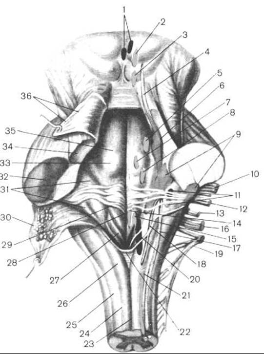
Fig. 249. Posterior surface of the pons and medulla oblongata, along with the PROJECTION OF THE cranial nerve nuclei onto the rhomboid fossa (after R. D. Sinelnikov)
1 — accessory (parasympathetic) nucleus of the Oculomotor nerve; 2 — nucleus of the oculomotor nerve; 3 — nucleus of the Trochlear nerve; 4 — nucleus of the mesencephalic tract of the trigeminal nerve; 5 — motor nucleus of the trigeminal nerve; 6 — pontine nucleus of the trigeminal nerve; 7 — nucleus of the Abducens nerve; 8 — nucleus of the Facial Nerve; 9 — nuclei of the Vestibulocochlear nerve; 10 — root of the facial nerve (VII pair); 11 — superior and inferior salivatory nuclei; 12 — vestibulocochlear nerve (VIII pair); 13 — Glossopharyngeal nerve (IX pair); 14 — hypoglossal nucleus; 15 — ambiguus nucleus; 16 — Vagus nerve (X pair); 17 — nucleus of the spinal tract of the trigeminal nerve; 18 — nucleus of the solitary tract; 19 — Accessory nerve (XI pair); 20 — dorsal nucleus of the vagus nerve; 21 — spinal nucleus of the accessory nerve; 22 — obex; 23 — posterior median sulcus; 24 — gracile fasciculus; 25 — cuneate fasciculus; 26 — tubercle of the gracile nucleus; 27 — vagal trigone; 28 — median sulcus of the rhomboid fossa; 29 — medullary striae; 30 — superior medullary velum; 31 — vestibular area; 32 — middle cerebellar peduncle; 33 — facial colliculus; 34 — superior cerebellar peduncle; 35 — median eminence; 36 — superior medullary velum
2. The sensory nucleus of the trigeminal nerve, which receives fibers from the sensory root of this nerve, consists of two parts:
a) the pontine nucleus of the trigeminal nerve (nucleus pontius nervi trigeminalis) lies lateral and slightly posterior to the motor nucleus; the projection of the pontine nucleus corresponds to the locus coeruleus;
b) the (inferior) spinal nucleus of the trigeminal nerve (nucleus spinalis (inferior) nervi trigeminalis) appears as a continuation of the previous nucleus, has an elongated shape, runs the entire length of the medulla oblongata, and extends into the upper segments of the spinal cord.
3. The Nucleus of the mesencephalic tract of the trigeminal nerve (nucleus mesencephalicus nervi trigeminalis) is located cranial to the motor nucleus of this nerve, adjacent to the cerebral aqueduct, reaching as far as the inferior colliculi of the corpora quadrigemina of the Midbrain.
The VI pair, the abducens nerve (n. abducens), has a single motor nucleus, the abducens nucleus (nucleus nervi abducentis), which is situated within the loop of the facial nerve genu, deep within the facial colliculus.
The VII pair, the facial nerve (n. facialis), has three nuclei:
1. The nucleus of the facial nerve (nucleus nervi facialis) is a large motor nucleus located quite deeply within the reticular Formation of the pons, lateral to the corresponding colliculus. The processes of the cells of this nucleus form the motor root.
2. The nucleus of the solitary tract (nucleus solitarius) is a general sensory nucleus common to the VII, IX, and X pairs of cranial nerves. It lies deep within the rhomboid fossa, projecting lateral and inferior to the hypoglossal trigone. Fibers conducting taste impulses terminate on the cells of this nucleus.
3. The superior salivatory nucleus (nucleus salivarius superior) is an autonomic (parasympathetic) nucleus located in the reticular formation of the pons, slightly dorsal and lateral to the motor nucleus of the facial nerve.
The VIII pair, the vestibulocochlear nerve (n. vestibulocochlearis), has two groups of nuclei: two cochlear (auditory) and four vestibular nuclei, which lie in the ventrolateral Regions of the pons and project within the vestibular area of the rhomboid fossa.
1. The ventral (anterior) cochlear nucleus (nucleus cochlearis ventralis (anterior)).
2. The dorsal (posterior) cochlear nucleus (nucleus cochlearis dorsalis (posterior)). The processes of Neurons from the spiral (cochlear) ganglion, which form the cochlear part of the nerve, terminate in synapses on the cells of these nuclei. These nuclei lie one ventromedial to the other and lateral to the vestibular nuclei.
The vestibular nuclei, which receive nerve impulses from the ampullary crests and maculae of the membranous labyrinth of the Inner ear, are:
1. the medial vestibular nucleus (nucleus vestibularis medialis), or Schwalbe's nucleus;
2. the lateral vestibular nucleus (nucleus vestibularis lateralis), or Deiters' nucleus;
3. the superior vestibular nucleus (nucleus vestibularis superior), or Bechterew's nucleus;
4. the inferior vestibular nucleus (nucleus vestibularis inferior), or Roller's nucleus.
The nuclei of the last four pairs of cranial nerves (IX, X, XI, and XII) lie in the lower triangle of the rhomboid fossa, which is formed by the dorsal part of the medulla oblongata.
The IX cranial nerve, the glossopharyngeal nerve (n. glossopharyngeus), has three nuclei.
1. The ambiguous nucleus (nucleus ambiguus) is motor, located in the reticular formation within the lateral regions of the lower half of the rhomboid fossa, and projects in the area of the caudal fossa. It is shared by the IX and X cranial nerves.
2. The solitary tract nucleus (nucleus solitarius) is sensory and shared by the VII, IX, and X cranial nerves.
3. The inferior salivatory nucleus (nucleus salivarius inferior) is autonomic (parasympathetic), located in the reticular formation of the medulla oblongata between the inferior olivary nucleus and the ambiguous nucleus.
The X cranial nerve, the vagus nerve (n. vagus), has three nuclei: motor, sensory, and autonomic (parasympathetic).
1. The ambiguous nucleus (nucleus ambiguus) is a motor nucleus shared by the IX and X cranial nerves.
2. The solitary tract nucleus (nucleus solitarius) is a sensory nucleus shared by the VII, IX, and X cranial nerves.
3. The dorsal nucleus of the vagus nerve (nucleus dorsalis nervi vagi) is parasympathetic and lies superficially in the region of the vagal trigone.
The XI cranial nerve, the accessory nerve (n. accessorius), features the motor nucleus of the accessory nerve (nucleus motorius nervi accessorii), which lies within the depth of the rhomboid fossa, inferior and lateral to the ambiguous nucleus, and extends into the Gray matter of the spinal cord at the level of the upper five to six segments.
The XII cranial nerve, the Hypoglossal nerve (n. hypoglossus), has a single nucleus situated in the inferior angle of the rhomboid fossa, deep within the hypoglossal trigone.
The nucleus of the hypoglossal nerve (nucleus nervi hypoglossi) is motor; its cellular processes participate in the Innervation of the Tongue Muscles and, together with the branches emerging from the Cervical plexus, in the innervation of the Muscles of the anterior cervical region.
Last update: 08/08/2026
Editorial and Educational Adaptation: This material has been compiled based on the primary/original source text. The project team performed an editorial review, corrected technical inaccuracies, structured sections, and adapted the content for an educational format.
What was processed:
- elimination of formatting defects (OCR errors, structural breaks, corrupted characters);
- editorial organization of content;
- standardization of terminology in accordance with academic sources;
- verification of factual statements against the original source text.
All mentions of the author, publication year, and origin of the primary text have been preserved in accordance with the source.