Human Anatomy - Kotsan I. Ya. 2009
Vestibulocochlear organ (organ of hearing and equilibrium)
Inner ear
The inner ear (auris interna) is located within the petrous part of the Temporal bone, between the tympanic cavity and the internal acoustic meatus. It consists of the osseous labyrinth and the membranous labyrinth enclosed within it (Fig. 272).
Class="center">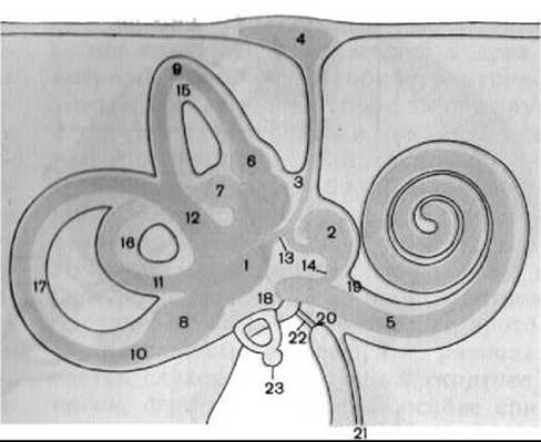
Fig. 272. Diagram of the osseous and membranous labyrinths (according to M. R. Sapin)
(the membranous labyrinth is indicated by a darker shade)
1 — utricle (elliptic sac); 2 — saccule (spherical sac); 3 — endolymphatic duct; 4 — endolymphatic sac; 5 — cochlear duct; 6 — anterior membranous ampulla; 7 — lateral membranous ampulla; 8 — posterior membranous ampulla; 9 — anterior semicircular duct; 10 — posterior semicircular duct; 11 — lateral semicircular duct; 12 — common membranous limb; 13 — utriculosaccular duct; 14 — uniting duct; 15 — anterior semicircular canal; 16 — lateral semicircular canal; 17 — posterior semicircular canal; 18 — vestibule; 19 — scala vestibuli; 20 — scala tympani; 21 — cochlear canal; 22 — secondary tympanic membrane; 23 — stapes
The osseous labyrinth (labyrinthus osseus) is a system of interconnected cavities of various shapes. The wall of the osseous labyrinth is formed by compact Bone tissue and lined with a thin Connective Tissue membrane. The length of the osseous labyrinth along its longitudinal axis is about 20 mm. The osseous labyrinth comprises the vestibule, semicircular canals, and cochlea.
The vestibule (vestibulum) is the central part of the osseous labyrinth, situated between the tympanic cavity and the internal acoustic meatus. Its posterior wall contains five openings of the semicircular canals; the anterior wall features a fairly wide opening of the cochlear canal; and the lateral wall presents the oval window (fenestra vestibuli) and the round window (fenestra cochleae), through which the osseous labyrinth communicates with the tympanic cavity. The medial wall of the vestibule bears two recesses (pits). The anterior one is rounded and called the spherical recess (recessus sphericus), which houses the saccule of the membranous labyrinth. The posterior recess is somewhat elongated and designated as the elliptical recess (recessus ellipticus), which contains the utricle of the membranous labyrinth and the small internal opening of the vestibular aqueduct (apertura interna aqueductus vestibuli). The vestibular aqueduct (aqueductus vestibuli) is a narrow canal passing through the bone substance of the petrous part of the temporal bone and terminating on its surface with the external opening of the vestibular aqueduct (apertura externa aqueductus vestibuli). The elliptical and spherical recesses are separated by the vestibular crest (crista vestibuli). Just below the posterior end of the vestibular crest lies a small cochlear recess (recessus cochlearis), which contains the beginning of the cochlear duct.
The osseous semicircular canals (canales semicirculares ossei) are three arcuate tubes positioned in three mutually perpendicular planes. The width of the lumen of each osseous semicircular canal in cross-section is approximately 2 mm.
The anterior or sagittal semicircular canal (canalis semicircularis anterior), 18–20 mm long, is oriented vertically at a right angle to the axis of the petrous part of the temporal bone. It lies superior to the other semicircular canals, and its upper point on the anterior surface of the petrous bone forms the arcuate eminence (eminentia arcuata).
The posterior semicircular canal (canalis semicircularis posterior) is the longest of the canals (22 mm in length), also vertical, and positioned almost parallel to the posterior surface of the petrous part.
The lateral semicircular canal (canalis semicircularis lateralis), 14–16 mm long, is situated horizontally. The convex part of the lateral semicircular canal projects into the tympanic cavity, forming the prominence of the lateral semicircular canal (prominentia canalis semicircularis lateralis) on the internal wall.
Each canal has two limbs; however, they open into the vestibule through only five apertures because the adjacent ends of the anterior and posterior semicircular canals merge into a common limb (crus commune). One limb of each canal expands before entering the vestibule, forming an osseous ampulla (ampulla ossea). The ampullated limb is termed the ampullary osseous limb (crus osseum ampullare), while the non-ampullated limb is called the simple osseous limb (crus osseum simplex).
The osseous cochlea (cochlea) is the anterior part of the osseous labyrinth, represented by a spirally winding cochlear canal (canalis spiralis cochleae) that makes two and a half turns around the cochlear axis. The first turn of the cochlea is called the basal turn, the second is the middle turn, and the final one is the apical turn.
The cochlea is conical in shape. It features a base (basis cochleae), which is 7–10 mm wide, and an apex or cupula of the cochlea (cupula cochleae). The distance from the base to the apex is 4–5 mm. The Base of the cochlea is directed medially toward the internal acoustic meatus, whereas the apex points toward the tympanic cavity.
The spiral canal of the cochlea is 28–30 mm long and ends blindly in the region of the apex of the petrous bone. The diameter of the canal is not uniform throughout: it is wide at its origin (6 mm) and gradually narrows toward the apex of the cochlea, reaching 2 mm.
The axis of the cochlea, lying horizontally, is the modiolus. The modiolus consists of spongy bone tissue and forms the internal wall of the spiral canal. Its broad part, or the base of the modiolus (basis modioli), faces the internal acoustic meatus and exhibits numerous openings. These openings lead into the longitudinal canals of the modiolus (canales longitudinales modioli), which transmit the fibers of the cochlear part of the Vestibulocochlear nerve.
Winding around the modiolus is the osseous spiral lamina (lamina spiralis ossea), which does not reach the opposite wall of the canal. The osseous spiral lamina begins on the inner wall of the vestibule near the cochlear window and, ascending toward the apex (cupula) of the cochlea, terminates in the region of the final turn with a curved margin—the hamulus of the spiral lamina (hamulus laminae spiralis)—which bounds an oval-shaped opening called the helicotrema (helicotrema). The base of the osseous spiral lamina is thicker than its free edge and contains throughout its length the spiral canal of the modiolus (canalis spiralis modioli), which lodges the cochlear nerve ganglion, namely the spiral ganglion of the cochlea (ganglion spirale cochleae). The spiral canal of the modiolus communicates via the longitudinal canals of the modiolus with the openings at the base of the cochlea, and through the spiral cleft running along the entire length of the spiral lamina, it communicates with the Cytology/practical/79.html">Spiral organ of Corti.
The osseous spiral lamina, together with the cochlear duct of the membranous labyrinth, divides the cavity of the spiral canal of the cochlea into two parts: the upper one, the scala vestibuli, and the lower one, the scala tympani.
The scala vestibuli begins in the anterior region of the vestibule, ascends along the upper surface of the spiral lamina to the apex of the cochlea, where it transitions into the scala tympani in the area of the hamulus of the spiral lamina. The transition point between the scala vestibuli and scala tympani is the helicotrema, through which the cavities of both scalae communicate with each other. The scala vestibuli communicates with the perilymphatic space of the vestibule.
The scala tympani begins at the helicotrema and runs along the lower surface of the spiral lamina toward the base of the cochlea. After making 2¼ to 2½ turns, the scala tympani ends blindly in the initial region of the basal turn of the cochlea. Here, on the outer wall of the scala tympani, lies the cochlear window (fenestra cochleae), which is closed by the secondary tympanic membrane (membrana tympani secundaria). The anterior margin of the cochlear window is bounded by the crest of the cochlear window (crista fenestrae cochleae), anterior to which, at the floor of the scala tympani, lies the internal opening of the cochlear canaliculus (apertura interna canaliculi cochleae). This canaliculus begins with a funnel-shaped expansion and, passing through the substance of the petrous part of the temporal bone, terminates on its inferior surface with the external opening of the cochlear canaliculus (apertura externa canaliculi cochleae) anterior to the jugular fossa.
The membranous labyrinth (labyrinthus membranaceus) is located inside the osseous labyrinth and largely mirrors its contours. The wall of the membranous labyrinth consists of a thin, semitransparent connective tissue membrane lined with squamous epithelium. The space between the osseous and membranous labyrinths is called the perilymphatic space (spatium perilymphaticum). It is filled with a clear Lymph-like fluid, the perilymph (perilympha), and is traversed by connective tissue trabeculae that support the membranous labyrinth. The total volume of the perilymph is approximately 80 mm3. Through the elastic and compliant cochlear and vestibular windows, the perilymphatic space communicates with the Middle ear. Furthermore, via the perilymphatic duct, which runs within the cochlear canaliculus, perilymph can drain into the subarachnoid space on the Inferior surface of the petrous part of the temporal bone.
The membranous labyrinth is filled with endolymph (endolympha). The total volume of endolymph is 2.76 mm3. The endolymphatic space is connected, via the endolymphatic duct, to the endolymphatic sac (saccus endolymphaticus), which lies within the dura mater on the posterior surface of the petrous part of the temporal bone. It serves as a relatively elastic reservoir that communicates with the internal space of the semicircular ducts and the labyrinth.
The membranous labyrinth consists of the vestibular labyrinth, three semicircular ducts, and the cochlear duct.
The vestibular labyrinth (labyrinthus vestibularis) comprises the elliptical and spherical sacs.
The elliptical sac, or utricle (utriculus), is elongated in shape, with a diameter of 2.5–3.5 mm, and is located in the elliptical recess of the osseous labyrinth of the vestibule. The semicircular ducts of the membranous labyrinth open into the cavity of the utricle.
The spherical sac (sacculus) is pear-shaped, measures 2–3 mm in diameter, and is located in the spherical recess of the bony labyrinth. The saccule communicates with the cochlear duct via the uniting duct (ductus reuniens).
The elliptical (utricle) and spherical sacs communicate with each other through the utriculosaccular duct (ductus utriculosaccularis), from which the endolymphatic duct (ductus endolymphaticus) originates. The endolymphatic duct passes through the vestibular aqueduct and ends blindly as the endolymphatic sac (saccus endolymphaticus), which is situated on the posterior wall of the petrous part of the temporal bone within a duplication of the dura mater.
On the inner wall of the elliptical and spherical sacs lie whitish receptor (static) spots (maculae). The spot of the elliptical sac, or utricle (macula utriculi), measures 2.3–3 mm and is located on the floor of the utricle. The spot of the spherical sac (macula sacculi), with a diameter of 1.5 mm, is positioned on the medial wall of the sac. In the region of the maculae, the walls of the utricle and saccule are lined with neuroepithelium, which consists of receptor and supporting Cells. The receptor cells bear hairs surrounded by a gelatinous otolithic membrane (membrana statoconium) and covered with small (up to 15 µm) solid particles containing calcium salts, known as statoconia (otoliths). The maculae perceive the static position of the body (HEAD) and its rectilinear translational movement without acceleration.
The semicircular ducts (ductus semicirculares) are located inside the bony semicircular canals, precisely mirroring their shape. Accordingly, the anterior, posterior, and lateral membranous semicircular ducts (ductus semicirculares anterior, posterior et lateralis) are distinguished, featuring membranous limbs (crura membranacea) and membranous ampullae (ampullae membranaceae) located within the corresponding bony structures. Projecting into the lumen of each membranous ampulla is the ampullary crest (crista ampullaris), the core of which is formed by connective tissue and nerve fibers. The ampullary crest is covered by a neuroepithelial layer of Hair and supporting cells and a gelatinous cupula into which the hairs of the Hair cells extend. The receptor apparatus of the ampullary crests perceives accelerated Movements of the body (head) in various directions.
The cochlear duct (ductus cochlearis) is situated within the spiral canal of the cochlea, beginning in the Vestibule of the bony labyrinth with the vestibular cecum (caecum vestibulare) and ending at the apex of the cochlea with the cupular cecum (caecum cupulare). The vestibular cecum connects via the uniting duct with the saccule. In cross-section, the cochlear duct is triangular. The lumen of the cochlear duct is bounded by three walls: one is the outer wall (paries externus ductus cochlearis), which joins the outer wall of the bony cochlea; the second is the vestibular wall of the cochlear duct (paries vestibularis ductus cochlearis), facing the scala vestibuli; and the third is the tympanic wall (paries tympanicus ductus cochlearis), located at the border with the scala tympani. It appears as a continuation of the osseous spiral lamina and is called the basilar lamina (lamina basilaris), or the spiral membranous lamina.
The outer wall of the cochlear duct is connected to the periosteum lining the inner surface of the bony cochlear cavity. It consists of three layers: the outer connective tissue layer, which is a continuation of the spiral ligament of the cochlea (lig. spirale cochleae) anchoring the basilar lamina to the outer wall of the cochlea; the middle vascular stria (stria vascularis), whose vessels produce endolymph; and the inner layer, represented by the epithelium lining the lumen of the cochlear duct.
The vestibular wall of the cochlear duct begins On the surface of the osseous spiral lamina facing the cavity of the scala vestibuli. Extending toward the outer wall of the cochlea, the vestibular membrane forms a 45° angle with the osseous spiral lamina. This wall of the cochlear duct is the smallest, consisting of a connective tissue foundation covered with epithelium.
The tympanic wall of the cochlear duct, or basilar lamina, is stretched between the free edge of the osseous spiral lamina and the outer wall of the cochlea, where it is secured by the spiral ligament of the cochlea. In addition to the basilar lamina, the outer division of the osseous spiral lamina, which enters the lumen of the cochlear duct, participates in The formation of this wall.
The edge of the osseous spiral lamina is thickened and supplemented here by the spiral limbus (limbus spiralis), which consists of connective tissue and epithelial elements. From it, a crest, or vestibular lip (labium limbi vestibulare), projects freely into the lumen of the cochlear duct and continues into the tectorial membrane (membrana tectoria). At the site where the basilar lamina connects with the osseous spiral lamina, the edge of the latter extends into the tympanic crest, or tympanic lip (labium limbi tympanicum). These two Lips are separated from each other by the spiral sulcus (sulcus spiralis). The edge of the tympanic lip is perforated by nerve foramina through which the spiral cleft of the osseous spiral lamina opens into the cochlear canal.
On the basilar lamina of the tympanic wall lies the spiral Organ of Hearing (organum spirale), which was first described in 1851 by the Italian anatomist and histologist A. Corti, and is therefore also referred to as the spiral organ of Corti.
The basilar lamina consists of A large number (24,000) of fibrous fibers (auditory strings) of varying lengths, stretched from the end of the osseous spiral lamina to the opposite wall of the spiral cochlear duct, spanning from its base to the cupola. The fibers consist of fine fibrils that anastomose with one another. The length of the fibers increases in the direction from the base of the cochlea to its apex (cupola). According to the well-known Helmholtz theory (1875), these fibers act as resonators that, through their vibrations, account for the perception of tones of different pitches; however, Electron Cell/15.html">Microscopy data show that they form an elastic network that collectively resonates with strictly graded vibrations.
Among the principal Cells of the spiral organ, external and internal supporting cells and external and internal receptor cells are distinguished. The receptor cells bear hairs—microvilli—on their surface. Located above the hair cells is the tectorial membrane (membrana tectoria) of a gelatinous consistency, one margin of which is attached to the osseous spiral lamina, while the other ends freely in the lumen of the cochlear duct slightly beyond the external receptor cells. The sensory hair cells of the spiral organ of Corti transform the mechanical energy of sound vibrations in the endolymph and perilymph into the energy of nerve impulses.
Last update: 08/08/2026
Editorial and Educational Adaptation: This material has been compiled based on the primary/original source text. The project team performed an editorial review, corrected technical inaccuracies, structured sections, and adapted the content for an educational format.
What was processed:
- elimination of formatting defects (OCR errors, structural breaks, corrupted characters);
- editorial organization of content;
- standardization of terminology in accordance with academic sources;
- verification of factual statements against the original source text.
All mentions of the author, publication year, and origin of the primary text have been preserved in accordance with the source.