Human Anatomy and Physiology (with Age-Related Characteristics of the Child's Body) - Sapin M.R., Sivoglazov V.I. 2002
Nervous system
Sensory organs
Organ of hearing and equilibrium (vestibulocochlear organ)
The Organ of Hearing and Equilibrium is paired. Within it, the Organ of Hearing is divided into the outer, middle, and Inner ear (Fig. 98).
The outer ear includes the auricle and the external acoustic meatus, separated from the Middle ear by the tympanic membrane. The auricle, adapted for collecting sound waves, is formed of elastic Cartilage covered with Skin. The lower part of the auricle (earlobe) is a skin fold that contains no cartilage. The auricle is attached to the Temporal bone by ligaments. The external acoustic meatus has cartilaginous and bony parts. At the junction where the cartilaginous part becomes the bony part, the acoustic meatus narrows and curves. In an adult, the length of the external acoustic meatus is about 33—35 mm, and its lumen diameter ranges from 0.8 to 0.9 cm. The external acoustic meatus is lined with skin containing tubular glands (modified Sweat Glands) that produce a yellowish secretion—cerumen (earwax).
The tympanic membrane separates the outer ear from the middle ear. It is a Connective Tissue plate covered externally by thin skin and internally, from the side of the tympanic cavity, by a mucous membrane. In the center of the tympanic membrane, There is a depression (the umbo of the tympanic membrane)—the site of attachment for one of the auditory ossicles, the malleus. The tympanic membrane is divided into an upper, thin, flaccid part that contains no Collagen fibers, and a lower, elastic, tense part. The membrane is positioned obliquely, forming an angle of 45—55 degrees with the horizontal plane, open laterally.
The middle ear is located within the petrous part of the temporal bone; it includes the tympanic cavity and the auditory tube, which connects the tympanic cavity to the Pharynx. The tympanic cavity, with a volume of about 1 cm3, lies between the tympanic membrane laterally and the inner ear medially. Within the tympanic cavity, which is lined with mucous membrane, are three auditory ossicles articulately joined to one another (the malleus, incus, and stapes) that transmit vibrations of the tympanic membrane to the inner ear. The movement of the auditory ossicles is regulated by tiny Muscles Attached to them—the stapedius Muscle and the tensor tympani muscle.
The tympanic cavity has six walls. The superior (tegmental) wall separates the tympanic cavity from the cranial cavity. The inferior (jugular) wall lies adjacent to the jugular fossa of the temporal bone. The medial (labyrinthine) wall separates the tympanic cavity from the inner ear. This wall contains the oval vestibular window, closed by the Base of the stapes, and the round cochlear window, closed by the secondary tympanic membrane. The lateral (membranous) wall is formed by the tympanic membrane and the surrounding PARTS OF THE temporal bone. On the posterior (mastoid) wall, there is an opening—the entrance to the mastoid antrum. Below this opening is the pyramidal eminence, which houses the stapedius muscle. The anterior (carotid) wall separates the tympanic cavity from the carotid canal. The tympanic opening of the auditory tube, which has bony and cartilaginous parts, opens on this wall. The bony part is the semicanal of the auditory tube, which is the lower division of the musculotubal canal. The upper semicanal contains the tensor tympani muscle.
Class="center">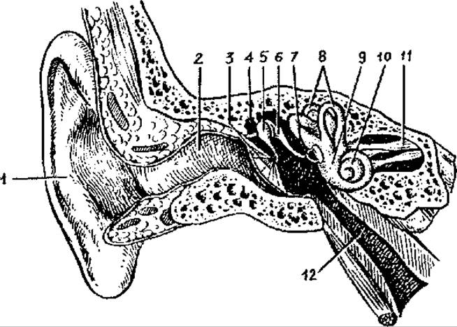
Fig. 98. Vestibulocochlear Organ:
1 — auricle, 2 — external acoustic meatus, 3 — tympanic membrane, 4 — tympanic cavity, 5 — malleus, 6 — incus, 7 — stapes, 8 — semicircular ducts, 9 — vestibule, 10 — cochlea, 11 — Vestibulocochlear nerve, 12 — auditory tube
The inner ear is located in the petrous part of the temporal bone between the tympanic cavity and the internal acoustic meatus. It is a system of narrow bony cavities (labyrinths) containing receptor apparatuses that perceive sound and changes in body position. Within the bony cavities, which are lined with periosteum, lies the membranous labyrinth, which mirrors the shape of the bony labyrinth. Between the membranous labyrinth and the bony walls, there is a narrow space—the perilymphatic space, filled with a fluid called perilymph.
The bony labyrinth consists of the vestibule, three semicircular canals, and the cochlea. The bony vestibule is an oval cavity that communicates with the semicircular canals. On the lateral wall of the bony vestibule, there is an oval-shaped vestibular window, closed by the base of the stapes. At the level of the beginning of the cochlea, there is the round cochlear window, closed by an elastic membrane,
The three bony semicircular canals lie in three mutually perpendicular planes. The anterior semicircular canal lies in the sagittal plane, the lateral in the horizontal plane, and the posterior in the frontal plane. Each semicircular canal has two limbs, one of which (the ampullary bony limb) expands to form an ampulla before entering the vestibule. The limbs of the anterior and posterior semicircular canals fuse to form a common bony limb. Thus, the three canals open into the vestibule through five orifices.
The bony cochlea makes 2.5 turns around a horizontally oriented central pillar. A bony spiral lamina is wound around the pillar like a screw thread and is pierced by fine canaliculi. The fibers of the cochlear part of the vestibulocochlear nerve pass through these canaliculi. At the base of the lamina lies the spiral canal, which contains the spiral ganglion. The lamina, together with the attached membranous cochlear duct, divides the cavity of the cochlear canal into two spirally wound passages—scalae (the scala vestibuli and scala tympani), which communicate with each other at the apex of the cochlea.
The walls of the membranous labyrinth are formed of connective tissue. The membranous labyrinth is filled with a fluid—endolymph, which drains through the endolymphatic duct, passing through the vestibular aqueduct, into the endolymphatic sac located within the dura mater on the posterior surface of the petrous part. From the perilymphatic space, perilymph drains via the perilymphatic duct, passing through the cochlear canaliculus, into the subarachnoid space on the Inferior surface of the petrous part of the temporal bone.
(vestibular apparatus of the inner ear)
The vestibular apparatus Functions to perceive the body's position in space and maintain balance. Any change in THE POSITION OF the body (or HEAD) stimulates the receptors of the vestibular apparatus. Impulses are transmitted to the Brain, which then sends nerve impulses to the appropriate muscles to correct BODY POSITION AND movements.
The vestibular apparatus consists of two parts: the vestibule and the semicircular ducts (channels). Within the bony vestibule lie two expansions of the membranous labyrinth: the elliptical sac (utricle) and the spherical sac (saccule). The spherical sac lies closer to the cochlea and communicates with the membranous cochlear duct via a connecting duct. The openings of the three membranous semicircular ducts—anterior, posterior, and lateral, oriented in three mutually perpendicular planes—open into the elliptical sac (utricle). The anterior (or superior) semicircular duct lies in the frontal plane, the posterior in the sagittal plane, and the lateral (external) in the horizontal plane. One end of each semicircular duct is dilated, forming an ampulla. On the inner surface of the spherical and elliptical sacs and the ampullae of the semicircular ducts, there are specialized regions containing sensory Hair Cells that perceive the body's position in space and disturbances of balance.
The elliptical and spherical sacs house a complex Structure known as the otolith apparatus, specifically called the maculae. The maculae of the sacs, oriented in the vertical and horizontal planes, consist of clusters of sensory hair cells. On the surface of these hair-bearing sensory cells lies a gelatinous otolithic membrane containing calcium carbonate crystals called otoliths or statoliths. The hairs of the receptor cells are embedded in the otolithic membrane. In the ampullae of the semicircular ducts, the receptor hair cells are located on the crests of folds, known as ampullary crests. Atop the hair Cells of the cristae sits a gelatinous, transparent cupula, which in shape is compared to a bell, though without a cavity.
Both the maculae of the sacs and the cristae of the semicircular duct ampullae are structures where sensory receptor hair cells respond very sensitively to any Changes in the position of the head (and body) in space. With any change in head position, the receptor hair cells detect changes in the state and movement of either the gelatinous otolithic membrane with its otoliths in the maculae, or the gelatinous cupula in the ampullary cristae. Any such action on the receptor hair cells generates a Nerve Impulse within them.
The sensory cells of the maculae perceive linear acceleration, gravity, and vibrational oscillations. In the normal, habitual position of the head, the otoliths exert pressure on specific hair cells. When the position changes, the otoliths exert pressure on other receptor cells, generating new nerve impulses that travel to the brain—specifically to the central Divisions of the vestibular analyzer—signaling a disturbance of the habitual equilibrium.
The sensory hair cells in the ampullary cristae generate nerve impulses during various rotational Movements of the head. These sensory cells are stimulated by the movement of the endolymph within the membranous semicircular ducts. Since the semicircular canals are oriented in three mutually perpendicular planes, any Rotation of the head inevitably sets the endolymph in motion within one canal or another, and its inertial pressure stimulates the receptor cells. The excitation generated in the receptor hair cells of the maculae and ampullary cristae is transmitted to the Nerve Cells of the vestibular ganglion, located at the bottom of the internal acoustic meatus. The axons of these cells form the vestibular part of the vestibulocochlear nerve (cranial nerve VIII), which exits through the internal acoustic meatus into the cranial cavity along with the cochlear part. In the cerebellopontine angle, the nerve fibers enter the brain substance and reach the vestibular nuclei located in the vestibular area on the floor of the Rhomboid fossa. The axons of the vestibular nuclei cells project to the fastigial nuclei of the Cerebellum via its inferior peduncle, to the Spinal Cord, and also as part of the medial longitudinal fasciculus of the Brainstem. From the cells of the vestibular nuclei, some fibers decussate and project to the thalamus, from where impulses are directed to the cortex of the parietal and temporal lobes (cortical centers of the statokinetic analyzer). In response to The stimulation of vestibular receptors, reflex reactions occur. Muscle tone is adjusted reflexively. To maintain and restore balance, the position of the head and the entire body is adjusted in the required direction.
It is well known that damage to the vestibular apparatus causes dizziness, and a person loses their balance. Hyperexcitability of the sensory cells of the vestibular apparatus causes motion sickness symptoms and other disorders.
Organ of hearing
(sound-perceiving apparatus of the inner ear)
The cochlea is a spirally coiled bony canal. The base of the cochlea faces the internal acoustic meatus. Inside the bony canal of the cochlea runs the membranous cochlear duct, which, like the bony cochlea, makes 2.5 turns and contains endolymph. The cochlear duct is formed by the bony wall (externally) and two connective tissue membranes—the more elastic basilar membrane and the thinner vestibular membrane. These two membranes divide the bony canal of the cochlea into three spiral passages: upper, middle, and lower. The middle passage is the cochlear duct, the upper is called the scala vestibuli (vestibular duct), and the lower is the scala tympani (tympanic duct). Both the scala vestibuli and scala tympani are filled with perilymph. The upper passage—the scala vestibuli—begins near the oval window, then winds spirally to the apex of the cochlea, where it connects to the lower passage—the scala tympani—via a narrow opening. The scala tympani, also winding spirally, ends at the round window, which is closed by the elastic secondary tympanic membrane.
Inside the endolymph-filled cochlear duct, resting on its basilar membrane adjacent to the scala tympani, lies the sensory apparatus—the spiral organ (of Corti). The spiral organ, which extends along all 2.5 turns of the cochlea, consists of 3—4 rows of receptor (hair) cells, totaling up to 24 000. Each receptor Cell has 30 to 120 fine hairs—microvilli—that end freely in the endolymph. Suspended above the hair cells along the entire length of the cochlear duct is the mobile tectorial membrane, with its free edge facing inward into the duct and the other edge attached to the basilar membrane.
Sound perception. Sound, which consists of air vibrations in the form of sound waves, enters the external auditory canal via the auricle and acts upon the tympanic membrane.
In this process, sound intensity depends on the amplitude of the sound wave vibrations perceived by the tympanic membrane. The greater the amplitude of the sound waves and, consequently, the vibration of the tympanic membrane, the louder the sound will be. Pitch depends on the frequency of the sound wave vibrations. A higher frequency of vibrations per unit of time is perceived by the auditory organ as higher tones (high-pitched, treble voices). A lower frequency of sound wave vibrations is perceived as low tones (deep, bass voices). The human ear can perceive sounds over a wide range, from 16 to 20 000 vibrations per second. Speech sound waves range from 150 to 2500 vibrations per second. As a person ages, their ear detects fewer sound wave vibrations. In elderly individuals, the ear can perceive no more than 15 000—13000 vibrations per second.
A person is able to determine the Location and direction of sounds through the coordinated function of both ears. An individual deaf in one ear must adapt to determine the direction of sound.
Vibrations of the tympanic membrane are transmitted to the auditory ossicles, whose movement (specifically of the stapes) causes the membrane of the oval window to vibrate (Fig. 99). The movement of the oval window sets the perilymph in motion within the scala vestibuli and scala tympani. These perilymph vibrations are easily transmitted to the endolymph in the cochlear duct. As the basilar membrane and endolymph move, the microvilli (hairs) of the receptor cells Touch the tectorial membrane with a specific frequency and force. Consequently, the sensory hair cells become excited, generating a receptor potential (nerve impulse). The receptor hair cells transduce mechanical stimuli into nerve impulses. The auditory nerve impulse is transmitted from the receptor cells to the next Neurons, whose cell bodies are located in the spiral ganglion. The axons of these cells form the cochlear part of the vestibulocochlear nerve. From there, impulses travel along the auditory nerve fibers to the brain, first reaching the subcortical auditory centers—the superior colliculi of the corpora quadrigemina and the lateral geniculate bodies. At the level of the subcortical auditory centers, reflex arcs are completed, mediating subconscious Reflexes (motor, secretory) in response to various auditory stimuli. Conscious perception of sounds, along with their higher analysis and synthesis, occurs in the cortical center of the Auditory Analyzer, located in the cortex of the superior temporal gyrus. Auditory nerve impulses arriving at the inferior colliculi of the corpora quadrigemina are transmitted to one of the extrapyramidal tracts—the tectospinal tract. Nerve impulses travel along this pathway to the motor nuclei of the anterior horns of the spinal cord, and through them, to the skeletal muscles.
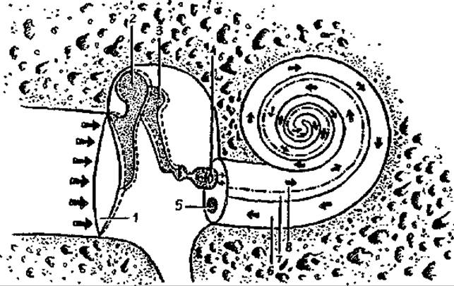
Fig. 99. Propagation of a sound wave (indicated by arrows) in the outer, middle, and inner ear:
1 — tympanic membrane, 2 — malleus, 3 — incus, 4 — stapes, 5 — round window, 6 — scala tympani, 7 — cochlear duct, 8 — scala vestibuli
Age-related Features of the organ of hearing and equilibrium
In the newborn, the auricle is flattened, its cartilage is soft, and the overlying skin is thin. The lobule of the auricle (earlobe) is small. The auricle grows most rapidly During the first 2 years of life and after the age of 10. It grows faster in length than in width. The external auditory canal in the newborn is narrow, long (about 15 mm), sharply curved, and has constrictions at the boundary between its widened medial and lateral parts. The walls of the external auditory canal are cartilaginous, except for the tympanic ring. The skin lining the external canal is thin and delicate. In a 1-year-old child, the length of the external auditory canal is about 20 mm, and in a 5-year-old child, it is 22 mm.
The tympanic membrane in the newborn is relatively large. Its height is 9 mm, and its width, as in adults, is 8 mm. The tympanic membrane in the newborn is more oblique than in the adult. The angle it forms with the inferior wall of the external auditory canal is 35—40°.
The tympanic cavity of a newborn differs little in size from that of an adult, but it appears narrow due to the thickened mucous membrane at this age. At birth, the tympanic cavity contains fluid which, with the onset of Respiration, passes through the auditory tube into the pharynx and is swallowed. The walls of the tympanic cavity are thin, especially the superior wall. The posterior wall has a wide opening leading to the mastoid antrum. Mastoid cells are absent in the newborn due to the poor Development of the mastoid process. The auditory ossicles are close in size to those of an adult. The auditory tube in the newborn is straight, wide, and short (17—21 mm). During the first year of life, the auditory tube grows slowly, and more rapidly during the second year. The length of the auditory tube is 20 mm in a 1-year-old child, 30 mm at 2 years, 35 mm at 5 years, and 35—38 mm in an adult. The lumen of the auditory tube narrows gradually: from 2.5 mm at 6 months to 2 mm at 2 years, and to 1—2 mm in a 6-year-old child.
The inner ear of the newborn is well developed, and its dimensions are close to those of an adult. The bony walls of the semicircular canals are thin and gradually thicken due to the fusion of ossification centers in the petrous part of the temporal bone.
Developmental Disorders of the receptor apparatus (spiral organ) and underdevelopment of the auditory ossicles that prevents their movement lead to congenital deafness. Defects in the position, shape, and STRUCTURE OF THE outer ear (malformations) are usually associated with underdevelopment of the Mandible (micrognathia) or even its absence (agnathia).
REVIEW AND SELF-control questions:
1. Describe the tympanic cavity and the Organs located within it. Name the walls of the tympanic cavity.
2. List the anatomical structures belonging to the inner ear.
3. Explain which structures of the inner ear perceive static and dynamic changes in body position, and how they do so.
4. Describe The structure of the cochlea, its scalae, and the cochlear duct.
5. Discuss the location and function of perilymph and endolymph.
6. Along which pathways and to which subcortical and cortical centers do nerve impulses from the vestibular and auditory receptors of the inner ear travel?
Last update: 10/08/2026
Editorial and Educational Adaptation: This material has been compiled based on the primary/original source text. The project team performed an editorial review, corrected technical inaccuracies, structured sections, and adapted the content for an educational format.
What was processed:
- elimination of formatting defects (OCR errors, structural breaks, corrupted characters);
- editorial organization of content;
- standardization of terminology in accordance with academic sources;
- verification of factual statements against the original source text.
All mentions of the author, publication year, and origin of the primary text have been preserved in accordance with the source.