Human Anatomy - H. I. Koliadenko 2009
Sense Organs (Analyzers)
Vestibulocochlear Organ
In the course of evolution, the vestibulocochlear organ achieved its highest development in mammals. In invertebrates, it is poorly developed and Functions mainly as an equilibrium organ, typically shaped like a pit containing sensory hairs inside. Salt crystals are deposited within this pit, shifting in response to changes in body position. The only exception is found in certain insects, where the Organ of Hearing is located on the first abdominal segment or the Tibia. The Progressive development of the vestibulocochlear organ begins in vertebrates. Fishes possess a well-developed Organ of Equilibrium and, only in its earliest developmental stages, an organ of hearing resembling a blind pouch called the lagena, which evolves into the cochlea. In amphibians, the cochlea already performs an auditory function. It becomes even more refined in birds and crocodiles, where the cochlea forms a spiral twist, yet it reaches its ultimate refinement in mammals.
Structure OF THE auditory organ. The Organ of hearing consists of three parts: the outer, middle, and Inner ear.
The outer ear comprises the auricle (pinna) and the external acoustic meatus (Fig. 192). The auricle is located in the temporal region and is formed by auricular Cartilage covered with Skin. The lower part of the auricle, which lacks cartilage, is called the lobule, or earlobe. In addition to this structure, the auricle features the helix, antihelix, tragus — a small projection shielding the external acoustic opening — and the antitragus. Several rudimentary Muscles lie beneath the skin of the auricle, making it virtually immobile. In most animals, however, the auricle is highly mobile thanks to the contraction of numerous muscles.
Class="center">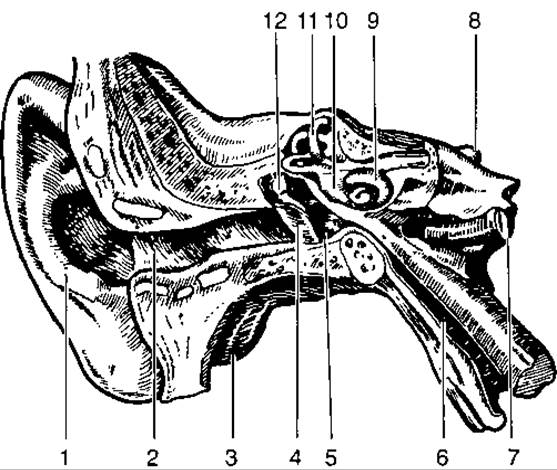
Fig. 192. The vestibulocochlear organ:
1 — auricle; 2 — external acoustic meatus; 3 — mastoid process of the Temporal bone; 4 — tympanic membrane; 5 — tympanic cavity; 6 — auditory tube; 7 — Internal Carotid Artery; 8 — Vestibulocochlear nerve; 9 — labyrinth of the inner ear; 10 — vestibule; 11 — semicircular canals; 12 — auditory ossicles
The external acoustic meatus begins deep within the auricle at the external acoustic opening. It is 24–30 mm long and up to 0.9 mm in diameter. Throughout its length, the auditory canal is lined with skin containing numerous Sebaceous Glands that secrete sebum and cerumen (earwax). The inner end of the acoustic meatus is sealed by the tympanic membrane, which separates the outer ear from the Middle ear.
The middle ear consists of the tympanic cavity with three auditory ossicles, the tympanic membrane, mastoid air Cells, and the auditory tube (Fig. 192). The tympanic cavity is a small space within the pyramid of the temporal bone featuring six walls: outer (membranous), inner (labyrinthine), anterior (carotid), posterior (mastoid), upper (tegmental), and lower (jugular). The outer wall is formed by the tympanic membrane. The inner wall connects the middle ear to the inner ear and contains two openings: the upper one, oval, covered by the Base of the stapes; and the lower one, round, sealed by the secondary tympanic membrane. The upper wall separates the tympanic cavity from the cranial cavity, the lower wall from the jugular fossa, the anterior wall from the carotid canal, and the posterior wall from the mastoid process.
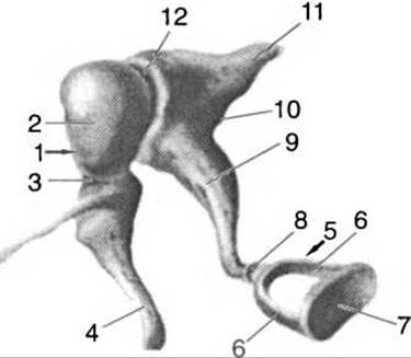
Fig. 193. Auditory ossicles and their joints:
1 — malleus; 2 — HEAD of malleus; 3 — neck of malleus; 4 — handle of malleus; 5 — stapes; 6 — crura of stapes; 7 — base of stapes; 8 — incudostapedial joint; 9 — long crus of incus; 10 — incus; 11 — short crus of incus; 12 — incudomalleolar joint
Posteriorly, the tympanic cavity communicates with the mastoid air cells. The cavity is lined with mucous membrane and filled with air. Three auditory ossicles are arranged in a chain within the tympanic cavity: the malleus, incus, and stapes (Fig. 193). The malleus consists of a head, neck, and handle. The handle is fused with the tympanic membrane, while the head lies above the tympanic membrane in a recess of the tympanic cavity. The incus features a body with a saddle-shaped depression that articulates with the head of the malleus, as well as two limbs — a short one and a long one. The short limb attaches to the wall of the tympanic cavity via a ligament, whereas the long limb articulates with the head of the stapes. The stapes comprises a head, two crura (limbs), and a base (footplate). The base of the stapes fits into the oval window, separating the middle ear from the inner ear.
The auditory tube connects the tympanic cavity to the Pharynx. It is a tube 3–4 cm long, composed of cartilaginous and bony parts. The auditory tube ventilates the middle ear with air and plays a vital role in equalizing atmospheric pressure on the tympanic membrane.
The inner ear is situated within the pyramid of the temporal bone and consists of the bony and membranous labyrinths (Fig. 194).
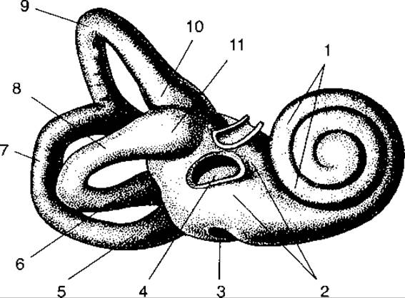
Fig. 194. Bony labyrinth of the auditory organ:
1 — cochlea; 2 — vestibule; 3 — cochlear window; 4 — vestibular window; 5, 6, 10, 11 — ampullae of semicircular canals; 7 — posterior semicircular canal; 8 — lateral semicircular canal; 9 — superior semicircular canal
The bony labyrinth, located within the petrous part of the temporal bone, comprises the vestibule, cochlea, and bony semicircular canals.
The vestibule occupies a central position and contains three recesses: the elliptical, spherical, and cochlear recesses, separated by the vestibular crest. The elliptical recess communicates via five openings with the bony semicircular canals, while the other two connect with the cochlear ducts. Small cribriform spots (maculae cribrosae) are found within these recesses, transmitting Branches of the vestibular division of the vestibulocochlear nerve. Two openings are located on the lateral wall of the vestibule: the vestibular window (oval window) and the cochlear window (round window), through which the vestibule connects to the tympanic cavity. The vestibular window is closed by a membrane, whereas the cochlear window is sealed by the stapes.
The bony semicircular canals lie in three mutually perpendicular planes (horizontal, frontal, and sagittal). The canals open into the vestibule via their limbs. Three of these limbs feature expansions known as bony ampullae, while the other three are smooth. The smooth limbs of the anterior and posterior semicircular canals unite into a common limb before entering the vestibule, meaning the semicircular canals open into the vestibule through a total of five openings.
The cochlea (Fig. 195) makes two and a half spiral turns around a central bony pillar called the modiolus. A bony spiral lamina extends from the modiolus, dividing the canal's cavity into two parts, or scalae. The upper scala, the scala vestibuli, begins at the vestibule and extends to the apex of the cochlea. At the apex, a small opening called the helicotrema connects the scala vestibuli with the scala tympani. The scala tympani runs from the cochlear apex back to its base, where it opens into the bony labyrinth via the cochlear window, which is sealed by the secondary tympanic membrane.
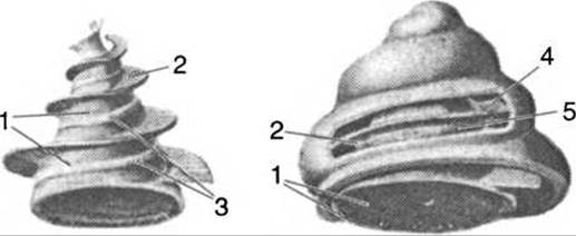
Fig. 195. Bony cochlea:
1 — modiolus; 2 — osseous spiral lamina; 3 — site of fusion between the modiolus and the edges of the cochlear turns; 4 — scala vestibuli; 5 — scala tympani
In addition to the cochlea and semicircular canals, the vestibular aqueduct opens into the vestibule and emerges on the posterior surface of the pyramid of the temporal bone.
Membranous labyrinth. Inside the osseous labyrinth lies the membranous labyrinth. A space filled with fluid, the perilymph, is formed between the walls of the osseous and membranous labyrinths. Endolymph is contained within the membranous labyrinth. The perilymph drains into the subarachnoid space, whereas the endolymph drains into the endolymphatic duct of the dura mater. The Vestibule of the osseous labyrinth houses two PARTS OF THE membranous labyrinth: the utricle and the saccule, which are connected by a duct. The semicircular ducts of the membranous labyrinth open into the utricle. The saccule is connected to the cochlea. The walls of the membranous labyrinth and semicircular canals consist of Connective Tissue, and their internal surface is lined with a single-layered squamous epithelium that transitions into the neuroepithelium of the maculae (elevations) located in the saccules and the ampullae of the semicircular canals. The maculae and cristae are covered with receptor Hair cells. Their surface abounds with otoliths—microscopic calcium carbonate crystals. When THE POSITION OF the head changes, the endolymph, along with the otoliths, shifts toward the sensory cells responsible for maintaining body equilibrium in space. The sensory Cells of the maculae and cristae connect with the peripheral nerve fibers of the vestibular nerve, which transmits excitation to the neural centers of the Brain.
Between the vestibular and tympanic scalae lies the triangular, spirally coiled cochlear duct. It begins blindly from the vestibular saccule—where the utriculosaccular duct empties—and likewise ends blindly at the apex.
The cochlear duct has three walls: external, upper, and lower. The lower wall is called the basilar lamina (spiral membrane) and fuses with the osseous spiral lamina. The basilar membrane consists of fibrous filaments stretched between the osseous spiral lamina and the wall of the osseous cochlear duct. These fibers vary in length and number approximately 24,000. They are set into motion by the vibrations of the perilymph and endolymph.
Depending on the pitch of a sound, the fibers whose length is adapted to perceive it will vibrate: the longer the fiber, the lower the tonal frequency that triggers its vibration; conversely, high-frequency sounds cause the shorter fibers of the basilar lamina to vibrate. Situated on the basilar lamina is the spiral organ (Organ of Corti), which comprises supporting and receptor cells. The receptor cells are known as hair cells, or sensory cells. Their surface features a delicate hair. These sensory hair cells synapse with the processes of the bipolar cells belonging to the first neuron of the Auditory pathway. Overhanging the spiral organ is the tectorial membrane, composed of a gelatinous substance. Sound perception occurs as follows: sound waves captured by the outer ear reach the tympanic membrane, the vibrations of which are transmitted to the auditory ossicles of the middle ear, and subsequently via the vestibular window to the perilymph of the scala vestibuli. At the apex of the cochlea, the scala vestibuli communicates with the scala tympani, through which the perilymph vibrations reach the cochlear window, which is sealed by the secondary tympanic membrane. The vibrations of the perilymph are transferred to the endolymph, which in turn causes the corresponding fibers on the basilar lamina to vibrate. As a result of the basilar lamina's oscillation, the sensory hair cells make contact with the tectorial membrane, which receives and relays the auditory stimuli.
The conducting pathway of the cochlear analyzer begins with the sensory Neurons of the spiral ganglion, located within the modiolus of the osseous cochlea, which is also the Water/144.html">Origin of the osseous spiral lamina. The dendrites of the spiral ganglion pass through the canaliculi of the basilar membrane to reach the sensory hair cells—the receptors of the spiral organ. The neurites of the spiral ganglion travel through the canals of the modiolus into the internal acoustic meatus, where they merge with the fibers of the vestibular nerve to form the ROOT of the VIII cranial nerve, directed toward the nerve nuclei situated in the Rhomboid fossa of the Fourth ventricle. From the Medulla Oblongata, the fibers of the auditory nerve extend to the inferior colliculi of the tectum of the Midbrain, then to the medial geniculate bodies, further to the thalamus (optic thalamus), and finally to the auditory center located in the middle part of the superior temporal gyrus of the Cerebral Cortex.
The conducting pathway of the vestibular analyzer originates from the vestibular ganglion, located within the internal acoustic meatus. Its dendrites extend to the receptors found in the maculae of the vestibule and the cristae of the ampullae of the semicircular canals, while its neurites run to the common root of the VIII cranial nerve leading to the medulla oblongata. The majority of vestibular nerve fibers terminate in the vestibular nuclei of the rhomboid fossa, whereas a smaller portion extends to the cortex of the cerebellar vermis. The cortical center of the vestibular analyzer is situated in the anterior region of the temporal lobe cortex of the cerebral hemispheres.
Last update: 08/08/2026
Editorial and Educational Adaptation: This material has been compiled based on the primary/original source text. The project team performed an editorial review, corrected technical inaccuracies, structured sections, and adapted the content for an educational format.
What was processed:
- elimination of formatting defects (OCR errors, structural breaks, corrupted characters);
- editorial organization of content;
- standardization of terminology in accordance with academic sources;
- verification of factual statements against the original source text.
All mentions of the author, publication year, and origin of the primary text have been preserved in accordance with the source.