Special Histology and Embryology: Practical Course - V. K. Napkhanyuk 2001
Sensory Organs
The Organ of Hearing and Equilibrium
The Organ of Hearing and Balance, or Vestibulocochlear Organ (organum vestibulocochleare), consists of the external, middle, and internal ear, which together perceive sound, gravitational, and vibrational stimuli, as well as linear and angular accelerations.
The External ear (auris externa) comprises the auricle (pinna), the external acoustic meatus, and the tympanic membrane.
The auricle is formed by a thin plate of elastic Cartilage, covered with Skin containing a few fine hairs and Sebaceous Glands, while Sweat Glands are very sparse.
The external acoustic meatus is formed by cartilage that is a continuation of the elastic cartilage of the auricle. The surface of the meatus is covered with thin skin containing hairs and associated sebaceous glands. Located deeper than the sebaceous glands are the ceruminous glands (glandulae ceruminosae), which secrete earwax (cerumen).
The tympanic membrane has an oval or elongated shape. In its central part, it consists of two layers formed by bundles of Collagen fibers with fibroblasts interspersed between them. The fibers of the outer layer run radially, while those of the inner layer run circularly.
The upper part of the tympanic membrane contains a smaller number of collagen fibers. Fine elastic fibers are present at the periphery and in the center of the membrane.
The outer surface is covered by a very thin (50–60 µm) layer of epidermis.
The inner surface, facing the Middle ear, is covered by a mucous membrane 20–40 µm thick, formed by a simple squamous epithelium.
The middle ear (auris media) consists of the tympanic cavity, auditory ossicles, and the auditory (Eustachian) tube.
The tympanic cavity is lined with a simple squamous epithelium, which transitions into a cuboidal or columnar epithelium in certain areas.
The medial wall of the tympanic cavity features two openings, or windows: the oval window contains the Base of the stapes, which is held in place by a thin annular ligament surrounding it; the round window lies posterior to the oval window and is closed by a fibrous membrane. It separates the tympanic cavity from the scala tympani of the cochlea.
The auditory ossicles are the malleus, incus, and stapes. Acting as a lever system, they transmit vibrations from the tympanic membrane of the external ear to the oval window, from which the vestibular scala of the Inner ear begins.
The auditory tube, which connects the tympanic cavity to the nasopharynx, has a well-defined lumen with a diameter of 1–2 mm. The lumen is lined with a pseudostratified ciliated columnar epithelium containing goblet mucous Cells. The auditory tube regulates air pressure within the tympanic cavity of the middle ear.
The internal ear (auris interna) consists of the bony labyrinth and the membranous labyrinth located within it.
In specific Regions of the membranous labyrinth, there are fibrous sensory epitheliocytes of the Auditory and Vestibular apparatus: the auditory receptor cells are located in the Cytology/practical/79.html">Spiral organ of the cochlea (cochlear labyrinth), whereas the receptor Cells of the Organ of Balance are found in the utricle and saccule, as well as the ampullary crests of the semicircular ducts (vestibular labyrinth).
The cochlear duct (ductus cochlearis) is a spiral Structure about 3.5 cm long, filled with endolymph and surrounded by perilymph. The cochlear duct and the surrounding cavities of the tympanic and vestibular scalae are filled with perilymph; these are enclosed within the bony cochlea, which makes 2.5 turns around a central bony axis (modiolus).
In cross section, the cochlear duct has a triangular shape, the sides of which are formed by:
a) the vestibular membrane (membrana vestibularis), which forms the upper-medial wall of the duct. This is a thin fibrillar Connective Tissue plate covered with a simple squamous epithelium facing the endolymph, and an endothelium facing the perilymph;
b) the stria vascularis, which lies on the lateral wall of the bony cochlea. Situated on the spiral ligament (ligamentum spirale), its epithelium is stratified and composed of flat, light basal cells and tall, branching, dark columnar cells rich in Cell/35.html">Mitochondria. The stria vascularis is believed to perform a secretory function;
c) the basilar lamina (lamina basilaris), upon which the spiral organ rests. This is a connective tissue plate that runs in a spiral along the entire length of the cochlear duct. The side facing the spiral organ is covered by the basal membrane of the epithelium of this organ. The foundation of the basilar lamina consists of fine collagen fibers (strings) that stretch as a continuous radial bundle from the osseous spiral lamina to the spiral ligament, projecting into the cavity of the bony cochlear canal. On the side of the scala tympani, the basilar lamina is covered by a layer of flat mesenchymal cells.
Sound perception takes place in the spiral organ, which is located along the entire length of the cochlear duct.
The spiral organ (organum spirale) consists of two groups of cells: 1) sensory cells; 2) supporting cells.
The sensory cells are subdivided into: inner sensory Hair epitheliocytes (epitheliocyti sensoriae pilosoe internae), which are flask-shaped cells with widened bases arranged in a single row. The surface of their slightly convex apices bears 30 to 60 short, deflectable microvilli—stereocilia—arranged in 3–4 rows; and outer sensory hair epitheliocytes (epitheliocyti sensoriae pilosoe externae), which have rounded bases and bear a cuticular plate with stereocilia on their apical surface. These Hair cells lie in three parallel rows, though humans may have 4–5 such rows.
Unlike sensory cells, the supporting epitheliocytes of the spiral organ rest directly with their bases on the basement membrane. The following types of supporting epitheliocytes are distinguished:
— inner phalangeal epitheliocytes (epitheliocyti phalangeae internae), which lie beneath the inner sensory hair epitheliocytes and possess thin finger-like processes (Phalanges);
— internal and external pillar cells (epitheliocyti pilaris internae et externae), which Touch at an acute angle to form a regular triangular canal — the internal tunnel (cuniculus internus), filled with endolymph;
— external phalangeal cells (epitheliocyti phalangeae externae) of prismatic shape, located on the basilar membrane in 3–4 rows near the external pillar cells;
— external limiting epitheliocytes (cellulae epitheliocyti limitans externae), situated on the basement membrane adjacent to the external phalangeal epitheliocytes and forming a continuous row of low epithelial cells;
— external supporting epitheliocytes (epitheliocyti sustentas externus), which are cubic in shape and gradually transition into the epithelium lining the stria vascularis.
Slides for Study
Slide 1. Cross-section of the cochlear duct (Fig. 9).
Low power. Three cavities (ducts) are visible in the slide. The middle cavity is triangular in shape; this is the cochlear duct (ductus membranacea). The upper and lower cavities belong to the bony cochlea: the upper is called the scala vestibuli, and the lower is the scala tympani. Both are filled with perilymph, while the cochlear duct contains endolymph.
The walls of the membranous duct have a complex structure. The upper-medial wall faces the scala vestibuli and is formed by the vestibular membrane (paries vestibularis). This is a thin fibrillar plate covered by a simple squamous epithelium facing the endolymph and an endothelium facing the perilymph. The outer wall is formed by the spiral ligament (lig. spirale cochleae), which is an extension of the periosteum covered by the vascular stria (stria vascularis) — a so-called stratified columnar epithelium. It consists of flat, clear basal cells and tall, branching, dark prismatic cells. Blood capillaries are located between the cells.
The lower wall of the membranous cochlear duct is represented by the basilar membrane (lamina basilaris), which is based on fine collagen fibers ("strings"). The membrane runs in a spiral along the entire length of the cochlear duct. The spiral (Corti's) organ (organum spirale) rests on the basilar membrane, with a basement membrane positioned between them.
Label on the drawing: 1) cochlear duct; 2) scala vestibuli; 3) scala tympani; 4) vestibular membrane; 5) spiral ligament; 6) basilar membrane; 7) spiral (Corti's) organ.
High power. Study and draw the spiral organ (Fig. 10). This organ is formed by supporting and sensory cells. These cells are subdivided into internal and external groups.
The tunnel serves as the boundary. The basal ends of the supporting cells rest on the basement membrane.
Among the supporting cells, the following are distinguished:
— pillar cells — internal and external (cellula piloris interna and externa), which are arranged in a single row and form a triangular channel between them, known as the tunnel.
— phalangeal cells, or Deiters' cells — external or internal (epiteliocyti phalangeae externae and internae). The external cells are arranged in 3–5 rows, and the internal cells in a single row;
— external limiting cells (cellulae epitheliocyti limitans externae), or Hensen's cells;
— external supporting cells (epitheliocyti sustentans externus), or Claudius' cells, which have a cubic shape.
Class="center">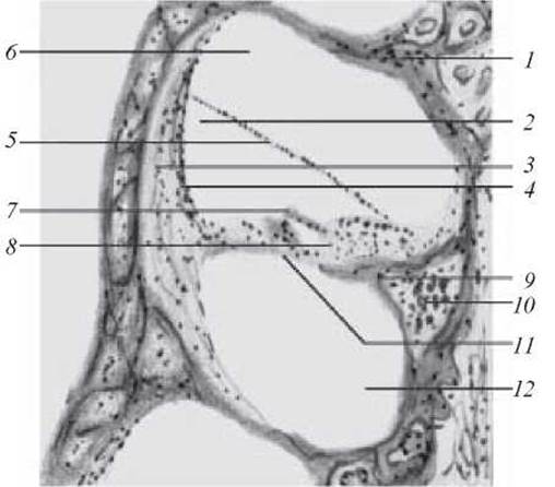
Fig. 9. Cross-section of the cochlear duct. Hematoxylin and eosin staining. x 56:
1 — bony wall of the cochlea; 2 — membranous cochlear duct; 3 — spiral ligament; 4 — stria vascularis; 5 — vestibular membrane; 6 — scala vestibuli; 7 — tectorial membrane; 8 — spiral organ; 9 — osseous spiral lamina; 10 — spiral ganglion; 11 — tympanic wall of the cochlear duct with the basilar membrane; 12 — scala tympani
Sensory, or hair, cells (cellulae sensoriae pilosae) are located on the phalangeal (Deiters') cells. The external hair cells are arranged in 3–5 rows, corresponding to the number of Deiters' cells. The internal hair cells are arranged in a single row.
The tectorial membrane (membrana tectoria) is located above the spiral organ.
Label on the drawing: 1) internal hair cell; 2) external hair cell; 3) internal pillar cells; 4) external pillar cells; 5) tunnel; 6) internal supporting cells; 7) external supporting cells; 8) limbus of the osseous spiral lamina; 9) vestibular lip of the limbus; 10) tympanic lip of the limbus; 11) spiral sulcus of the limbus.
The vestibular part of the membranous labyrinth consists of two sacs — the utricle (utriculus) and the saccule (sacculus), which are connected by a narrow duct and communicate with three semicircular canals positioned in three mutually perpendicular planes within the bony canals. At the point where these canals join the utricle, they feature expansions called ampullae. In the regions of the utricle, saccule, and ampullae, the wall of the membranous labyrinth contains areas with sensory cells. In the sacs, these areas are called maculae: the macula of the utricle (macula utriculi) and the macula of the saccule (macula sacculi), while in the ampullae, they are known as crests, or cristae ampullares.
The wall of the vestibular part of the membranous labyrinth consists of a simple squamous epithelium, except for the regions of the cristae of the semicircular canals and the maculae, where it transitions into a cuboidal and columnar epithelium (micrograph 6).
The maculae of the saccule are lined with an epithelium resting on a basement membrane and consisting of sensory and supporting cells. The epithelial surface is covered by a specialized gelatinous otolithic membrane in which crystals of calcium carbonate—otoliths, or statoconia—are embedded.
The sensory hair cells (cellulae sensoriae pilosae) bear numerous hairs on their apical surfaces facing the labyrinth cavity. The base of The Cell is in contact with afferent and efferent nerve endings (micrograph 7).
Structurally, Two Types of cells are distinguished: 1) pear-shaped cells, with a broad rounded base embraced by a nerve ending that forms a chalice-like sheath establishing focal synaptic contacts with the receptor cell; 2) columnar cells of prismatic shape, whose outer surface features a cuticle giving rise to 60–80 non-motile hairs—stereocilia about 40 µm in length—and a single motile cilium—kinocilium—which shares The structure of a standard motile cilium.
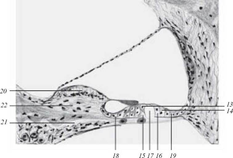
Fig. 10. Spiral (Corti's) organ. Hematoxylin and eosin staining. X 400:
For designations 1–12 see Fig. 9. 13 — inner hair cell; 14 — outer hair cell; 15 — inner pillar cells; 16 — outer pillar cells; 17 — inner tunnel; 18 — inner supporting cells; 19 — outer supporting cells; 20 — vestibular lip of the limbus; 21 — tympanic lip of the limbus; 22 — spiral sulcus of the limbus
The round macula contains approximately 18,000 receptor cells, whereas the oval macula contains about 33,000.
Supporting cells (epitheliocyti sustentans) are interspersed among the sensory cells and are distinguished by dark oval nuclei and an Abundance of mitochondria. Numerous fine cytoplasmic microvilli are found on their apical surfaces.
The macula of the utricle serves as the site for the perception of linear accelerations, namely gravity, acting as a gravity receptor associated with modulating the Muscle tone that determines body posture.
The macula of the saccule also Functions as a gravity receptor while simultaneously detecting vibrational stimuli.
Ampullary crests, or cristae ampullares: each ampullary dilatation of a semicircular canal contains cristae in the form of transverse folds.
The ampullary crest is lined with sensory hair cells and supporting epitheliocytes.
The apical part of these cells is enveloped by a gelatinous, transparent cupola (cupula gelatinosa) approximately 1 mm in length. The gelatinous cupola acts as a receptor for angular accelerations.
Slide 2. Cross section through the utricle of the vestibule and the ampulla of the semicircular canal (Fig. 11).
High magnification. Examine the slide and identify the macula of the saccule (it is a flattened structure based on periosteum). Supporting and sensory hair cells are located upon it.
The hairs of the sensory cells are covered by a specialized gelatinous otolithic membrane containing crystals of calcium carbonate—otoliths, or statoconia.

Fig. 11. Cross section through the utricle of the vestibule and the ampulla of the semicircular canal. Hematoxylin and eosin staining. x 200:
1 — cavity of the utricle; 2 — connective tissue base of the utricle and its lining epithelium; 3 — macula of the utricle (a — hair and supporting cells; b — gelatinous substance with otoliths); 4 — ampullary cavity; 5 — membranous part of the ampulla and its lining epithelium; 6 — crista ampullaris (a — sensory hair cells; b — supporting cells; c — cupola); 7 — vestibular nerve ganglion

Electronic micrograph 6. Hair cell of the ampullary crest. x 20,000:
1 — Nucleus of the hair cell; 2 — cuticle; 3 — mitochondria; 4 — Ribosomes; 5 — calyx-like nerve ending (encompassing the hair cell); 6 — mitochondria within the nerve ending (after Friedman)
Control questions
1. Embryonic sources of The Organ of Hearing and Equilibrium.
2. The external ear.
3. Middle ear.
4. Inner ear.
5. STRUCTURE OF THE cochlear duct of the membranous labyrinth.
6. Spiral organ and its structure.
7. Structural features and function of sensory epithelial cells.
8. Structural features and function of supporting epithelial cells.
9. Structure of the vestibular part of the membranous labyrinth.
10. Structure and function of the maculae of the sacculus and utriculus.
11. Structure and function of the ampullary crests.
12. Microscopic and ultramicroscopic Structural Features of receptor and supporting cells of the balance organ.
Situational problems
1. What function will be impaired if the receptor cells of the ampullary crests in the semicircular canals of the membranous labyrinth are damaged? What are these cells called?
2. As a result of a chronic inflammatory process, the spiral ganglion is affected. What functional changes will be detected?

Electron micrograph 7. Hair cells of the utricular macula. x 20,000:
1 — cylindrical hair cells (type II receptor cells); 2 — chalice-shaped hair cells (type I receptor cells); 3 — nerve endings on type I cells; 4 — nerve endings on type II cells; 5 — bundle of static hairs; 6 — stereocilia (non-motile hairs); 7 — kinocilia (motile hairs with a typical fibrillar structure); 8 — supporting cells; 9 — desmosomes (according to A. A. Kronshtein and G. A. Pyatkina)
3. A person has damage to the cells of the spiral organ in the lower turns of the cochlea. The perception of which sounds will be impaired?
Sample examination questions
1. Sources of development, structure, and histophysiology of the Organ of Hearing.
2. Development, structure, and histophysiology of the organ of balance.
Last update: 10/08/2026
Editorial and Educational Adaptation: This material has been compiled based on the primary/original source text. The project team performed an editorial review, corrected technical inaccuracies, structured sections, and adapted the content for an educational format.
What was processed:
- elimination of formatting defects (OCR errors, structural breaks, corrupted characters);
- editorial organization of content;
- standardization of terminology in accordance with academic sources;
- verification of factual statements against the original source text.
All mentions of the author, publication year, and origin of the primary text have been preserved in accordance with the source.