Human Anatomy and Physiology - N. I. Fedyukovich 2003
Sensory Organs
Organ of hearing and balance
The Organ of Hearing and Balance, the Vestibulocochlear Organ (organum vestibulocochleare) in humans, has a complex Structure, perceives sound wave vibrations, and determines the orientation of the body in space.
The vestibulocochlear organ (Fig. 148) is divided into three parts: the outer, middle, and Inner ear. These parts are closely integrated anatomically and functionally. The outer and Middle ear conduct sound vibrations to the inner ear, thus acting as a sound-conducting apparatus. The inner ear, which consists of the bony and membranous labyrinths, forms the actual Organ of Hearing and balance.
Class="center">
Fig. 148. Vestibulocochlear organ (organ of hearing and balance):
1 — superior semicircular canal; 2 — vestibule; 3 — cochlea; 4 — auditory nerve; 5 — carotid artery; 6 — auditory tube; 7 — tympanic cavity; 8 — tympanic membrane; 9 — external acoustic meatus; 10 — external acoustic opening; 11 — auricle; 12 — malleus
The outer ear includes the auricle, the external acoustic meatus, and the tympanic membrane, which are designed to capture and conduct sound vibrations. The auricle consists of elastic Cartilage, has a complex configuration, and is covered externally by Skin. Cartilage is absent in the lower part, known as the lobule of the auricle or earlobe. The free margin of the auricle is folded and called the helix, while the parallel ridge running along it is the antihelix. Near the anterior margin of the auricle, a projection stands out—the tragus, with the antitragus located behind it. The auricle is attached to the Temporal bone by ligaments and has rudimentary Muscles, which are well developed in animals. The auricle is structured to concentrate sound vibrations as much as possible and direct them into the external acoustic opening.
The external acoustic meatus is an S-shaped tube that opens externally at the acoustic opening, ends blindly deep inside, and is separated from the middle ear cavity by the tympanic membrane. The length of the acoustic meatus in an adult is about 36 mm; its diameter reaches 9 mm at the beginning and 6 mm at its narrowest point. The cartilaginous part, which is a continuation of the auricular cartilage, makes up 1/3 of its length, while the remaining 2/3 is formed by the bony canal of the temporal bone. At the transition between these two parts, the external acoustic meatus is narrowed and curved. It is lined with skin and is rich in Sebaceous Glands, as well as ceruminous glands that secrete earwax.
The tympanic membrane is a thin, translucent, oval plate measuring 11x9 mm, located at the boundary between the outer and middle ear. It is positioned obliquely, forming an acute angle with the lower wall of the acoustic meatus. The tympanic membrane consists of two parts: a larger lower part—the pars tensa, and a smaller upper part—the pars flaccida. Externally, it is covered with skin, its core is formed by Connective Tissue, and internally it is lined with a mucous membrane. In the center of the tympanic membrane, There is a depression—the umbo, which corresponds to the attachment of the handle of the malleus on the inner side.
The middle ear includes the air-filled tympanic cavity (with a volume of about 1 cm³), which is lined with mucous membrane, and the auditory (Eustachian) tube. The middle ear cavity connects to the mastoid antrum and, through it, to the mastoid Cells of the mastoid process.
The tympanic cavity is located within the petrous part of the temporal bone, between the tympanic membrane laterally and the bony labyrinth medially. It has six walls:
1) the superior tegmental wall — separates it from the cranial cavity and is located on the superior surface of the petrous part of the temporal bone; 2) the inferior jugular wall — separates the tympanic cavity from the external Base of the Skull, is located on the Inferior surface of the petrous part of the temporal bone, and corresponds to the jugular fossa; 3) the medial labyrinthine wall — separates the tympanic cavity from the bony labyrinth of the inner ear. On this wall is the oval window (fenestra vestibuli), closed by the base of the stapes; slightly higher on this wall is the prominence of the facial canal, and below is the round window (fenestra cochleae), closed by the secondary tympanic membrane, which separates the tympanic cavity from the scala tympani; 4) the posterior mastoid wall — separates the tympanic cavity from the mastoid process and has an opening leading into the mastoid antrum, which in turn connects to the mastoid cells; 5) the anterior carotid wall — borders the carotid canal. Here is the tympanic opening of the auditory tube, through which the tympanic cavity connects to the nasopharynx; 6) the lateral membranous wall — formed by the tympanic membrane and the surrounding PARTS OF THE temporal bone.
Within the tympanic cavity, there are three auditory ossicles covered with mucous membrane, as well as Ligaments and Muscles. The auditory ossicles (Fig. 149) are small. Articulating with each other, they form a chain extending from the tympanic membrane to the oval window. All ossicles are connected by joints and covered with mucous membrane. The handle of the malleus is attached to the tympanic membrane, while its HEAD articulates with the incus, which in turn is movably connected to the stapes. The base of the stapes closes the oval window.
There are two muscles in the tympanic cavity: one runs from the canal of the same name to the handle of the malleus, and the other—the stapedius Muscle—passes from the posterior wall to the posterior limb of the stapes. Contraction of the stapedius muscle alters the pressure exerted by the base of the stapes on the perilymph.
The auditory tube has an average length of 35 mm and a width of 2 mm; it serves to allow air to pass from the Pharynx into the tympanic cavity, maintaining equal pressure with the external environment, which is crucial for the normal functioning of the sound-conducting apparatus. The auditory tube has cartilaginous and bony parts and is lined with ciliated epithelium. The cartilaginous part of the auditory tube begins at the pharyngeal opening on the lateral wall of the nasopharynx, runs downward and laterally, then narrows to form the isthmus. The bony part is smaller than the cartilaginous part, lies in the semicanal of the same name in the petrous part of the temporal bone, and opens into the tympanic cavity via the tympanic opening of the auditory tube.
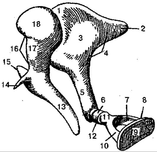
Fig. 149. Auditory ossicles:
1 — incudomalleolar joint; 2 — short limb of the incus; 3 — body of the incus; 4 — incus; 5 — long limb of the incus; 6 — lenticular process; 7 — posterior limb of the stapes; 8 — stapes; 9 — base of the stapes; 10 — anterior limb of the stapes; 11 — head of the stapes; 12 — incudostapedial joint; 13 — handle of the malleus; 14 — anterior process of the malleus; 15 — lateral process of the malleus; 16 — malleus; 17 — neck of the malleus; 18 — head of the malleus
The inner ear is located within the petrous part of the temporal bone, separated from the tympanic cavity by its labyrinthine wall. It consists of a bony labyrinth and a membranous labyrinth housed within it.
The bony labyrinth consists of the cochlea, vestibule, and semicircular canals. The vestibule is a small, irregularly shaped cavity. On its lateral wall, there are two openings: the oval window and the round window. On the medial wall of the vestibule, the vestibular crest is located, which divides the vestibular cavity into two recesses—the anterior spherical recess and the posterior elliptical recess. Through an opening in the posterior wall, the vestibular cavity connects with the bony semicircular canals, and through an opening in the anterior wall, the spherical recess of the vestibule connects with the bony spiral canal of the cochlea.
The cochlea is the anterior part of the bony labyrinth; it is a coiled spiral canal of the cochlea (Fig. 150) that makes 2.5 turns around the cochlear axis. The base of the cochlea is directed medially toward the internal acoustic meatus, while the apex of the cochlear cupula is directed toward the tympanic cavity. The axis of the cochlea lies horizontally and is called the modiolus. A bony spiral lamina winds around the modiolus, partially dividing the spiral canal of the cochlea. At the base of this lamina is the spiral canal of the modiolus, which houses the spiral ganglion of the cochlea.
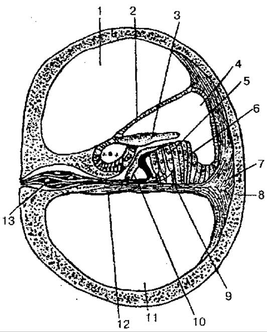
Fig. 150. Cochlear canal (cross section):
1 — scala vestibuli; 2 — vestibular membrane of the cochlear duct; 3 — tectorial membrane; 4 — cochlear duct; 5 — Hair cells with cilia; 6 — supporting cells; 7 — spiral ligament; 8 — bony tissue of the cochlea; 9 — supporting Cell; 10 — pillar cells of Corti; 11 — scala tympani; 12 — basilar membrane; 13 — Nerve Cells of the spiral ganglion
The bony semicircular canals are three arch-shaped, thin tubes lying in three mutually perpendicular planes. In cross-section, the width of each bony semicircular canal is about 2 mm. The anterior (sagittal, superior) semicircular canal lies higher than the other canals, and its highest point on the anterior surface of the petrous part forms the arcuate eminence. The posterior (frontal) semicircular canal is located parallel to the posterior surface of the petrous part of the temporal bone. The lateral (horizontal) semicircular canal projects slightly into the tympanic cavity. Each semicircular canal has two ends—bony limbs. One of them is the simple bony limb, and the other is the ampullary bony limb. The semicircular canals open into the vestibule cavity by five openings, with the adjacent limbs of the anterior and posterior canals fusing to form a common bony limb that opens via a single opening.
The membranous labyrinth is located inside the bony labyrinth and replicates its contour. The walls of the membranous labyrinth consist of a thin connective tissue lamina covered with squamous epithelium. Between the bony and membranous labyrinths, there is a space—the perilymphatic space, filled with a fluid called perilymph. From this space, via the perilymphatic duct which passes through the cochlear canaliculus, the perilymph drains into the subarachnoid space of the Meninges. The membranous labyrinth is filled with endolymph and contains the utricle and saccule, three semicircular ducts, and the cochlear duct. The utricle is located in the recess of the same name and connects with the saccule. Both sacs are connected by the utriculosaccular duct, from which the endolymphatic duct arises. The utricle connects with the semicircular ducts via five openings, and the saccule connects with the cochlear duct. On the inner surface of the saccule and utricle, and on the walls of the membranous ampullae of the semicircular ducts, there are hair (sensory) cells covered with a gelatinous substance. These cells perceive endolymph vibrations during rectilinear movement, acceleration, rotation, and head tilts. Stimulation of these cells is transmitted to the sensory endings—cells of the vestibular ganglion of the VIII cranial nerve, and then to the vestibular nuclei of the Medulla Oblongata and Cerebellum.
The cochlear duct (membranous labyrinth of the cochlea) begins blindly in the vestibule and extends within the spiral canal of the cochlea. In cross-section, it is triangular in shape. Three walls of the cochlear duct are distinguished: the outer wall fuses with the periosteum of the outer wall of the spiral canal of the cochlea; the tympanic wall of the cochlear duct is a continuation of the osseous spiral lamina and separates the cochlear duct from the scala tympani; the vestibular wall is represented by a membrane that runs obliquely upward from the spiral lamina to the outer wall of the cochlear duct. The upper part of the spiral canal of the cochlea is represented by the scala vestibuli, and the lower part by the scala tympani. Near the apex of the cochlea, both scalae communicate with each other through the helicotrema; they contain perilymph. At the base of the cochlea, the scala tympani ends at the window closed by the secondary tympanic membrane. The scala vestibuli communicates with the perilymphatic space of the vestibule, the oval window of which is closed by the footplate of the stapes.
Inside the cochlear duct, on the spiral membrane, lies the spiral organ (organ of Corti). At the base of the spiral organ is the basilar membrane, which contains up to 2400 thin Collagen fibers (strings) that attach to the opposite wall of the spiral canal of the cochlea and act as resonator strings. Located on the basilar membrane are supporting and receptor hair (sensory) cells, which detect mechanical vibrations of the perilymph in the scala vestibuli and scala tympani. Sound vibrations in the air, captured by the tympanic membrane, are transmitted via the auditory ossicles to the perilymph of the scala vestibuli, and then to the perilymph of the scala tympani, which is closed at the base of the cochlea by the secondary tympanic membrane. Sound vibrations of the perilymph in the scala tympani are transmitted to the basilar membrane, which Supports the spiral (auditory) organ, and to the endolymph in the cochlear duct. Subsequently, the vibrations of the endolymph and the basilar membrane activate the sound-perceiving apparatus, whose hair (sensory, receptor) cells convert mechanical movement into a Nerve Impulse. This impulse is received by the endings of bipolar cells, whose bodies lie in the spiral ganglion, while their central processes form the cochlear part of the Vestibulocochlear nerve (cranial nerve VIII). They then travel through the internal acoustic meatus to the Brain, reaching the anterior and posterior cochlear nuclei located in the Pons in the vestibular area of the Rhomboid fossa. Here, the impulse is transmitted to the next neuron, the cells of the auditory nuclei. The processes of the anterior Nucleus cells form a bundle of nerve fibers (the trapezoid body). The axons of the posterior nucleus then plunge into the brain substance and join the fibers of the trapezoid body. On the opposite side of the pons, the fibers of the trapezoid body curve, giving rise to the lateral lemniscus, and proceed to the subcortical auditory centers—the medial geniculate body and the inferior colliculus of the tectal plate of the Midbrain. Next, the processes of the cells of the medial geniculate body pass through the internal capsule and head toward the auditory center (the cortical end of the Auditory Analyzer). The latter is located in the cortex of the superior temporal gyrus (transverse temporal gyri). In this region, the nerve impulses coming from the sound-perceiving apparatus are analyzed.
The human ear can perceive a fairly wide range of sound frequencies: from 16 to 20,000 Hz. Frequencies below 16 Hz are called infrasound, and those above 20,000 Hz are called ultrasound. Each frequency is perceived by specific areas of auditory receptors that respond to a particular pitch. The highest sensitivity of the auditory analyzer is observed in the mid-frequency range (from 1000 to 4000 Hz). Speech utilizes sounds within the range of 150–2500 Hz. The auditory ossicles form a lever system that improves the transmission of sound vibrations from the air of the ear canal to the perilymph of the inner ear. Due to the difference in size between the small area of the stapes footplate and the large area of the tympanic membrane, as well as the specific lever-like Articulation of the ossicles, the pressure on the membrane of the oval window increases by 20 times or more compared to that on the tympanic membrane, which helps amplify the sound. Furthermore, the auditory ossicle system can modify the force of high sound pressures. As soon as the sound wave pressure approaches 110–120 dB, the movement of the ossicles changes significantly, reducing the pressure of the stapes on the oval window of the inner ear, which protects the auditory receptor apparatus from prolonged sound overload. This pressure change is achieved by the contraction of the middle ear muscles (the tensor tympani and stapedius muscles) and a decrease in the vibration amplitude of the stapes. The auditory analyzer is capable of adaptation. Prolonged exposure to sounds leads to a decrease in the sensitivity of the auditory analyzer (adaptation to sound), while the absence of sound leads to an increase in sensitivity (adaptation to silence). With the help of the auditory analyzer, the distance to a sound source can be determined relatively accurately. The most precise estimation of the distance to a sound source occurs at a distance of about 3 m. Sound direction is determined thanks to binaural hearing. The ear closer to the sound source perceives it earlier and, consequently, with greater intensity. At the same time, the delay time on the way to the other ear is also determined. It is known that the thresholds of the auditory analyzer are not strictly constant and fluctuate significantly in humans depending on the functional state of the body and environmental factors.
Two Types of sound transmission are distinguished: air conduction and bone conduction. In air conduction, sound waves are captured by the auricle and transmitted through the external auditory canal to the tympanic membrane, and then via The system of auditory ossicles to the perilymph and endolymph. Through air conduction, humans are capable of perceiving sounds ranging from 16 to 20,000 Hz. Bone conduction of sound occurs through the BONES OF THE skull, which also conduct sound. Air conduction is more efficient than bone conduction.
The receptors of the vestibular apparatus are stimulated by the tilting or movement of the head. This triggers reflex muscle contractions that help right the body and maintain an appropriate posture. The receptors of the vestibular apparatus enable the perception of the head's position in space and body movement. It is known that sensory cells are embedded in a gelatinous mass containing otoliths, which consist of small crystals of calcium carbonate. In a normal body position, gravity causes the otoliths to exert pressure on specific hair cells. If the head is tilted crown-down, the otolith hangs from the hairs; with a lateral head tilt, one otolith presses on the hairs while the other hangs. The change in otolith pressure excites the sensory hair cells, which signal THE POSITION OF the head in space. The sensory cells of the cristae in the ampullae of the semicircular canals are excited by movement and acceleration. Since the three semicircular canals are situated in three different planes, head movement in any direction causes the movement of endolymph. Stimulation of the sensory hair cells is transmitted to the sensory endings of the vestibular part of the vestibulocochlear nerve. The Cell bodies of these Neurons are located in the vestibular ganglion, which lies at the bottom of the internal acoustic meatus, and their central processes, as part of the vestibulocochlear nerve, enter the cranial cavity and then travel to the brain's vestibular nuclei. The processes of the vestibular nuclei cells (the next neuron) project to the cerebellar nuclei and the Spinal Cord, forming the vestibulospinal tract. They also enter the medial longitudinal fasciculus of the Brainstem. Some fibers of the vestibular part of the vestibulocochlear nerve bypass the vestibular nuclei and go directly to the cerebellum.
When the vestibular apparatus is stimulated, numerous motor reflex responses occur, altering the function of Internal Organs, alongside various sensory reactions. An example of such reactions is the appearance of rapid, repetitive Eyeball movements (nystagmus) after a rotation test: a person makes rhythmic Eye Movements in the direction opposite to the rotation, followed by very rapid movements in the direction of the rotation. Changes in Cardiac Activity, vasoconstriction or vasodilation, a decrease in Blood pressure, and increased gastrointestinal peristalsis may also occur. Stimulation of the vestibular apparatus can cause dizziness, environmental disorientation, and nausea. The vestibular apparatus is also involved in the regulation and redistribution of muscle tone.
Last update: 08/08/2026
Editorial and Educational Adaptation: This material has been compiled based on the primary/original source text. The project team performed an editorial review, corrected technical inaccuracies, structured sections, and adapted the content for an educational format.
What was processed:
- elimination of formatting defects (OCR errors, structural breaks, corrupted characters);
- editorial organization of content;
- standardization of terminology in accordance with academic sources;
- verification of factual statements against the original source text.
All mentions of the author, publication year, and origin of the primary text have been preserved in accordance with the source.