Human Anatomy and Physiology - N. I. Fedyukovich 2003
Sensory Organs
Organ of Vision
The Organ of Vision is one of the primary Sensory Organs, playing a crucial role in perceiving the environment. In diverse human activities, especially in performing highly delicate tasks, The Organ of vision is of paramount importance. Having reached perfection in humans, the visual organ captures light flux, directs it onto specialized photoreceptor Cells, perceives black-and-white and color images, and perceives objects in three dimensions and at various distances.
The organ of vision is located in the Orbit and consists of THE EYE AND the accessory apparatus (Fig. 144).
Class="center">
Fig. 144. Structure OF THE eye (diagram):
1 — sclera; 2 — choroid; 3 — retina; 4 — fovea centralis; 5 — blind spot; 6 — Optic nerve; 7 — conjunctiva; 8 — ciliary ligament; 9 — cornea; 10 — pupil; 11, 18 — optic axis; 12 — anterior chamber; 13 — lens; 14 — iris; 15 — posterior chamber; 16 — ciliary Muscle; 17 — vitreous body
The eye (oculus) consists of the Eyeball and the optic nerve with its sheaths. The eyeball is rounded, having anterior and posterior poles. The former corresponds to the most prominent part of the outer fibrous tunic (cornea), and the latter to the most prominent part located lateral to the exit of the optic nerve from the eyeball. The line connecting these points is called the external axis of the eyeball, while the line connecting a point on the inner surface of the cornea with a point on the retina is called the internal axis of the eyeball. Alterations in The ratio of these lines cause impaired image focusing on the retina, leading to nearsightedness (myopia) or farsightedness (hypermetropia).
The eyeball consists of the fibrous and vascular tunics, the retina, and the core of the eye (aqueous humor of the anterior and posterior chambers, the lens, and the vitreous body).
The fibrous tunic is the outer dense coat that performs protective and light-conducting Functions. Its anterior part is called the cornea, and its posterior part is the sclera. The cornea is the transparent, avascular part of the tunic, resembling a watch Glass in shape. The diameter of the cornea is 12 mm, and its thickness is about 1 mm.
The sclera consists of dense Fibrous Connective Tissue, about 1 mm thick. At the junction with the cornea, within the thickness of the sclera, There is a narrow channel—the scleral venous sinus. The extraocular Muscles attach to the sclera.
The vascular tunic contains A large number of Blood Vessels and pigment. It consists of three parts: the choroid proper, the ciliary body, and the iris. The choroid proper forms the largest part of the vascular tunic, lining the posterior part of the sclera and loosely fusing with the outer tunic; a narrow cleft-like perichoroidal space lies between them.
The ciliary body resembles a moderately thickened section of the vascular tunic, lying between the choroid proper and the iris. The Base of the ciliary body consists of loose connective tissue rich in vessels and smooth muscle cells. The anterior section has about 70 radially arranged ciliary processes, which form the ciliary crown. Radially arranged fibers of the ciliary zonule attach to the latter, then extend to the anterior and posterior surfaces of the lens capsule. The posterior section of the ciliary body—the ciliary ring—resembles thickened circular bands that merge into the choroid. The ciliary Muscle consists of intricately interwoven bundles of smooth muscle cells. Their contraction alters the dimensions of the lens and its refractive power, allowing adjustment for clear vision of objects (accommodation).
The iris is the most anterior part of the vascular tunic, shaped like a disc with an aperture (the pupil) in the center. It consists of vascularized connective tissue, pigment cells that determine eye color, and radially and circularly arranged muscle fibers.
The iris has an anterior surface, which forms the posterior wall of the anterior chamber of the eye, and a pupillary margin, which bounds the pupillary aperture. The posterior surface of the iris forms the anterior wall of the posterior chamber of the eye, while its ciliary margin connects to the ciliary body and sclera via the pectinate ligament. The muscle fibers of the iris, by contracting or relaxing, decrease or increase the diameter of the pupil.
The inner (sensory) tunic of the eyeball—the retina—is closely apposed to the vascular tunic. The retina has a large posterior optic part and a smaller anterior 'blind' part, which combines the ciliary and iridial PARTS OF THE retina. The optic part consists of the inner pigment and inner neural parts. The latter contains up to 10 layers of Nerve Cells. The inner part of the retina includes cells with processes shaped like cones and rods, which are the photoreceptive elements of the eyeball. Cones perceive light rays in bright (daylight) conditions and also serve as color receptors, while rods function in dim light and act as receptors for twilight vision. The remaining nerve cells play an associative role; the axons of these cells bundle together to form the nerve that exits the retina.
In the posterior region of the retina lies the exit site of the optic nerve—the optic disc, and lateral to it is the macula lutea. This area contains the highest concentration of cones and is the site of greatest visual acuity.
The core of the eye includes the anterior and posterior chambers, filled with aqueous humor, the lens, and the vitreous body. The anterior chamber of the eye is the space between the cornea anteriorly and the anterior surface of the iris posteriorly. The circumferential area where the margins of the cornea and iris meet is bounded by the pectinate ligament. Located between the bundles of this ligament are the spaces of the iridocorneal angle (spaces of Fontan). Through these spaces, aqueous humor drains from the anterior chamber into the scleral venous sinus (canal of Schlemm) and then enters the anterior ciliary Veins. The anterior chamber communicates with the posterior chamber of the eyeball through the pupillary aperture. The posterior chamber, in turn, communicates with the spaces between the fibers of the lens and the ciliary body. Around the periphery of the lens lies a belt-like space (canal of Petit) filled with aqueous humor.
The lens is a biconvex lens located behind the Chambers of the eye, possessing light-refracting power. It has anterior and posterior surfaces and an equator. The substance of the lens is colorless, transparent, dense, and devoid of Vessels and nerves. Its inner part—The Nucleus—is much denser than the peripheral part. Externally, the lens is covered by a thin, transparent, elastic capsule to which the ciliary zonule (zonule of Zinn) is attached. Contraction of the ciliary muscle alters the dimensions of the lens and its refractive power.
The vitreous body is a jelly-like transparent mass, devoid of vessels and nerves, and enclosed by a membrane. It is located in the vitreous chamber of the eyeball, behind the lens, and is closely apposed to the retina. On the side of the lens, there is a depression in the vitreous body called the hyaloid fossa. The refractive index of the vitreous body is close to that of the aqueous humor filling the chambers of the eye. In addition, the vitreous body performs supportive and protective functions.
Accessory Organs of the eye. The accessory Organs of the eye include the extraocular muscles (Fig. 145), Orbital Fasciae, eyelids, eyebrows, Lacrimal Apparatus, orbital fat body, conjunctiva, and the Sheath of the eyeball.
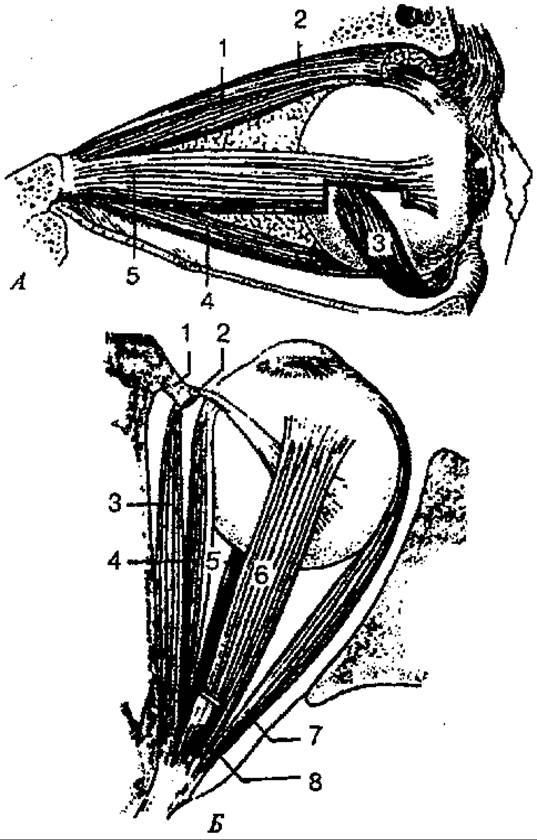
Fig. 145. Muscles of the eyeball:
A — lateral view: 1 — superior rectus muscle; 2 — levator palpebrae superioris muscle; 3 — inferior oblique muscle; 4 — inferior rectus muscle; 5 — lateral rectus muscle; B — superior view: 1 — trochlea; 2 — tendon sheath of superior oblique muscle; 3 — superior oblique muscle; 4 — medial rectus muscle; 5 — inferior rectus muscle; 6 — superior rectus muscle; 7 — lateral rectus muscle; 8 — levator palpebrae superioris muscle
The motor apparatus of the eye is represented by six muscles. These muscles originate from the common tendinous ring around the optic nerve deep within the orbit and insert into the eyeball. There are four rectus muscles of the eyeball (superior, inferior, lateral, and medial) and two oblique muscles (superior and inferior). The muscles act in such a way that both eyes turn coordinately and are directed at the same point. The levator palpebrae superioris muscle also originates from the common tendinous ring. The extraocular muscles are striated muscles and contract voluntarily.
The orbit, which houses the eyeball, is lined by the periorbita, which fuses with the dura mater of the Brain in the region of the optic canal and the superior orbital fissure. The eyeball is enclosed by a sheath (or Tenon's capsule) that is loosely connected to the sclera, forming the episcleral space. Between this sheath and the periorbita lies the orbital fat body, which acts as an elastic cushion for the eyeball.
The eyelids (superior and inferior) are structures situated in front of the eyeball, covering it from above and below, and completely closing it when shut. The eyelids have anterior and posterior surfaces and free margins. The latter, joined by commissures, form the medial and lateral angles of the eye. The lacrimal lake and lacrimal caruncle are located in the medial angle. On the free margin of the upper and lower eyelids, near the medial angle, a small elevation is visible—the lacrimal papilla, with an opening at its apex that marks the beginning of the lacrimal canaliculus.
The space between the margins of the eyelids is called the palpebral fissure. Eyelashes are located along the anterior margin of the eyelids. The core of the eyelid is formed by the tarsus, which is covered by Skin on the outside and by the palpebral conjunctiva on the inside, which then transitions into the bulbar conjunctiva. The recess formed by the reflection of the palpebral conjunctiva onto the eyeball is called the conjunctival sac. Besides their protective function, the eyelids reduce or block the incoming light.
At the border between the forehead and the upper eyelid is the eyebrow, which is a Hair-covered ridge that serves a protective function.
The lacrimal apparatus consists of the lacrimal gland with its excretory ducts and the lacrimal passages. The lacrimal gland is located in the lacrimal fossa in the lateral angle, near the superior wall of the orbit, and is covered by a thin connective tissue capsule. The excretory ducts (about 15 of them) of the lacrimal gland open into the conjunctival sac. Tears bathe the eyeball and constantly moisten the cornea. The movement of tears is facilitated by blinking. Then, tears drain along the capillary space near the eyelid margins into the lacrimal lake. The lacrimal canaliculi originate here and open into the lacrimal sac. The latter is located in the lacrimal fossa in the inferomedial angle of the orbit. Inferiorly, it transitions into a fairly wide nasolacrimal duct, through which the lacrimal fluid enters the Nasal cavity.
Visual pathway of the visual analyzer (Fig. 146). Light entering the retina first passes through the transparent refractive media of the eye: the cornea, the aqueous humor of the anterior and posterior chambers, the lens, and the vitreous body. The light beam is regulated along its path by the pupil. The refractive media direct the light beam to the most sensitive part of the retina—the area of sharpest vision—the macula with its fovea centralis. Passing through all layers of the retina, light triggers complex photochemical transformations of visual pigments. As a result, a Nerve Impulse is generated in the photosensitive cells (rods and cones), which is then transmitted to the next retinal Neurons—bipolar cells (neurocytes), and then to the neurocytes of the ganglion layer, the ganglion neurocytes. The processes of the latter run toward the optic disc and form the optic nerve. Entering the Skull through the optic canal along the Inferior surface of the brain, the optic nerve forms an incomplete optic chiasm. The optic tract begins at the optic chiasm and consists of nerve fibers from the ganglion Cells of the retinal eyeball. These fibers then travel along the optic tract to the subcortical visual centers: the lateral geniculate body and the superior colliculi of the Midbrain tectum. In the lateral geniculate body, the fibers of the third neuron (ganglion neurocytes) of the visual pathway terminate and synapse with the cells of the next neuron. The axons of these neurocytes pass through the internal capsule and reach the cells of the occipital lobe near the calcarine sulcus, where they terminate (the cortical end of the visual analyzer). Some axons of the ganglion cells bypass the geniculate body and enter the superior colliculus via its brachium. From the Gray matter of the superior colliculus, impulses travel to the oculomotor nucleus and the accessory oculomotor nucleus, from which the oculomotor muscles, pupillary Constrictor muscles, and ciliary muscle are innervated. These fibers carry impulses in response to light stimulation, causing the pupils to constrict (pupillary reflex) and the eyeballs to turn in the required direction.

Fig. 146. Diagram of The structure of the visual analyzer:
1 — retina; 2 — uncrossed fibers of the optic nerve; 3 — crossed fibers of the optic nerve; 4 — optic tract; 5 — cortical analyzer
The Mechanism of Photoreception is based on the step-by-step transformation of the visual pigment rhodopsin under the action of light quanta. The latter are absorbed by a group of atoms (chromophores) of specialized molecules—chromolipoproteins. The chromophore that determines the degree of Light absorption in visual pigments is retinal, an aldehyde of vitamin A alcohol. Retinal is always in the form of 11-cis-retinal and normally binds to the colorless protein opsin, thereby forming the visual pigment rhodopsin, which, through a series of intermediate stages, is again split into retinal and opsin. During this process, the molecule loses its color, a phenomenon known as bleaching. The pathway of rhodopsin molecule transformation is represented as follows.

The process of visual excitation occurs during the interval between The formation of lumirhodopsin and metarhodopsin II. Once light exposure ceases, rhodopsin is immediately resynthesized. First, with the participation of the enzyme retinal isomerase, all-trans-retinal is completely converted into 11-cis-retinal, which then combines with opsin to form rhodopsin once again. This continuous process underlies dark adaptation. In complete darkness, it takes about 30 minutes for all rods to adapt and for the eyes to reach maximum sensitivity. Image formation in the eye occurs with the participation of the optical systems (the cornea and the lens), which produce an inverted and reduced image of the object on the retinal surface. The adaptation of the eye for clear vision of objects at various distances is called accommodation. The Mechanism of ocular accommodation is associated with the contraction of the ciliary muscles, which alter the curvature of the lens.
When viewing objects at a close distance, convergence acts simultaneously with accommodation, meaning that the axes of both eyes are brought together. The visual lines converge more the closer the object being viewed is.
The refractive power of The Optical System of the eye is expressed in diopters ("D" — dptr). One diopter (1 D) is defined as the power of a lens with a focal length of 1 m. The refractive power of the human eye is 59 dptr when viewing distant objects and 70.5 dptr when viewing close ones.
There are three main refractive errors of the eye: nearsightedness, or myopia; farsightedness, or hypermetropia; and age-related farsightedness, or presbyopia (Fig. 147). The primary cause of all eye defects is a mismatch between the refractive power and the length of the eyeball, unlike in a normal eye. In nearsightedness (myopia), light rays converge in front of the retina within the vitreous body, and a blur circle is formed on the retina instead of a point; the eyeball in this case is longer than normal. Concave lenses with negative diopters are used for vision correction.

Fig. 147. Path of light rays in a normal eye (A), in myopia
(B1 and B2), in hypermetropia (C1 and C2), and in astigmatism (D1 and D2):
B2, C2 — biconcave and biconvex lenses for correcting myopia and hypermetropia defects; D2 — cylindrical lens for astigmatism correction; 1 — zone of clear vision; 2 — zone of blurred image; 3 — corrective lenses
In farsightedness (hypermetropia), the eyeball is short, and therefore parallel rays coming from distant objects focus behind the retina, resulting in an unclear, blurred image of the object on the retina. This defect can be compensated for by using the refractive power of convex lenses with positive diopters.
Presbyopia (age-related farsightedness) is associated with reduced elasticity of the lens and weakened tension of the zonule of Zinn (ciliary zonule) with a normal eyeball length.
This refractive error can be corrected using biconvex lenses. Vision with one eye gives us an idea of an object in only one plane. Depth perception and a correct understanding of the relative positions of objects are only possible when viewing with both eyes simultaneously. Binocular vision provides The ability to fuse the separate images received by each eye into a single whole.
Visual acuity characterizes the spatial resolving power of the eye and is determined by the smallest angle at which a person can distinguish two separate points. The smaller the angle, the better the vision. Normally, this angle is 1 arcminute, or 1 unit.
To determine visual acuity, special charts are used that display letters or symbols of various sizes.
The field of vision is the space perceived by one eye when it is stationary. Changes in the visual field can be an early sign of certain eye and brain diseases.
Color perception is the ability of the eye to distinguish colors. Thanks to this visual function, humans can perceive about 180 color shades. Color Vision is of great practical importance in A number of professions, especially in art. Like visual acuity, color perception is a function of the retinal cone system. Color vision deficiencies can be congenital (inherited) or acquired.
Color vision deficiency is commonly referred to as color blindness and is determined using pseudoisochromatic plates, which present a collection of colored dots forming a symbol. A person with normal vision easily distinguishes the outlines of the symbol, whereas a color-blind person cannot.
Last update: 08/08/2026
Editorial and Educational Adaptation: This material has been compiled based on the primary/original source text. The project team performed an editorial review, corrected technical inaccuracies, structured sections, and adapted the content for an educational format.
What was processed:
- elimination of formatting defects (OCR errors, structural breaks, corrupted characters);
- editorial organization of content;
- standardization of terminology in accordance with academic sources;
- verification of factual statements against the original source text.
All mentions of the author, publication year, and origin of the primary text have been preserved in accordance with the source.