Human Anatomy - Kotsan I. Y. 2009
Sense Organs
Visual Organ (Eye)
Accessory Organs of the Eye
The Accessory structures of the eye include the eyelids, eyelashes, eyebrows, conjunctiva, Lacrimal Apparatus, Orbital Fasciae, and extrinsic eye Muscles (Fig. 267).
Class="center">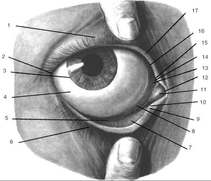
Fig. 267. Eyelids, right eye (after R. D. Sinelnikov)
1 — upper eyelid; 2 — lateral angle of the eye; 3 — corneal limbus; 4 — bulbar conjunctiva; 5 — inferior conjunctival fornix; 6 — lower eyelid; 7 — palpebral conjunctiva; 8 — semilunar fold of the conjunctiva; 9 — inferior lacrimal papilla; 10 — inferior lacrimal punctum; 11 — lacrimal caruncle; 12 — medial angle of the eye; 13 — lacrimal lake; 14 — superior lacrimal punctum; 15 — superior lacrimal papilla; 16 — posterior margin of the eyelid; 17 — anterior margin of the eyelid
The eyelids (palpebrae) are Skin folds that cover the Eyeball anteriorly. They protect it from drying out and from foreign dust particles reaching its surface, as well as regulate the intensity of light falling on the eyeball.
The upper and lower eyelids (palpebra superior et palpebra inferior) are distinguished. The upper eyelid is somewhat larger than the lower one. When closed, the eyelids completely cover the eyeball. When open, a palpebral fissure (rima palpebralis) remains between their margins, through which the anterior surface of the eyeball is visible. Meeting at the sides, the eyelids form the lateral and medial angles of the eye (angulus oculi lateralis et angulus oculi medialis). The lateral angle of the eye is sharper, whereas the medial one is rounded and borders a depression on the medial side known as the lacrimal lake (lacus lacrimalis). Also in the medial angle of the eye is a small elevation called the lacrimal caruncle (caruncula lacrimalis). At the junction of the upper and lower eyelids at the angles of the eye, the medial and lateral palpebral commissures (comissura palpebralis medialis/lateralis) are formed.
The anterior surface of the eyelids (facies anterior palpebrae) is convex, covered with thin skin containing short vellus Hair, Sebaceous Glands, and Sweat Glands. The posterior surface of the eyelids (facies posterior palpebrae), facing the eyeball, is concave and lined with the conjunctiva (tunica conjunctiva).
The framework of each eyelid consists of a Connective Tissue plate that resembles Cartilage in density and is therefore traditionally called the tarsal plate (tarsus). The superior tarsal plate (tarsus superior) is wider than the inferior tarsal plate (tarsus inferior). The width of the upper tarsal plate is 10 mm, and the lower one is 5 mm. The length of each of these plates reaches 20 mm, with a thickness of 1 mm.
Seemingly continuing the tarsal plates toward the conjunctival fornices is a layer of smooth Muscle fibers forming the superior and inferior tarsal muscles (m. tarsalis superior et m. tarsalis inferior), which comprise the structural Base of the eyelids above the superior tarsal plate and below the inferior tarsal plate, respectively. A thin and broad tendon of the levator palpebrae superioris muscle attaches to the upper margin and anterior surface of the superior tarsal plate.
The free margins of the eyelids are about 2 mm thick and are demarcated from the eyelid surfaces by two paired margins. The anterior palpebral margins (limbi palpebrales anteriores) are somewhat rounded and covered with skin, while the posterior palpebral margins (limbi palpebrales posteriores) are somewhat pointed and lined with conjunctiva. Along the anterior palpebral margins lie 2–3 rows of curved hairs, the eyelashes (cilia). Opening into the hair follicles of the eyelashes are the ciliary glands (glandulae ciliares), also known as the glands of Moll, and sebaceous glands (glandulae sebaceae), or the glands of Zeis. Along the posterior palpebral margin, the ducts of the tarsal glands (glandulae tarsales) open via small orifices. The tarsal glands, or Meibomian glands, are tubular glands whose secretory parts are located within the thickness of the tarsal plates (30–40 glands in the upper plate and 20–30 in the lower). The tarsal glands produce an oily sebum via holocrine secretion, which lubricates the eyelid margin and prevents tears from overflowing the conjunctival sac.
On the free margin of the upper and lower eyelids, near the medial angle of THE EYE AND lateral to the lacrimal lake, there is an elevation called the lacrimal papilla (papilla lacrimalis) with an opening at its apex—the lacrimal punctum (punctum lacrimalis), which marks the beginning of the lacrimal canaliculus (see below).
The eyebrows (supercilia) are elongated folds of skin located above the upper eyelids along the lower margin of the forehead and covered with hair. The eyebrows serve a protective function by diverting streams of sweat and raindrops from the forehead away from the palpebral fissure.
The conjunctiva (tunica conjunctiva) is a pink connective tissue membrane structurally similar to a mucous membrane. It is subdivided into the palpebral conjunctiva (tunica conjunctiva palpebralis), which covers the posterior surface of the eyelids, and the bulbar conjunctiva (tunica conjunctiva bulbaris), which covers the anterior surface of the eyeball. Where the conjunctiva transitions from the upper and lower eyelids onto the eyeball, recesses are formed called the superior and inferior conjunctival fornices (fornix conjunctivae superior et fornix conjunctivae inferior). The superior fornix is deeper than the inferior one. The fornices act as reserve folds of the conjunctiva necessary for the Movements of the eyeball and eyelids. A similar role is played by the semilunar fold of the conjunctiva (plica semilunaris conjunctivae), located in the region of the medial angle of the eye lateral to the lacrimal caruncle. Morphologically, it is a rudimentary nictitating membrane (third eyelid) of reptiles. The entire space lying anterior to the eyeball and bounded by the conjunctiva is called the conjunctival sac (saccus conjunctivalis), which opens anteriorly along the line of the palpebral fissure. Under normal conditions, a small amount of tear fluid is present in the conjunctival sac.
Last update: 08/08/2026
Editorial and Educational Adaptation: This material has been compiled based on the primary/original source text. The project team performed an editorial review, corrected technical inaccuracies, structured sections, and adapted the content for an educational format.
What was processed:
- elimination of formatting defects (OCR errors, structural breaks, corrupted characters);
- editorial organization of content;
- standardization of terminology in accordance with academic sources;
- verification of factual statements against the original source text.
All mentions of the author, publication year, and origin of the primary text have been preserved in accordance with the source.