Human Anatomy Part 1 - K. A. Dyubenko, A. K. Kolomiyytsev, Yu. B. Chaykovsky 2002
Special Section
Respiratory system, systema respiratorium [respiratory apparatus, apparatus respiratorius] - General Information
Constrictor muscles
1. The lateral cricoarytenoid Muscle, musculus cricoarytenoideus lateralis, is a paired muscle that originates from the arch of the cricoid Cartilage and inserts into the muscular process of the arytenoid cartilage. Upon contraction, it pulls the muscular process forward and downward. This brings the vocal processes closer together and narrows the rima glottidis.
2. The thyroarytenoid muscle, musculus thyroarytenoideus, is a paired muscle originating from the internal surface of the thyroid cartilage lamina, extending upward and backward to insert into the muscular process of the arytenoid cartilage. When both Muscles contract, the portion of the laryngeal cavity above the vocal cords narrows. Simultaneously, the arytenoid cartilages are pulled forward, slightly relaxing the vocal cords.
3. The transverse arytenoid muscle, musculus arytenoideus transversus, is an unpaired muscle connecting both arytenoid cartilages. Contraction of this muscle draws the cartilages together and narrows the rima glottidis.
4. The oblique arytenoid muscle, musculus arytenoideus obliquus, is a paired muscle that arises from the muscular process of the arytenoid cartilage, courses obliquely upward, decussates with the contralateral muscle of the same name, and inserts into the apex of the opposite arytenoid cartilage. Upon contraction, it constricts the posterior part of the rima glottidis.
5. The aryepiglottic muscle, musculus aryepiglotticus, is a paired muscle originating from the apex of the arytenoid cartilage as a continuation of the preceding muscle. Running within the aryepiglottic fold, it inserts into the lateral margin of the epiglottis. It narrows the laryngeal inlet and pulls the epiglottis downward.
Class="center">
Fig. 284. Muscles of the Larynx, musculi laryngis: A — left view; B — posterior view
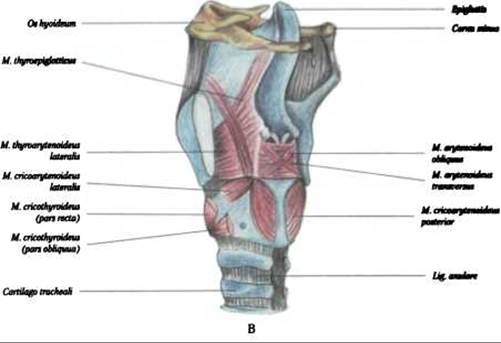
Fig. 284. Muscles of the larynx, musculi laryngis:
C — lateral view (the lamina of the thyroid cartilage is sectioned)
Last update: 08/08/2026
Editorial and Educational Adaptation: This material has been compiled based on the primary/original source text. The project team performed an editorial review, corrected technical inaccuracies, structured sections, and adapted the content for an educational format.
What was processed:
- elimination of formatting defects (OCR errors, structural breaks, corrupted characters);
- editorial organization of content;
- standardization of terminology in accordance with academic sources;
- verification of factual statements against the original source text.
All mentions of the author, publication year, and origin of the primary text have been preserved in accordance with the source.