Review of Medical Physiology - William F. Ganong 2002
Functions of the Nervous System
Vision
Mechanism of Photoreception
Origin of electrical impulses
The potential difference that initiates the generation of an excitation potential is caused by The Effect of light on the light-sensitive elements of rods and cones. The absorption of light rays by these elements induces Conformational Changes in their molecules, initiating a series of sequential transformations that activate Neurons.
The eye is a unique organ because the action potentials of photoreceptor Cells, like the electrical impulses of the vast majority of its neuronal elements, are local and graded; only the generation of ganglion Cell potentials, which are capable of being transmitted over considerable distances, follows the "all-or-none" principle.
The response to stimulation in rods, cones, and horizontal cells is hyperpolarization (Fig. 8-14), whereas the response of bipolar cells is either hyperpolarization or depolarization. Amacrine cells, in turn, generate depolarizing potentials and spikes that can serve as triggers for the excitation of ganglion cells.
When comparing action potentials in cones and rods, it can be observed that cone action potentials have a sharp onset and termination, whereas rod action potentials have a sharp onset and a gradual termination. The curves relating Action Potential amplitudes to stimulus intensity are similar in rods and cones, but rods are significantly more sensitive. Therefore, the rod response is proportional to stimulus intensity when illumination levels are below the cone threshold. Conversely, the cone response is proportional to stimulus intensity at high illumination levels, once the rod response has reached a maximum and can no longer change. This is why cones generate an adequate response to changes in illumination intensity above a certain level, but poorly reflect absolute illumination, whereas rods perceive absolute illumination.
Ionic Basis of photoreceptor potentials
Sodium channels in the outer segments of rods and cones are open in the dark, and There is a continuous flow of ions from the inner to the outer segments (Fig. 8-15). A flow of ions also moves toward the synaptic terminal of the photoreceptor cell; the inner segment Na+-K+-ATPase maintains ionic equilibrium. The release of the synaptic neurotransmitter is steady in the dark. When light strikes the outer segment, the resulting reactions close a fraction of the Na+ channels, resulting in a hyperpolarizing receptor potential. The hyperpolarization reduces the release of the synaptic neurotransmitter, which in turn acts as a signal that ultimately leads to the generation of action potentials by ganglion cells. These action potentials travel to the Brain.
Light-sensitive substances
The light-sensitive substances of the eyes of humans and most mammals consist of a protein component, opsin, and retinal, an aldehyde of vitamin A1. The term retinal1 is used to distinguish this substance from retinal2, which is found in the eyes of certain animal species. Retinal molecules are aldehydes, which is why they are often called retinals. Group A Vitamins are alcohols and are called retinols.
Rhodopsin
The light-sensitive pigment of rods is called rhodopsin, or visual purple. The opsin within the rhodopsin molecule is called scotopsin. Rhodopsin exhibits maximum sensitivity to light with a wavelength of 505 nm. Human rhodopsin has a Molecular Weight of 41,000. It is localized in the membranes of rod discs, accounts for up to 90% of the protein in these membranes, and is one of many G protein-coupled receptors with a folded molecular shape (see Fig. 1-40). Retinal is positioned parallel to the membrane surface (Fig. 8-16) and is attached to a Lysine residue at position 296 of the seventh transmembrane domain.
Class="center">
Fig. 8-14. Intracellular recording of retinal cell responses to light stimulation. Synaptic contacts between cells are also shown. Rod (R) on the left perceives a flash of light, whereas the rod on the right perceives continuous moderate illumination. H, horizontal cell; B, bipolar cell; A, amacrine cell; G, ganglion cell (reproduced with permission from Dowling JE: Organization of vertebrate retinas. Invest Ophthalmol 1970;9:655).
In the dark, rhodopsin retinal1 is in the 11-cis configuration. The effect of light is to change the shape of the retinal molecule, converting it to the all-trans isomer (Fig. 8-17). This, in turn, affects the configuration of opsin; this shape change activates the attached heterotrimeric G protein known as Transducin, or Gt1. This protein exchanges GDP for GTP, after which its α-subunit dissociates and retains its intrinsic GTPase activity as long as GTP Hydrolysis takes place. The termination of transducin action is accelerated by its binding of ß-arrestin (see Chapter 4); the α-subunit activates cGMP phosphodiesterase, which converts cGMP to 5'-GMP (Fig. 8-18); cGMP keeps Na+ channels open, whereas a decrease in cytoplasmic cGMP concentration leads to the closure of some of these channels. This triggers a hyperpolarizing potential.

Fig. 8-15. Effect of light on ion transport in photoreceptor cells. In the dark, outer segment Na+ channels are open due to cGMP. Light causes The conversion of cGMP to 5'-GMP, leading to the closure of some channels and hyperpolarization of the photoreceptor synaptic terminal.

Fig. 8-16. Schematic representation of rhodopsin Structure, showing the localization of retinal1 R in the rod disc membrane.
The cycle of reactions described above occurs very rapidly and amplifies the light signal. This Amplification effect explains the extremely high sensitivity of rod photoreceptors, which are capable of generating a response even to a single photon of light.
Upon acquiring the all-trans configuration, retinal1 dissociates from opsin (a process known as bleaching). Part of the rhodopsin undergoes direct regeneration; in the presence of NADH, part of the retinal is reduced by the enzyme Alcohol dehydrogenase to vitamin A1, which subsequently interacts with scotopsin to form rhodopsin (see Fig. 8-17). All of these reactions, with the exception of The formation of the all-trans isomer of retinal1, are independent of illumination intensity and proceed equally well in light and darkness. The amount of rhodopsin in receptors is inversely proportional to illumination intensity.
Cone pigments
Primates have Three types of cones. These receptors provide Color Vision and are maximally sensitive to light rays with wavelengths of 440, 535, and 565 nm (see below). Each of them contains retinal1 and opsin. Cone opsin resembles that found in rod rhodopsin. Its molecule traverses the cone membrane seven times and has specific features in each cone type. As noted, The Plasma Membrane of a cone forms invaginated saccules, unlike the discs characteristic of rods. The response of cones to light stimulation generally resembles that of rods. Light activates retinal1, which in turn activates the Gt2 protein, which differs somewhat from rod transducin; Gt2 activates phosphodiesterase, catalyzing the conversion of cGMP to 5'-GMP. The result is the closure of Na+ channels between the extracellular fluid and the cone Cytoplasm, a decrease in intracellular Na+ concentration, and hyperpolarization of the synaptic terminal.

Fig. 8-17. Top: Structure of retinal1 showing the 11-cis configuration (solid lines) and the light-induced transition to the all-trans molecular configuration (dashed lines). Bottom: Effect of light on rhodopsin.

Fig. 8-18. Cytology/cytology/16.html">Early stages of light Transduction in rods. Light activates rhodopsin, which stimulates GTP binding by transducin. This, in turn, activates phosphodiesterase, which catalyzes the conversion of cGMP to 5’-GMP. Consequently, the decrease in the intracellular concentration of cGMP leads to the closure of cGMP-Gated Ion Channels.
The sequence of reactions whereby light stimulation of photoreceptor cells generates a signal in the subsequent neural element of the retina is summarized in Fig. 8-19.
Resynthesis of cyclic GMP
Light decreases the concentration of Ca2+ and Na+ in photoreceptors. The reduction in Ca2+ levels activates guanylyl cyclase, which drives The production of cGMP. It also inhibits the light-activated phosphodiesterase. Both of these effects facilitate the return of photoreceptors to their resting state by opening Na+ channels.
Retinal Synaptic Transmitters
A wide variety of synaptic transmitters have been identified in the retina, including acetylcholine, glutamate, dopamine, serotonin, GABA, Glycine, substance P, Somatostatin, thyrotropin-releasing hormone, gonadotropin-releasing hormone, enkephalins, ß-endorphins, cholecystokinin, vasoactive intestinal polypeptide, neurotensin, and Glucagon (see Chapter 4). Kainate receptors mediate synaptic connections between cones and a specific type of bipolar cell. Amacrine cells are unique among retinal cells in their ability to synthesize acetylcholine. In some vertebrate species, cells located at the border of the inner nuclear and inner plexiform layers (see Fig. 8-2) release dopamine, which diffuses throughout the retina. One of the effects of dopamine is its action on Gap Junctions, which permit the free passage of ionic currents between horizontal cells in the dark, thereby increasing the size of the photoreceptor receptive fields. Light reduces ionic currents and uncouples the connections between horizontal cells, an effect driven by increased dopamine production in daylight.
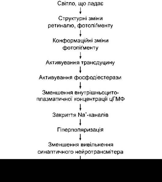
Fig. 8-19. Sequence of light transduction events in rods and cones.
Image Formation
Visual information Processing in the retina involves the formation of three distinct images. The first image, formed by the action of light rays on photoreceptors, is converted into a second image in bipolar cells, which is subsequently transformed into a third image in ganglion cells. During the Formation of the second image, signals are modified by horizontal cells, and during the third, by amacrine cells. Because signal modifications during transmission through the lateral geniculate bodies are minimal, the third image ultimately reaches the occipital areas of the Cerebral Cortex.
A characteristic feature of bipolar and ganglion cells (as well as Cells of the lateral geniculate bodies and layer IV of the visual cortex) is that they respond best to small, circular stimuli, and that within a single receptive field, a ring of light surrounding a dark center (annular illumination) inhibits the response of the central point (Fig. 8-20). The center may be excitatory with inhibitory responses in the periphery ("on-center" cell) or inhibitory with excitatory responses in the periphery ("off-center" cell). The inhibition of the central response by adjacent stimulated areas presumably occurs via a negative feedback mechanism between neighboring photoreceptors, mediated by horizontal cells. For example, activation of adjacent photoreceptors by a ring of light triggers hyperpolarization of horizontal cells, which, in turn, inhibits the response of central photoreceptors. Inhibition of the central response by increasing peripheral illumination is an example of lateral, or afferent, inhibition—a specialized form of inhibition whereby the Activation of a particular neural element is coupled with the suppression of adjacent elements. This is a fundamental operational principle of mammalian Sensory systems that sharpens stimulus perception and enhances pattern recognition.
Electroretinogram
Electrical activity of the eye is studied by recording potential differences between an electrode inside THE EYE AND another electrode on its posterior surface, or, in human subjects, between the corneal surface and the scalp. A flash of light elicits a characteristic sequence of waves: the rapid a- and b-waves, associated with retinal electrical activity, and the slow c-wave, generated by the retinal pigment epithelium. An electroretinogram can aid in diagnosing ocular diseases when direct visualization of the retina is precluded by opacities of the ocular media. This diagnostic modality also enables the detection of hereditary retinal dystrophies in cases where ophthalmoscopy proves ineffective.
Last update: 10/08/2026
Editorial and Educational Adaptation: This material has been compiled based on the primary/original source text. The project team performed an editorial review, corrected technical inaccuracies, structured sections, and adapted the content for an educational format.
What was processed:
- elimination of formatting defects (OCR errors, structural breaks, corrupted characters);
- editorial organization of content;
- standardization of terminology in accordance with academic sources;
- verification of factual statements against the original source text.
All mentions of the author, publication year, and origin of the primary text have been preserved in accordance with the source.