Review of Medical Physiology - William F. Ganong 2002
Functions of the Nervous System
Vision
Mechanism of Image Formation
The eyes convert the energy of visible light rays into action potentials in the Optic nerve. The wavelengths of the visible spectrum range from 397 to 723 nm. Images of objects in the external environment are focused onto the retina. Light rays hitting the retina trigger the generation of action potentials by rods and cones. The impulses arising in the retina travel to the Cerebral Cortex, where visual sensations are formed.
Principles of Optics
Light rays are refracted when passing from a medium of one density into a medium of a different density, except when they strike the interface perpendicularly. Parallel light rays falling on a biconvex lens (Fig. 8-9) are refracted to a point behind the lens, known as the principal focus. The principal focus lies on a line passing through the centers of curvature of the lens—the so-called principal optical axis. The distance between the lens and the principal focus is called the principal focal length. For practical purposes, light rays coming from an object located more than 6 m away from the lens are considered parallel. Rays from an object closer than 6 m diverge before striking the lens and are therefore focused behind the principal focus (see Fig. 8-9). Biconcave lenses cause light rays to diverge.
The greater the curvature of a lens, the greater its refractive power, which is usually measured in diopters. The number of diopters is the reciprocal of the principal focal length expressed in meters. For example, a lens with a principal focal length of 0.25 m has a refractive power of 1/0.25, or 4 diopters. At rest, the human eye has a refractive power of about 60 diopters.
Class="center">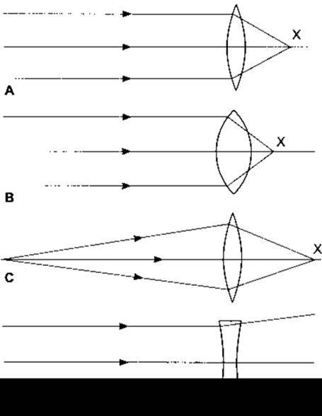
Fig. 8-9. Refraction of light rays by lenses. A — biconvex lens; B — biconvex lens with a higher refractive power than in A; C — lens as in A, illustrating The Effect of a closer object distance; D — biconcave lens. The central line in each diagram represents the principal optical axis. Point X indicates the principal focus.
Accommodation
When the ciliary Muscle is relaxed, parallel light rays entering an optically normal (emmetropic) eye are focused on the retina. If objects at a distance of less than 6 m are viewed with a relaxed ciliary muscle, the rays will be focused behind the retina, and the outlines will blur. Structure/149.html">The problem of focusing images of near objects on the retina can be solved by increasing either the distance between the lens and the retina or the curvature (refractive power) of the lens. In teleost fish, this problem is solved by elongating the Eyeball—similar to a camera, where the image of a close object is focused on the film by moving the lens away from it; in mammals, the curvature of the lens changes.
The process by which the curvature of the lens is increased is called accommodation. At rest, the lens is held in a stretched state by the suspensory ligament. Because the lens structure is subjected to tension and its capsule is highly elastic, the tension of the suspensory ligament causes the lens to flatten. When the gaze is shifted to a near object, the ciliary muscle contracts, which reduces the distance between the edges of the ciliary body and slackens the fibers of the suspensory ligament, allowing the lens to assume a more convex shape. In young individuals, changes in lens curvature can add up to 12 diopters of refractive power to the eye. The relaxation of the suspensory ligament fibers occurs partly through a sphincter-like contraction of the circular fibers of the ciliary muscle, and partly through the contraction of longitudinal muscle fibers attached anteriorly at the corneoscleral junction. When these muscle fibers contract, they pull the ciliary body forward and inward, bringing its edges closer together. Changes in lens curvature during accommodation primarily affect its anterior surface (Fig. 8-10). This fact was proven many years ago by a simple experiment. If an object is held in front of the eyes of a person looking into the distance, three reflected images can be seen in their eyes: a small, clear, upright image from the cornea; a larger, fainter, and also upright image from the anterior surface of the lens; and a small inverted image from the posterior surface of the lens. When the gaze is shifted to a near object, the large, blurred, upright image decreases in size and shifts closer to the other upright image, while the inverted image changes very little. The size change of the second image is caused by an increase in the curvature of the anterior surface of the lens (see Fig. 8-10). The constancy of the small upright image and the minimal Changes in the inverted image indicate that during accommodation, the curvature of the cornea does not change, and the curvature of the posterior surface of the lens changes very little.

Fig. 8-10. Accommodation. Solid lines show the shape of the lens, iris, and ciliary body in the relaxed state; dashed lines show these same structures during accommodation.
Near Point of Vision
Accommodation is an active process that requires muscular effort and can therefore be tiring. The ciliary muscle is one of the most frequently used Muscles in The Human Body. The degree to which the lens curvature can increase is generally limited, so light rays from a very close object cannot be focused on the retina even with maximum exertion. The point closest to the eye from which a clear image can still be obtained through accommodation is called the near point of vision. Throughout life, the near point of vision recedes—slowly at first, and then increasingly faster—from 9 cm at age ten to approximately 83 cm at age 60. This recession is primarily caused by an increase in lens rigidity, resulting in a loss of accommodation due to a gradual decrease in the possible degree of lens deformation (Fig. 8-11). Typically, at ages 40–45, the loss of accommodation causes difficulty with reading and close work. This condition, known as presbyopia, is usually corrected with convex lenses.
Near Response
In addition to accommodation, viewing near objects involves the convergence of the optical axes and the constriction of the pupils. This triple response—accommodation, convergence, and pupillary constriction—is called the near response.
Other Pupillary Reflexes
When light is directed into one eye, the pupil of that eye constricts (pupillary light reflex); simultaneously, the pupil of the other eye also constricts (consensual light reflex). The optic nerve fibers transmitting the impulses that cause these effects diverge from the optic tracts near the lateral geniculate bodies. On each side, they enter the Midbrain via the brachium of the superior colliculus and terminate in the pretectal Nucleus. From this nucleus, second-order Neurons project to the ipsilateral and contralateral Edinger-Westphal nuclei. Third-order neurons run from there to the ciliary ganglion as part of the Oculomotor nerve, and fourth-order neurons run from this ganglion to the ciliary body, respectively. This pathway runs more dorsally than the near-response pathway. Therefore, in some cases of light-near dissociation, the accommodative response remains intact (Argyll Robertson pupil). One cause of this pathology is CNS Syphilis, though the Argyll Robertson pupil can also occur in other diseases selectively affecting the midbrain.

Fig. 8-11. Decrease in the amplitude of accommodation with age. Different symbols represent data from various authors (reproduced with permission from Fisher RF: Presbyopia and the changes with age in the human crystalline lens. J Physiol 1973;228:765).
Retinal Image
The eyeball refracts light rays through the anterior surface of the cornea and both the anterior and posterior surfaces of the lens. The refraction process can be represented graphically without significant error by assuming that all refraction occurs solely at the anterior surface of the cornea. Figure 8-12 illustrates such a simplified, schematic eye. In this diagram, the nodal point (the optical center of the eye) coincides with the junction of the middle and posterior thirds of the lens, located 15 mm from the retina. This is the point through which light rays pass without being refracted. All other rays passing through the pupil from any point on an object are refracted and focused on the retina.
If the height of the observed object (AB) and its distance from the observer (Bn) are known, the size of the image on the retina can be calculated because triangles AnB and anb in Fig. 8-12 are similar. The angle AnB is called the visual angle for the object AB. Note that an inverted image is formed on the retina. Characteristically, this inverted image is perceived and transmitted to the visual cortex of the contralateral half of the Brain as the correct orientation of the object. This light perception is innate and present in infants. If the retinal image is inverted using special lenses, objects will likewise be perceived as upside down.

Fig. 8-12. Schematic eyeball: n — nodal point; AnB and anb — similar triangles. In this simplified eye, the nodal point is located 15 mm from the retina. It is conventionally assumed that all rays are refracted at the corneal surface, 5 mm from the nodal point, between a medium of density 1.000 (air) and a medium of density 1.333 (Water). Dashed lines represent the path of light rays originating from point A and, after corneal refraction, focusing at point a on the retina.
Common Image-Forming Defects
In some individuals, the eyeball is shorter than normal, causing parallel light rays to focus behind the retina. This condition is known as hyperopia, or farsightedness (Fig. 8-13). Constant accommodation, even when viewing distant objects, may partially compensate for this defect; however, the persistent tension of the ciliary muscles leads to fatigue and can cause headaches and blurred vision. Prolonged convergence of the visual axes associated with accommodation may lead to strabismus (squint) (see below). This defect is corrected using convex lenses, which increase the refractive power of THE EYE AND shorten the focal length.
In myopia (nearsightedness), the anteroposterior diameter of the eyeball exceeds normal values. Myopia is considered to be hereditary. In animal experiments, myopia can be induced by altering the refractive power of the eye during development. In humans, a link has been found between The Development of myopia and children sleeping in a lighted room up to the age of two. Consequently, the shape of the eyeball is partly determined by its light-refracting conditions. Strenuous near-work at a young age, such as during studying, accelerates the progression of myopia. This defect is corrected using biconcave lenses, which cause parallel rays to diverge before entering the eye.
Astigmatism is a fairly common visual disorder caused by uneven curvature of the cornea. If the corneal curvature along one meridian differs from that along another, light rays are focused differently, causing PARTS OF THE image to lose clarity. A similar defect can occur with a displaced or unevenly curved crystalline lens, though such pathology is rare. Astigmatism is typically corrected with cylindrical lenses positioned to balance light refraction across all meridians. Presbyopia was discussed earlier.
When the coordination between the left and right Components of the extraocular motor system is impaired, the image of an object is projected onto disparate (non-identical) retinal areas. This occurs in cases of damage to individual eye muscles and oculomotor nuclei, which disrupts the convergence of the visual axes. In such cases, the person perceives a double image (diplopia).

Fig. 8-13. Common optical defects of the eye. In hyperopia, the eyeball is shorter than normal, and light rays are focused behind the retina. A biconvex lens corrects this defect by increasing the refractive power of the eye. In myopia, the eyeball is longer than normal, and light rays are focused in front of the retina. A biconcave lens causes light rays to diverge before striking the cornea, thereby achieving proper focus on the retina.
Last update: 10/08/2026
Editorial and Educational Adaptation: This material has been compiled based on the primary/original source text. The project team performed an editorial review, corrected technical inaccuracies, structured sections, and adapted the content for an educational format.
What was processed:
- elimination of formatting defects (OCR errors, structural breaks, corrupted characters);
- editorial organization of content;
- standardization of terminology in accordance with academic sources;
- verification of factual statements against the original source text.
All mentions of the author, publication year, and origin of the primary text have been preserved in accordance with the source.