BIOCHEMISTRY - L. Stryer - 1984
VOLUME 3
PART V. MOLECULAR PHYSIOLOGY
CHAPTER 37. EXCITABLE MEMBRANES AND SENSORY SYSTEMS
37.17. Light Decreases Cyclic GMP Concentration by Activating Phosphodiesterase
As the experimental data indicate, the excitability of rod Cells depends on both Ca2+ and cGMP. The interplay between these agents may play a crucial role in visual excitation. Regarding the Molecular Mechanism of the light-induced release of Ca2+ into the Cytosol, many questions remain unresolved. However, significant progress has been made in recent years in understanding the Light regulation of cGMP levels in the outer segments of rod cells. Light appears to have no substantial effect on guanylyl cyclase, the enzyme that catalyzes cGMP synthesis:
Class="center">
In contrast, light exerts a remarkably potent effect on phosphodiesterase, the enzyme that hydrolyzes cGMP:

Illumination increases phosphodiesterase activity by several hundred-fold. The stimulation of this enzyme by photolyzed rhodopsin is mediated by a regulatory protein called Transducin. In the dark, transducin contains a tightly bound GDP molecule. Upon illumination, photolyzed rhodopsin forms a complex with GDP-transducin and catalyzes the exchange of GDP for GTP (Fig. 37.34). The resulting GTP-transducin complex activates phosphodiesterase. Crucially, a single photolyzed rhodopsin molecule catalyzes the exchange of GDP for GTP on several hundred transducin molecules, which in turn activates hundreds of phosphodiesterase molecules. Consequently, if the turnover number of phosphodiesterase is approximately 103 s-1, more than 105 cGMP molecules per photolyzed rhodopsin molecule are hydrolyzed per second in the light. The system returns to its initial dark state owing to an intrinsic GTPase activity. The bound GTP undergoes slow Hydrolysis to form GDP-transducin, which is incapable of activating phosphodiesterase. Thus, this entire cycle is driven by the Free energy of GTP hydrolysis. Here we see an example of how ~P is utilized for signal Amplification. The described cascade of reactions regulating cGMP levels (Fig. 37.35) is strikingly similar to the cascade mediating the action of β-adrenergic Hormones, particularly epinephrine (Section 35.4).
Fig. 37.33. Molecular model of cyclic GMP

Fig. 37.34. Photolyzed rhodopsin (Rh*) catalyzes The formation of a GTP-transducin complex; this complex in turn activates cyclic GMP phosphodiesterase

Fig. 37.35. Proposed cascade of reactions regulating cGMP levels in the retina. Abbreviations: Rh - rhodopsin, Rh* - photolyzed rhodopsin, T - transducin, PDEi - inactive phosphodiesterase, PDE - active phosphodiesterase

37.18. Color Vision Is Mediated by Three Types of Photoreceptors
In 1802, Thomas Young suggested that color perception is mediated by three primary receptors. As spectrophotometric studies of the intact retina performed more than 150 years ago demonstrated, there are Three types of cone cells in the eye, namely cells that absorb blue, green, and red light. To obtain the absorption spectra of these three photoreceptor pigments, the cones were illuminated with a beam of light
1 μm in diameter (Fig. 37.36). In addition, microelectrodes were inserted into many of the cones. Action spectra, based on Cell/33.html">Plasma Membrane hyperpolarization, fall into three groups with maxima in the blue, green, and red Regions of the visible spectrum, respectively. In the goldfish, the absorption maxima of the three color receptors are at 455, 530, and 625 nm, whereas rhodopsin is characterized by a maximum at 500 nm.
Fig. 37.36. Absorption spectra of the three color receptors

The chromophore in all three types of cones is 11-cis-retinal. In the absence of protein, the protonated Schiff base has an absorption maximum at 380 nm. Consequently, various groups on opsin exert a profound effect on the chromophoric Properties of the bound 11-cis-retinal. The dependence of the SPECTRAL PROPERTIES OF this chromophore on its protein environment is a specific manifestation of a general principle: protein interaction exerts a modulating influence on The properties of the prosthetic group. Another example of this is the function of heme as an oxygen carrier in Hemoglobin, an electron carrier in cytochrome c, and a catalyst in peroxidase.
Most forms of color blindness (daltonism) are caused by a sex-linked recessive mutation. About 1% of males are red-blind, and about 2% are green-blind. As spectral studies on the intact eye have shown, these individuals either completely lack photoreceptor molecules that perceive red or green light, or they possess an altered pigment with a shifted absorption spectrum. Thus, color blindness results from the absence or defect of one of the cone opsin types.
37.19. 11-cis-Retinal Is the Chromophore of All Known Visual Organs
Only in three animal phyla—Mollusks, Arthropods, and vertebrates—do eyes form image-projecting Organs. Anatomically, the eyes of these three groups are organized completely differently and appear to have evolved independently. Yet, in all three cases, 11-cis-retinal serves as the chromophore in the photoreceptor molecules. This is a striking example of convergent evolution. What is so special about 11-cis-retinal? First, this compound possesses an intense absorption band that is readily shifted into the visible region of the spectrum. Second, upon Light absorption, 11-cis-retinal undergoes rapid isomerization. Moreover, The rate of dark isomerization is extremely low. Third, the isomerization induces major structural changes. As a result, the absorbed light is transduced into atomic motions on a scale capable of triggering a Nerve Impulse. Finally, the ultimate precursors of 11-cis-retinal are carotenes (Section 20.27), which are widespread in living nature.
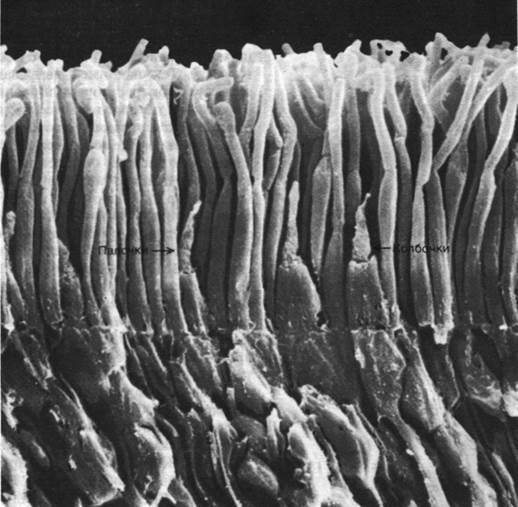
Fig. 37.37. Scanning electron micrograph of rod and cone cells in the photoreceptor layer of the retina
Last update: 06/08/2026
Editorial and Educational Adaptation: This material has been compiled based on the primary/original source text. The project team performed an editorial review, corrected technical inaccuracies, structured sections, and adapted the content for an educational format.
What was processed:
- elimination of formatting defects (OCR errors, structural breaks, corrupted characters);
- editorial organization of content;
- standardization of terminology in accordance with academic sources;
- verification of factual statements against the original source text.
All mentions of the author, publication year, and origin of the primary text have been preserved in accordance with the source.