BIOCHEMISTRY - L. Stryer - 1984
VOLUME 3
PART V. MOLECULAR PHYSIOLOGY
CHAPTER 37. EXCITABLE MEMBRANES AND SENSORY SYSTEMS
37.14. Light Triggers the Isomerization of 11-cis-Retinal
The primary event in visual excitation is well established. As George Wald demonstrated, light causes the isomerization of the 11-cis-retinal moiety of rhodopsin into all-trans-retinal. This isomerization drastically alters the geometry of retinal (Fig. 37.27). The Schiff base formed by retinal moves by approximately 5 Å relative to the chromophore ring region. In essence, the absorbed photon is converted into atomic motion.
Class="center">Fig. 37.27. The primary event in light excitation is the isomerization of the 11-cis isomer of the retinal Schiff base into the all-trans form.
The double bond between C-11 and C-12 is shown in green
Substantial isomerization of retinal occurs within the first few picoseconds following photon absorption, as indicated by the appearance of a new absorption band after intense laser pulse irradiation. The resulting photolysis intermediate, called bathorhodopsin (or prelumen-rhodopsin), contains a strained all-trans conformation of the chromophore. Subsequently, the protein continues to change its conformation, leading to a series of intermediates with distinct spectral properties (Fig. 37.28). The transition from metarhodopsin I to metarhodopsin II, which takes about a millisecond, involves the deprotonation of the Schiff base. The deprotonated Schiff base in metarhodopsin II is hydrolyzed within approximately one minute to yield opsin and all-trans-retinal; the latter diffuses away from opsin because it no longer fits the 11-cis isomer binding site. In the dark, all-trans-retinal isomerizes back into 11-cis-retinal, which binds to opsin, thereby regenerating rhodopsin. Unlike the rhodopsin photolysis reactions, the Hydrolysis of the Schiff base proceeds too slowly to play any role in Nerve Impulse generation.
Fig. 37.28. Intermediate stages of rhodopsin photolysis. The wavelengths corresponding to the absorption maxima of each compound are given, along with the time constant for each conversion
37.15. Light Causes Hyperpolarization of the Rod Outer Segment Plasma Membrane
The cis-to-trans isomerization of retinal and the subsequent Conformational Changes in rhodopsin are the primary events of visual excitation. The next crucial step required for nerve impulse generation was uncovered through electrophysiological studies of the intact retina. A quantum of light causes a transient hyperpolarization of the outer segment Cell/33.html">Plasma Membrane (Fig. 37.29). The kinetics of hyperpolarization depend on the light beam intensity and the steady Background illumination level. The response time to a single photon is about one second, whereas for intense incident light it is a few milliseconds. Action potentials do not occur in rods; their response to light is graded. The magnitude of the signal traveling from the outer segment to the synapse depends on the number of absorbed photons. In fully sensitive, dark-adapted rods, a half-maximal hyperpolarization level is observed upon the absorption of only 30 photons by an outer segment containing 40 • 106 rhodopsin molecules (Fig. 37.30). The absorption of a single photon by a dark-adapted rod elicits a hyperpolarization of about 1 mV, which is detected by the synapse and transmitted to other retinal Neurons. Exceptional sensitivity is not the only remarkable property of rods. Their second striking feature is that the response of the photoreceptor system to pulsed illumination depends on the background illumination level. Considerably more photons are required to excite rods exposed to continuous light than those kept in the dark (Fig. 37.30). Thanks to this property, known as adaptation, retinal rods are able to function across background illumination levels spanning many orders of magnitude.
Fig. 37.29. Light exposure leads to the hyperpolarization of retinal rods
Fig. 37.30. The sensitivity of retinal rods to pulsed illumination depends on the background light level
What is the ionic mechanism behind light-induced hyperpolarization? In the dark, The Plasma Membrane of the rod outer segment is highly permeable to Na+. This fact, coupled with a steep transmembrane Na+ concentration gradient, causes sodium ions to rapidly enter the outer segment in the dark. This gradient is maintained by the (Na+ + K+)-ATPase localized in the inner segment plasma membrane. Thus, in the dark, sodium ions enter the outer segment, diffuse into the inner segment, and are subsequently pumped out using ATP energy. Light somehow blocks the Na+ channels in the outer segment plasma membrane. As a result, the inward Na+ current decreases, and the inner face of the membrane becomes more electronegative. In other words, upon illumination, the Membrane Potential of the rods shifts toward the K+ equilibrium potential. Subsequently, the light-induced hyperpolarization near the illuminated discs is passively conducted along the plasma membrane to the synaptic body.
37.16. Mediators Transmit the Signal from Photolyzed Rhodopsin to the Plasma Membrane
The change in plasma membrane permeability to Na+ and the subsequent hyperpolarization are highly amplified Responses of the outer segment to light. Indeed, the absorption of just a single photon by a dark-adapted rod blocks the flow of over a million sodium ions. How does such massive Amplification arise? First of all, it should be noted that the disk membranes, which contain the bulk of rhodopsin molecules, do not Touch the rod plasma membrane and are not electrically coupled to it. Furthermore, a rhodopsin molecule that has absorbed a photon may be located several thousand angstroms away from the plasma membrane sodium channel. All this rules out direct interaction between the discs and the plasma membrane. There is virtually no doubt that the signal from photolyzed rhodopsin (Rh*) in the disk membranes is transmitted to the plasma membrane via diffusing mediators. Moreover, to achieve the high degree of amplification actually observed, the photolysis of a single rhodopsin molecule must be accompanied by the generation (or breakdown) of A large number of mediator molecules.
Fig. 37.31. Schematic representation of the hypothesis regarding The Role of Calcium Ions as mediators in visual excitation
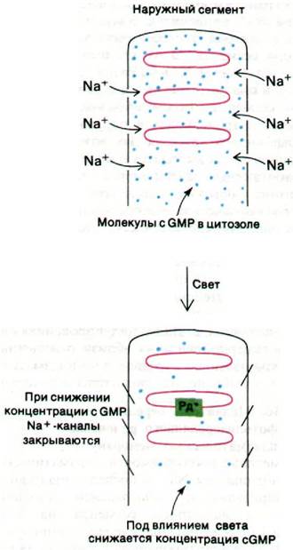
The exact Nature of the mediator has not yet been definitively established; research has put forward two highly probable candidates for this role: Ca2+ ions (Fig. 37.31) and cyclic GMP (Fig. 37.32). The following experimental data support Ca2+ as a mediator.
Fig. 37.32. Schematic representation of the hypothesis regarding the role of cGMP as a mediator in visual excitation. Rh* denotes photolyzed rhodopsin
1. Sodium channels in the plasma membrane close when the Ca2+ concentration in the Cytosol increases, and open when it decreases.
2. Microinjection of Ca2+-chelating agents into the cytosol decreases the light sensitivity of rod Cells. This desensitization indicates that the photolysis of a single rhodopsin molecule leads to the release of several hundred Ca2+ ions into the cytosol.
3. Following a light pulse, a significant amount of Ca2+ is extruded from the outer segments of rod cells.
However, judging by the results of other experiments, the mediator might be cGMP. The following data are crucial for this Conclusion.
1. Sodium channels in the plasma membrane open when the cGMP concentration in the cytosol increases and close when the concentration of this nucleoti
de decreases.
2. The cGMP content is regulated by light. Specifically, light activates a phosphodiesterase that hydrolyzes cGMP, as will be discussed in more detail below.
3. The photolysis of a single rhodopsin molecule leads to the rapid hydrolysis of 105 cGMP molecules.
Last update: 06/08/2026
Editorial and Educational Adaptation: This material has been compiled based on the primary/original source text. The project team performed an editorial review, corrected technical inaccuracies, structured sections, and adapted the content for an educational format.
What was processed:
- elimination of formatting defects (OCR errors, structural breaks, corrupted characters);
- editorial organization of content;
- standardization of terminology in accordance with academic sources;
- verification of factual statements against the original source text.
All mentions of the author, publication year, and origin of the primary text have been preserved in accordance with the source.