BIOCHEMISTRY - L. Stryer - 1984
VOLUME 3
PART V. MOLECULAR PHYSIOLOGY
CHAPTER 37. EXCITABLE MEMBRANES AND SENSORY SYSTEMS
37.11. Catecholamines and γ-Aminobutyric Acid (GABA) Also Function as Neurotransmitters
In addition to acetylcholine, other Neurotransmitters are known. A substance is considered a neurotransmitter if it satisfies the following criteria. First, microinjections of the putative neurotransmitter into the synaptic cleft must elicit a response identical to The stimulation of the presynaptic nerve. Second, the substance must be present in high concentrations within presynaptic nerve terminals. In this regard, the most compelling criterion is the isolation of synaptic vesicles containing the substance. Third, the postulated mediator must be released from the presynaptic nerve at the appropriate time and in sufficient quantity to affect the postsynaptic nerve.
A number of catecholamines satisfy these criteria. For instance, norepinephrine serves as the neurotransmitter in smooth Muscle junctions innervated by sympathetic nerves (in contrast to parasympathetic junctions, where acetylcholine is the neurotransmitter). The catecholamines epinephrine and dopamine are two other catecholaminergic neurotransmitters. These catecholamines are synthesized from Tyrosine in sympathetic nerve terminals and in the Adrenal Glands (Fig. 37.18). The first step of synthesis—the rate-limiting reaction of the entire process—is the hydroxylation of tyrosine to yield 3,4-dihydroxyphenylalanine (DOPA). This reaction is catalyzed by tyrosine hydroxylase, an enzyme analogous to phenylalanine hydroxylase. Tetrahydrobiopterin, a cofactor of the enzyme, serves as the activator of molecular oxygen in this process. The second step is the decarboxylation of DOPA, catalyzed by DOPA decarboxylase (a Pyridoxal phosphate-containing enzyme) to yield 3,4-dihydroxyphenylethylamine (dopamine). Next, dopamine is hydroxylated to norepinephrine in the presence of a copper-containing hydroxylase. Finally, epinephrine is formed from norepinephrine via methylation; the enzyme carrying out this reaction is a transmethylase that uses S-adenosylmethionine as the methyl group donor.
Class="center">Fig. 37.18. Biosynthetic pathway of catecholaminergic neurotransmitters

The inactivation of catecholamine neurotransmitters is achieved by the methylation of the 3-OH group of the catechol ring. The reaction is catalyzed by catechol-O-methyltransferase, which utilizes S-adenosylmethionine as the methyl group donor. Another pathway for the inactivation of these neurotransmitters is the removal of the amino group through oxidation by monoamine oxidase (Fig. 37.19).
Fig. 37.19. Inactivation of norepinephrine

Yet another neurotransmitter has been identified among Amino Acid Derivatives. This is γ-aminobutyrate, also referred to as γ-aminobutyric acid (GABA). GABA increases the permeability of postsynaptic membranes to K+, thereby driving the Membrane Potential further away from the threshold level required to trigger an Action Potential; thus, GABA Functions as an inhibitory neurotransmitter. GABA is produced by the decarboxylation of glutamate in a reaction catalyzed by glutamate decarboxylase (Fig. 37.20). As one might predict, the prosthetic group of this decarboxylase is pyridoxal phosphate. γ-Aminobutyrate is inactivated via Transamination to yield succinic semialdehyde, which is subsequently oxidized to succinate.
Fig. 37.20. Synthesis and inactivation of γ-aminobutyrate

37.12. A Single Photon Is Sufficient to Excite a Rod Cell in the Retina
Let us now examine excitable receptors activated by light. Humans possess Two Types of photoreceptor Cells, known as rods and cones based on their shape. Cones function in bright light and are responsible for Color Vision, whereas rods perceive dim light but do not distinguish colors. The human retina contains 3 million cones and 1 billion rods. These photoreceptor cells convert light energy into atomic motion and subsequently into a Nerve Impulse. Rods and cones form synapses with bipolar cells, which in turn interact with other Nerve Cells in the retina. The electrical signals generated by the photoreceptor cells are processed as they travel through a complex network of retinal nerve cells and are then transmitted to the Brain via the Optic nerve fibers. Thus, the retina performs two functions: it transforms light into nerve impulses and integrates visual information.
Fig. 37.21. Scanning electron micrograph of retinal rod cells

In 1938, Selig Hecht discovered that a single photon is sufficient to excite a human retinal rod Cell. Let us consider the molecular basis for the exceptionally high sensitivity of these cells. Rods are thin, elongated structures typically 1 µm in diameter and 40 µm in length. The Main Functions of this cell are sharply segregated spatially (Fig. 37.22). The outer segment of the rod is specialized for photoreception. It contains approximately 1000 stacked discs (Fig. 37.23). The discs are closed, flattened sacs about 160 Å thick. These membrane structures are densely packed with photoreceptor molecules. The disc membranes and The Plasma Membrane of the outer segment do not Touch. A narrow cilium connects the outer segment to the inner segment, which is rich in Mitochondria and Ribosomes. ATP is produced at a very high rate in the inner segment, and active Protein Synthesis takes place there. The discs of the outer segment have a lifespan of only 10 days and are constantly renewed. The inner segment adjoins The Nucleus, which is located close to the synaptic body. The synaptic body, containing numerous synaptic vesicles, forms a synapse with bipolar cells.
Fig. 37.22. Schematic representation of a retinal rod

Fig. 37.23. Electron micrograph of the outer segment of a retinal rod, showing the stacked discs

37.13. Rhodopsin Is the Photoreceptor Protein of Rods
Stimulation of photoreceptor cells requires the absorption of light. The absorption of a light photon must trigger structural Changes in the light-absorbing group (the chromophore). The photosensitive pigment of rod cells is rhodopsin, which consists of the protein opsin and a prosthetic group represented by 11-cis-retinal (Fig. 37.24). Rhodopsin is a transmembrane protein with a molecular mass of 38 kDa. Its N-terminus is located in the aqueous phase inside the disk, while the C-terminus is on the opposite side of the disk membrane, in the Cytosol. The N-terminal region of rhodopsin contains two oligosaccharide units covalently attached to an aspartate side chain.
Fig. 37.24. Structures of 11-cis-retinal, all-trans-retinal, and all-trans-retinol (vitamin A)

These sugars apparently play a crucial role in the directed transport of rhodopsin from the inner segment to the disks. The fact is that rhodopsin, like other eukaryotic Membrane Proteins, is synthesized on ribosomes attached to The Endoplasmic reticulum. Newly synthesized rhodopsin enters the Golgi apparatus and only then reaches the plasma membrane. New disks are formed at the Base of the inner segment by invagination of the plasma membrane, which is why the carbohydrate units of rhodopsin end up localized inside the disk, even though initially, as part of the plasma membrane, they faced the extracellular space (Fig. 37.25).
Fig. 37.25. Formation of disks by invagination of the plasma membrane. The arrows indicate the polarity of the rhodopsin molecules

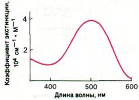
Opsin, like other proteins lacking prosthetic groups, does not absorb visible light. The color of rhodopsin and its sensitivity to light are determined by the presence of 11-cis-retinal, which serves as a highly efficient chromophore. Due to 11-cis-retinal, rhodopsin exhibits a broad absorption band in the visible spectrum with a maximum at 500 nm, which matches solar radiation remarkably well. The intensity of visible Light absorption by rhodopsin is also noteworthy. The extinction coefficient of rhodopsin at 500 nm is very high, specifically 4 • 104 cm-1 • M-1 (Fig. 37.26). The total absorption strength of rhodopsin for visible light approaches the maximum values found in Organic compounds. The superior chromophoric properties of 11-cis-retinal stem from the fact that it is a polyene. The alternation of six single and double (unsaturated) bonds within it creates an extended unsaturated system for electron transfer.
Fig. 37.26. Absorption spectrum of rhodopsin
11-cis-retinal is attached to rhodopsin via a Schiff base, which is formed by the linkage of the aldehyde group of 11-cis-retinal with the ε-amino group of a specific Lysine residue in opsin. The SPECTRAL PROPERTIES OF rhodopsin indicate that the Schiff base is in a protonated form.

The precursor of 11-cis-retinal is all-trans-retinol (vitamin A), which cannot be synthesized de novo in the mammalian body. All-trans-retinol (Fig. 37.24) is converted into 11-cis-retinal in two steps. First, the alcohol group is oxidized to an aldehyde group in the presence of retinol dehydrogenase and NADP+ as an electron acceptor. Then, under the action of retinal isomerase, the double bond between C-11 and C-12 is isomerized from the trans to the cis configuration. Vitamin A deficiency leads to night blindness (nyctalopia) and ultimately to damage of the rod outer segments.
Last update: 06/08/2026
Editorial and Educational Adaptation: This material has been compiled based on the primary/original source text. The project team performed an editorial review, corrected technical inaccuracies, structured sections, and adapted the content for an educational format.
What was processed:
- elimination of formatting defects (OCR errors, structural breaks, corrupted characters);
- editorial organization of content;
- standardization of terminology in accordance with academic sources;
- verification of factual statements against the original source text.
All mentions of the author, publication year, and origin of the primary text have been preserved in accordance with the source.