Biological Membranes - A. N. Ogurtsov 2012
Electrogenesis of Biomembranes
Intracellular Signaling
Calcium Ions
In the synapse between a pain receptor and an associative pain neuron (Figure 137), glutamic acid (glutamate) Functions as a neurotransmitter. When an Action Potential reaches the proximal axon terminal of the pain receptor, the regulatory exocytosis mechanism of glutamic acid is triggered in the presynaptic region as described above (Figure 125).
The depolarization of the axon membrane from -70 mV to +30 mV opens voltage-gated calcium channels within the membrane, causing the concentration of calcium ions to surge from 100 nM/L (at rest) to 1 µM/L, which prompts synaptic vesicles to release their contents into the synaptic cleft. In the synapse connecting a sensory neuron to an associative neuron, these vesicles are packed with sodium glutamate.
In this case, much like at the Neuromuscular Junction (see section 16.4), calcium ions act as an intracellular messenger—a substance whose elevated cytosolic concentration stimulates a Cell-specific response.
The cytosolic concentration of calcium can rise not only in response to an action potential, but also when signaling molecules—often referred to as Transmitters—appear in the extracellular environment.
The presence of such transmitter molecules is detected by integral membrane receptor Proteins specific to each molecular type. Upon binding a specific Ligand, these receptors trigger a defined metabolic cascade that results in the release of calcium from the smooth Endoplasmic reticulum into the Cytosol.
For example, in Blood Platelets, such a release of Ca2+ ions from The endoplasmic reticulum into the platelet cytosol stimulates the blood-clotting process.
Calcium exits the endoplasmic reticulum through Inositol trisphosphate-regulated calcium channels, whereas inositol trisphosphate (IP3) is produced by the β-isoform of the enzyme phospholipase C (PLCβ) via the Enzymatic Hydrolysis of The Plasma Membrane phospholipid phosphatidylinositol bisphosphate (PIP2).
For platelets, the chemical signal indicating that the clotting process must be initiated is the appearance of ADP molecules in the blood (Figure 139).
Class="center">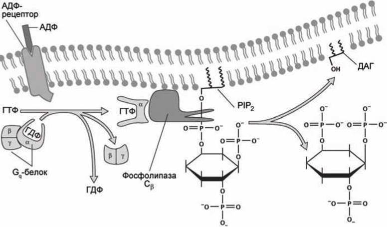
Figure 139 - Synthesis of inositol trisphosphate in response to ADP binding to a membrane receptor
ADP molecules can enter the bloodstream solely As a result of mechanical cell damage. The binding of ADP to a membrane GPCR receptor on the platelet surface activates this receptor, transforming it into a GEF protein (Guanine nucleotide Exchange Factor, Figure 79) for the trimeric Gq protein. This protein possesses GTPase activity and, in turn, activates the β-isoform of phospholipase C (precisely termed phosphoinositide phospholipase C).
Unlike the Ran GTPase, which directs Protein transport into the Cell Nucleus (Figure 80), the trimeric Gq protein consists of three subunits: the GTPase α subunit and two subunits, β and γ. These dissociate from the α subunit when it is bound to ATP, and reassemble into the trimeric protein after ATP is hydrolyzed into ADP.
The activated form of phospholipase C directly catalyzes the aforementioned hydrolysis of phosphatidylinositol bisphosphate (PIP2), yielding inositol trisphosphate (IP3) in the cytosol and diacylglycerol (DAG) in the membrane.
Thus, in response to the appearance of ADP molecules in the exoplasm, the concentration of IP3 increases within the Cytoplasm. These molecules then activate inositol trisphosphate-gated calcium channels (Figure 140).
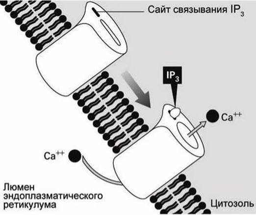
Figure 140 - Inositol trisphosphate-gated calcium channel
The surge in cytosolic calcium ion concentration alters platelet shape, causing the platelets to become "sticky" and clump together. This forms a blood clot and initiates coagulation at the site of the blood vessel injury.
The combination of signaling pathway components—comprising phosphatidylinositol bisphosphate PIP2, the trimeric Gq protein, the β-isoform of phospholipase C (PLCβ), and inositol trisphosphate-gated calcium channels—is present in virtually all Eukaryotic Cells. However, only those cells whose Plasma Membranes feature ADP receptors exhibit an increase in cytosolic calcium ion concentration when exposed to ADP. Other cells respond to different signaling molecules for which they possess specific Membrane Receptors, with over a hundred such receptors having been discovered.
A sharp rise in cytosolic calcium ion concentration serves as a universal intracellular signal, though the specific metabolic process triggered by this signal depends on The Cell type. In secretory cells (such as salivary gland cells or axon terminals), secretion is induced, whereas Muscle cells undergo sarcomere contraction. In all cases, calcium ions bind to calcium-binding proteins, and these calcium-Structure/178.html">Protein Complexes activate the corresponding cellular process.
In Skeletal Muscle cells, besides (1) Muscle contraction, calcium ions also stimulate (2) Glycogen hydrolysis via Glycogen phosphorylase kinase, and (3) enhance The activity of Krebs cycle Enzymes in Mitochondria by promoting the synthesis of NADP and ATP.
Many cells feature specialized calcium channels within the endoplasmic reticulum membrane known as ryanodine receptors. These channels were initially identified by their ability to bind the plant alkaloid ryanodine instead of IP3.
In skeletal muscle cells, these ryanodine receptors are physically linked to the voltage-gated calcium channels of the plasma membrane (Figure 141).
When the voltage-gated channel in the plasma membrane opens, the ryanodine receptor in the endoplasmic reticulum membrane opens as well, releasing calcium into the cell cytosol.

Figure 141 - Ryanodine receptors of skeletal muscle cells
In other cell types, no such mechanical coupling between the plasma Membrane Channels and the ryanodine receptors of the endoplasmic reticulum has been found; instead, these receptors are activated when the cytosolic calcium concentration exceeds a critical threshold.
For instance, in cardiomyocytes (Heart muscle cells), plasma membrane depolarization triggers the opening of plasma membrane calcium channels. Calcium ions entering the cell from the extracellular space then bind to the calcium-binding sites on the ryanodine receptors of the endoplasmic reticulum membrane. This induces the opening of the ryanodine receptor channels as well, releasing calcium from the smooth endoplasmic reticulum into the cytosol (Figure 142).
Once the external stimulus ceases, the cytosolic calcium concentration returns to its baseline (resting) level. Calcium ions are pumped out of the cytosol by (1) Ca2+-ATPases (sec. 7.2) and (2) 3-Na+/1-Ca2+ exchangers (sec. 11.2).

Figure 142 - Ryanodine receptors in cardiomyocytes
Last update: 13/08/2026
Editorial and Educational Adaptation: This material has been compiled based on the primary/original source text. The project team performed an editorial review, corrected technical inaccuracies, structured sections, and adapted the content for an educational format.
What was processed:
- elimination of formatting defects (OCR errors, structural breaks, corrupted characters);
- editorial organization of content;
- standardization of terminology in accordance with academic sources;
- verification of factual statements against the original source text.
All mentions of the author, publication year, and origin of the primary text have been preserved in accordance with the source.