Biological Membranes - A. N. Ogurtsov 2012
Electrogenesis of Biomembranes
Intracellular Signaling
Intracellular Calcium Oscillations
The interplay between Cell/33.html">Plasma Membrane and internal membrane Ca2+ channels, calcium pumps, and calcium-binding Proteins located in both membranes and the Cell Cytoplasm leads to so-called Ca2+ oscillations—periodic fluctuations in its cytoplasmic concentration (Figure 143).
In non-excitable Cells, the primary trigger for calcium oscillations is Inositol trisphosphate, which is produced from phosphatidylinositol bisphosphate when phospholipase C is activated by Hormones or growth factors (Figure 139). Inositol trisphosphate is capable of diffusing from The Plasma Membrane, where it is generated, to the membranes of The Endoplasmic reticulum within tens of seconds. The amount and concentration of the resulting IP3 are high enough to bind to all corresponding receptors, yet Ca2+ release occurs only at designated hot spots.
Hot spots refer to transient localized areas within The Cell that emerge, much like bubbles in boiling Water, at various sites on the endoplasmic reticulum membrane due to a high local concentration of Ca2+, inositol trisphosphate, or its receptor.
According to current models, the Ca2+ released into the cytoplasm at a hot spot diffuses along the reticulum, increasing the sensitivity of the receptor to inositol trisphosphate in its membranes and facilitating channel opening, thereby driving the Propagation of the Ca2+ wave front.
Class="center">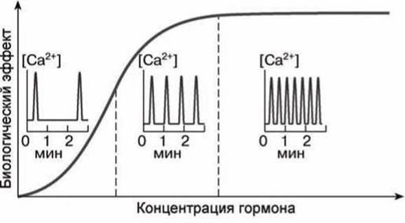
Figure 143 - Dependence of the fluctuation frequency of cytoplasmic Ca2+ ion concentration and the magnitude of the biological effect induced by Ca2+ ions on hormone concentration
A local rise in Ca2+ concentration at this membrane site above the threshold value leads to the inactivation of calcium channels, causing the hot spot to extinguish, while Ca2+ diffusion generates new sites of Ca2+ release from the reticulum. Calcium pumps also participate in extinguishing hot spots by transporting Ca2+ from the Cytosol back into the endoplasmic reticulum or the extracellular space.
In excitable Tissues, the primary influx of Ca2+ into the cell occurs via voltage-gated Ca2+ channels that are functionally coupled to ryanodine receptors. At the junction between these Two Types of channels, Ca2+ release drives the spread of a Ca2+ wave of ryanodine receptor activation along the reticulum membranes. Behind the Ca2+ wave front, the Ca2+ level drops because calcium is already depleted in that region of the reticulum, meaning Ca2+ no longer exits through the channels; conversely, calcium pumps actively clear Ca2+ from the cytoplasm. In excitable cells, the frequency of Ca2+ oscillations can be enhanced by IP3, cyclic ADP-ribose, or caffeine.
In many cell types, the calcium oscillation wave propagates outward from the Cell Nucleus and can take the form of expanding spheres or complex spirals.
In certain tissues (such as The Heart and Brain), calcium oscillations originating in a single cell can stimulate Ca2+ oscillations in neighboring cells at the exact same frequency as in the initiating cell.
Apparently, in these tissues, the Ca2+ wave can propagate through intercellular Gap Junctions (see Section 16.1), which possess high ionic conductance.
Within a single cell's cytoplasm, hormones and growth factors have virtually no effect on the amplitude of the Ca2+ concentration increase, but they do increase the frequency of its fluctuations (Figure 143). Typically, cytoplasmic Ca2+ levels range from 10 to 5·10-7 M, with frequencies ranging from one oscillation per minute to several oscillations per second.
The Ca2+-dependent effects of hormones and growth factors are directly proportional to the frequency of cytoplasmic Ca2+ fluctuations (Figure 143). This can be explained by the fact that higher oscillation frequencies increase the probability of calcium-binding proteins becoming saturated with calcium.
While dissociation of Ca2+ from high-affinity sites on calcium-binding proteins takes minutes, cytoplasmic Ca2+ oscillates much faster; consequently, calcium-binding proteins perceive this frequency information and—much like AC-to-DC converters—translate it into a slowly developing metabolic, morphological, or functional shift in the cell over the course of minutes or hours.
Calcium-binding proteins may contain multiple Ca2+-binding sites that exhibit positive cooperativity. Upon Ca2+ binding, the Protein Structure may feature an increased number of $\alpha$-helices, frequently exposing functional groups On the surface of the protein globule that mediate the interaction between the calcium-binding protein and downstream effector proteins. In this manner, Ca2+ induces Protein-Protein Interactions that alter their activity or cellular localization.
Ca2+ activates numerous catabolic processes (Glycolysis, lipolysis, proteolysis) and also stimulates Protein Synthesis, Muscle contraction and non-muscle motility, exocytosis, ion transport, and neurotransmitter secretion.
Last update: 13/08/2026
Editorial and Educational Adaptation: This material has been compiled based on the primary/original source text. The project team performed an editorial review, corrected technical inaccuracies, structured sections, and adapted the content for an educational format.
What was processed:
- elimination of formatting defects (OCR errors, structural breaks, corrupted characters);
- editorial organization of content;
- standardization of terminology in accordance with academic sources;
- verification of factual statements against the original source text.
All mentions of the author, publication year, and origin of the primary text have been preserved in accordance with the source.