Practical Guide to Zoology: Textbook - T. A. Dauda 2014
Multicellular Animals
Arthropods
Subphylum Tracheata - Class Insecta
The subphylum Tracheata comprises terrestrial arthropods that breathe via tracheae. Most tracheates possess a well-defined HEAD. Their head appendages are represented by only a single pair of antennae, three pairs of jaws (mandibles), and two pairs of maxillae.
The subphylum Tracheata includes two classes: Myriapoda and Insecta.
We will examine exclusively the Representatives of the Class Insecta.
The class Insecta encompasses all tracheate arthropods possessing three pairs of legs. Their bodies are distinctly divided into a head, Thorax, and abdomen. The majority of representatives within this class possess The ability to fly. The practical importance and diversity of insects have long established entomology as an independent scientific discipline.
The objective of this practical course is to provide a General Overview of insects as one of the classes of arthropods. Morphological and anatomical Research Methods are demonstrated using the oriental cockroach (Blatta orientalis) as a model Organism. Furthermore, a comparative Analysis of the mouthparts of various insects is presented, and preparations of representatives of the order Diptera are examined.
Order Cockroaches (Blattoidea)
Oriental cockroach (Blatta orientalis).
The oriental cockroach is closely associated with human habitation. It is nocturnal, hiding by day in wall cracks, floor crevices, and similar sheltered spots. Extremely voracious, it feeds on foodstuffs (bread, flour, pastry products, porridges, etc.) found in human dwellings, canteens, and bakeries. Crawling from garbage bins and spittoons onto human food supplies, it contaminates them by mechanically carrying pathogens of infectious diseases (typhoid fever, dysentery, cholera, tuberculosis, diphtheria, etc.) and helminth eggs on its tarsi and body.
Materials and Equipment. Captured cockroaches can be maintained (in winter, in a warm Location) in special breeding cages with wire-mesh walls or in boxes featuring a tight-fitting mesh lid, with the bottom lined with several layers of paper and wood shavings.
They are fed Water-soaked bread supplemented with a small amount of raw vegetables, and a small water container is provided for drinking.
Other closely related species may be used in place of the oriental cockroach: the German cockroach (Blattella germanica) or the American cockroach (Periplaneta americana).
The Study of external Morphology is carried out using dry or alcohol-preserved specimens. Chloroform-anesthetized animals are used for dissection.
Required tools and materials: a Microscope; a dissecting microscope; a hand lens; dissection instruments (a dissecting tray, scalpels, scissors, forceps, needles, pins); a sheet of white paper; Glass slides and coverslips; pipettes; water in a beaker or jar; freshly anesthetized cockroaches (males and females); cockroach oothecae and nymphs.

Fig. 98 External Anatomy of the oriental cockroach (Blatta orientalis):
A — male in dorsal view; B — female; 1-10 — abdominal segments; 11 — antenna; 12 — maxillary palp; 13 — eyes; 14 — wings; 15 — cerci; 16 — styli; 17 — anal opening; 18 — tarsus; 19 — Tibia; 20 — Femur.
Assignment. Examine the external anatomy of the oriental cockroach and make a dorsal-view drawing of it. Dissect and examine the cockroach's legs, drawing one of them. Dissect the cockroach, expose its Internal Organs, examine, and illustrate them.
Examination of External Morphology. Place the cockroach on a sheet of white paper inside a dissecting tray.
Examine the anesthetized cockroach using a hand lens. Note that the body is covered with a dark brown chitinous cuticle (an outer Skeleton, or exoskeleton) and consists of three distinct regions: the head, thorax, and abdomen. The body is flattened dorsoventrally, which enables the cockroach to squeeze into narrow crevices. Learn to distinguish males from females: males are smaller than females, possess more developed wings (Fig. 98), and have a longer and narrower abdomen.
Locate a pair of long, segmented antennae (or feelers) on the head, which function as tactile and olfactory organs. Upon examining an antenna under a lens, it is easy to observe that it consists of numerous segments. Using a hand lens, find a pair of simple ocelli at the Base of the antennae and the compound (faceted) eyes on the sides of the head. Lift the cockroach's head with forceps or a dissecting needle—since it is bent ventrally and largely concealed by the first thoracic segment—and locate the slender neck by which the head articulates with the thorax. The mouthparts of the cockroach, which are of the chewing type, are visible on the lower part of the head. Note the forward-projecting maxillary palps (do not mistake them for antennae).
Examine the thorax of the cockroach; it consists of the prothorax, mesothorax, and metathorax (three segments). The wide, triangular first thoracic segment is frequently mistaken for the entire thorax, but it represents merely the prothorax. The integument of each segment is formed by four movably articulated chitinous plates: the dorsal plate (tergite), the ventral plate (sternite), and two lateral plates (Pleura). Each segment bears a pair of legs. The dorsal surface of the mesothorax and metathorax each bears a pair of wings. All three thoracic segments are visible in females because they bear only wing buds. In the male, only the first segment is visible; its long wings conceal not only the remaining thoracic segments but also a portion of the abdominal segments. Only by removing the wings of the male is it possible to examine all the thoracic and abdominal segments.
Count all the abdominal segments—there are ten—and note that they lack appendages. Each segment is covered by two chitinous plates connected laterally by a soft membrane: the dorsal plate is the tergite and the ventral plate is the sternite. Not all abdominal segments are clearly visible; for instance, the 8th and 9th segments are concealed beneath the 7th. To examine and count the abdominal rings, pull the cockroach's antennae with the fingers of your right hand while holding the posterior abdominal segment with forceps. The segments will pull apart and become clearly distinguishable. At the posterior end of the abdomen in both sexes, a pair of finely segmented appendages—known as cerci—is visible. They are covered with sensory hairs and function as tactile organs. Additionally, males possess further appendages at the tip of the abdomen called styli. The cerci and styli are vestigial abdominal legs. Locate and examine with a hand lens how the sternite of the ultimate abdominal segment in the female is longitudinally cleft. It serves to hold the egg capsule (ootheca).
Using a hand lens, locate the stigmata, or spiracles (openings leading into the tracheae, or respiratory tubes), on the sides of the initial abdominal segments.
Examine the wings of the male oriental cockroach. There are two pairs, formed by thin folds of integument flattened into a lamina. The first pair is termed the tegmina (or elytra), and the second pair constitutes the true wings. The tegmina are stiffer and thicker, whereas the true wings are more delicate and membranous. In females, the wings are reduced to small, leaf-like plates.
Using forceps, pluck off the wings, place them on a glass slide, and examine them with a dissecting magnifier. You can see thickenings (Veins) running across and along the wing, which provide structural rigidity.
Turn the cockroach ventral side up.
Examine the legs of the cockroach—they are adapted for running. Each leg consists of 5 segments: the coxa, trochanter, femur, tibia, and tarsus.
The coxae are equipped with powerful Muscles that actuate the leg. The five-segmented tarsus terminates in a pair of claws with a suction pad between them; the insect uses these to grip uneven surfaces.
Grasp the coxa attached to the thoracic segment with forceps, use scissors to cut off the third leg of the cockroach, place it on a glass slide, and make a drawing while examining it under a dissecting magnifier.
Note the numerous spines of various sizes covering the femur and tibia, as well as the first tarsal segment, which is significantly longer than the others.
Dissecting preserved cockroaches stored in alcohol is difficult because they become brittle and fragile. Instead, specimens anesthetized with ether or chloroform are used.
Hold the cockroach in your left hand and carefully use sharp scissors to cut along the lateral margins of the body from the anus to the head. Make a transverse cut through the chitinous exoskeleton on the pronotum.
Pin the cockroach, dorsal side up, to the wax-bottomed dissecting dish using two pins (one through the head and one through the last abdominal segment). While pinning, stretch the body slightly. Pour water into the dish until the insect is completely submerged. Carefully lift the posterior edge of the dorsal exoskeleton with fine forceps, and using a sharp scalpel or a finely honed dissecting needle, detach and remove the cut exoskeleton from the abdomen and thorax, pinning it to the bottom of the dish with the inner side facing up. The entire dorsal portion must be removed. This exposes the body cavity (mixocoel). It is filled with internal organs, and the spaces between them contain a loose, whitish tissue that stores nutrient reserves—fats, CARBOHYDRATES, and Proteins. This is known as the fat body. It must be plucked out with forceps and rinsed away with a stream of water from a pipette.
Isolate the gut as shown in Figure 99. Unravel the loops of the intestine using fine forceps and dissecting needles, push it aside, pin it to the bottom of the dish, and examine all PARTS OF THE Digestive System. It begins with the Mouth opening, which leads into the Pharynx and Esophagus. Locate the esophageal dilation—the crop, which serves as a food storage reservoir—followed by the next section, the muscular gizzard where food is ground down. Touch the walls of the crop and gizzard with a needle. The pharynx, esophagus, crop, and gizzard constitute the foregut. Food is digested and absorbed in the midgut. Locate several blind caeca opening into the midgut, which serve to increase the digestive surface area.
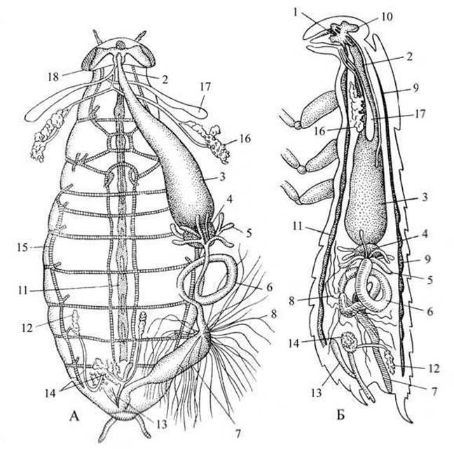
Fig. 99 Internal Structure of the oriental cockroach:
A — dorsal view; B — lateral view; 1 — pharynx; 2 — esophagus; 3 — crop; 4 — muscular gizzard; 5 — pyloric caeca; 6 — midgut; 7 — hindgut; 8 — Malpighian tubules; 9 — dorsal vessel (Heart); 10 — supraesophageal ganglion (Brain); 11 — ventral nerve cord; 12 — Testis; 13-14 — accessory glands; 15 — tracheal system; 16 — salivary gland; 17 — salivary gland reservoir; 18 — common salivary duct.
The hindgut consists of the colon and the rectum, which opens via the anus on the terminal segment. While untangling the gut and clearing away the fat body, two Salivary Glands can be observed in the thoracic region. Each consists of the gland proper and a reservoir. The ducts of the glands merge into a common duct that opens beneath the labium.
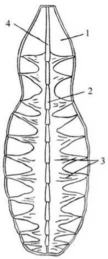
Fig. 100 Heart of the oriental cockroach:
1 — dorsal body wall severed during dissection; 2 — heart; 3 — alary muscles; 4 — aorta.
Locate the numerous fine tubules—the Malpighian tubules—situated at the junction of the Midgut and Hindgut. The Malpighian tubules function as excretory organs. Bathed in hemolymph, they absorb Metabolic waste products (primarily urates) from it and discharge them into the gut. The fat body also participates in excretion; its Cells accumulate excretory products converted into an insoluble state. In this form, they are harmless to the organism. The fat body Functions as a storage Kidney.
The Circulatory system OF insects is open and poorly developed. Its central organ is The Heart. Using a hand lens, examine the cockroach's heart on the inner surface of the excised dorsal body wall (Fig. 100).
Note that the Blood (hemolymph) is colorless. It consists of plasma and white Blood Cells—phagocytes.
The heart takes the form of an elongated tube composed of 13 chambers. It is closed posteriorly and continues anteriorly into the cephalic aorta, which opens into the body cavity. Blood flows out of the aorta into the body cavity, where it mixes with the body fluid (hemolymph). The heart is enclosed within a thin-walled chamber—the pericardial sinus—which communicates with the body cavity through numerous openings. Each heart chamber is equipped with a pair of lateral openings called ostia.
The cardiac chambers contract sequentially, pumping hemolymph into the cephalic aorta, from which it empties into the body cavity, circulates through the spaces between the organs, and then enters the pericardial sinus and flows back into the heart via the ostia. Pulsation of the heart chambers is driven by the contractility of the muscular heart walls aided by alary muscles, which are attached by one end to the heart walls and by the other to the body wall. Using a hand lens, examine the alary muscles located on the left and right sides of each chamber.
Make a drawing of the cockroach's heart.
In contrast to the circulatory system, the respiratory organs are exceptionally well developed. Respiration is carried out by a system of air-conducting tubes—tracheae—which branch throughout the body, envelop all organs, permeate all Tissues, and penetrate individual cells. Air enters the tracheal system through specialized openings called spiracles, located along the sides of the body.
Rinse a small piece of the fat body with water and locate the tracheae within it, or use a dissecting needle to isolate a silvery tube—a Trachea—from among other such tubes entwining the gut. Excise a small fragment of the trachea and examine it under a microscope, first at low and then at high magnification, in a drop of water beneath a coverslip. To better observe these transparent tubes, slightly dim the field of view. The internal walls of the tracheae are lined with cuticle forming spiral thickenings, which give the tracheal tubes exceptional elasticity. Air circulates through the tracheae driven by the respiratory Movements of the insect's abdomen. Compression of the abdomen forces air out of the tracheae, while abdominal expansion draws air inward through the spiracles into the tracheal system.
Insects are gonochoric (dioecious). The reproductive organs are located in the posterior part of the abdomen and are concealed by the fat body. Remnants of the fat body must be washed away with a jet of water from a pipette and removed using forceps. Examine the Male Reproductive System (Fig. 101). Locate the two Testes. They are easily observed in young males. In adult males, once their sperm reserve is depleted, the testes atrophy and become overgrown with the fat body.
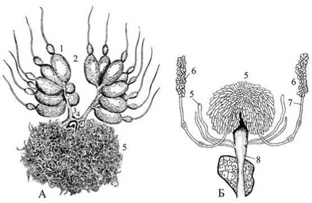
Fig. 101 Reproductive Organs of the Oriental cockroach:
A — female; B — male; 1 — ovariole; 2 — mature egg; 3 — oviduct; 4 — spermatheca; 5 — accessory glands; 6 — testes; 7 — vas deferens; 8 — ejaculatory duct.
The paired vas deferens, Seminal Vesicles, and the unpaired ejaculatory duct, which opens externally below the anus, are not visible. Clearly noticeable at the initial section of the ejaculatory duct are the white, mushroom-shaped accessory glands that secrete the fluid medium for spermatozoa.
Examine the FEMALE REPRODUCTIVE ORGANS in the dissected female. The female reproductive organs (Fig. 101) are represented by a paired Ovary, short paired oviducts, an unpaired Vagina, a spermatheca, and accessory glands. Locate the paired ovary. Each ovary is formed by eight ovarioles in which egg cells grow and mature. Other details of the FEMALE REPRODUCTIVE SYSTEM are difficult to observe.
The Central Nervous system consists of the supraesophageal ganglion (brain), connected by lateral commissures to the subesophageal ganglion, and the ventral nerve cord formed by nine ganglia. Dissection of the brain is too complex for routine Practical Classes, but the ventral nerve cord can be observed. To do this, completely remove the digestive organs, reproductive system, and fat body, and carefully clean the tissues at the bottom of the body cavity using a dissecting needle. This reveals a thin white thread with ganglionic nodes extending from the head to the posterior end of the body. Using a hand lens, examine the nerve cord with its ganglia and the extremely fine lateral nerves branching off to all parts of the body.
Make a drawing of the internal anatomy of the cockroach.
The Oriental cockroach reaches reproductive maturity at the age of five years. The female develops a whitish sac—an egg capsule (ootheca)—which is retained for some time in the depression of the last abdominal sternite. The capsule gradually turns brown, darkens, and drops off. White, wingless larvae hatch from the eggs. They molt several times while increasing in size, their integument gradually darkens, and they transform into adults. This type of development is called incomplete metamorphosis (hemimetabolously). It is characteristic of Orthoptera (grasshoppers, crickets), cockroaches, and true bugs.
Comparison of mouthparts in various insects
The insect mouthparts include the labrum (upper lip), a pair of mandibles (upper jaws), a pair of maxillae (lower jaws), and the labium (lower lip). The labrum is a fold of the chitinous head capsule. The labium is formed by the fusion of the second pair of maxillae. All mouthpart components, except for the labrum, represent modified limb pairs.
Depending on their lifestyle and diet, insects have developed various types of mouthparts. The chewing (mandibulate) type is adapted primarily for feeding on solid food, whereas the chewing-lapping type is adapted for liquid food. In addition, there are variations from these two main types (piercing-sucking, chewing-sponging, sponging, etc.) associated with diverse feeding conditions.
The chewing type of mouthparts is found in beetles, cockroaches, orthopterans, as well as beetle and butterfly larvae. The mouthparts of this type are adapted for seizing, biting, and chewing food. The piercing-sucking type is found in Diptera, lice, and Hemiptera (true bugs). These mouthparts are adapted for sucking blood or plant sap. The siphoning mouthparts of butterflies and moths are adapted for sucking floral nectar.
Materials required: microscope; dissecting stereomicroscope; hand lens; dissection tools (dissecting trays, pointed forceps, scissors, needles, pins); glass slides and coverslips; 1/2 sheet of white paper; cockroaches preserved in 70% alcohol; microscopic mounts of mouthparts of the bed bug, honey bee, and butterfly.
Task. Dissect the mouthparts of the Oriental cockroach. Place the mouthparts on a glass slide, examine them, and make a drawing. Examine and draw the head of a bed bug with its mouthparts, as well as the heads of a bee and a butterfly with their respective mouthparts.
Preparation of the cockroach mouthpart slide. Bend back the head of the Oriental cockroach (which is pressed against the thorax) using forceps, locate the slender neck, and cut it with scissors. Place a sheet of white paper on the table or in a dissecting tray. Position the severed head on the paper with the cut neck pointing upward and secure it in place by inserting a pin through the cut. Using a dissecting needle or fine forceps, separate the labium and place it on a glass slide. Next, detach the maxillae and place them on either side of the labium, observing the process through a hand lens. Detach the mandibles and position them above the previously isolated mouthparts. Carefully detach the labrum (as it is fragile). Place the labrum above and between the mandibles.

Fig. 102 Mouthparts of the Oriental cockroach:
1 — labrum; 2 — mandibles; 3-7 — Maxilla (3 — maxillary palp; 4-5 — galea and lacinia; 6 — stipes; 7 — cardo); 8-12 — labium (8 — submentum; 9 — mentum; 10 — glossa; 11 — paraglossa; 12 — labial palps).
Study of the cockroach mouthpart slide. Verify the arrangement of the isolated mouthparts using the diagram (Fig. 102). Examine them using a dissecting microscope or a compound microscope at low magnification. Note that the maxillae consist of two basal segments: the cardo and the stipes. The stipes bears the outer and inner lobes (galea and lacinia). Situated lateral to them are the five-segmented, Hair-setose palps. The outer lobes and five-segmented palps function as tactile organs. The inner lobes perform a masticatory function and serve as an auxiliary part of the chewing mouthparts.
The labium consists of a basal part—the submentum, an upper plate—the mentum (corresponding to the stipes of the maxillae), glossae (masticatory lobes), and three-segmented palps. All these parts mirror The structure of the maxillae, since the labium was formed by the fusion of the second pair of maxillae. The labrum covers the mouthparts from the front and has the shape of a thin, longitudinal semi-oval. The mandibles are hard, undivided, heavily chitinized plates whose inner margin bears robust Teeth. Insects use their mandibles to bite off and grind food.

Fig. 103 Piercing-sucking type of mouthparts (bed bug):
A — DORSAL VIEW OF the mouthparts; B — LATERAL VIEW OF the proboscis; 1 — labrum; 2 — labium; 3 — mandibles; 4 — maxillae; 5 — antennae.
Make a drawing of the cockroach mouthparts.
Studying the slide of a bedbug's head. Examine the bedbug's head under low magnification of the microscope and make a drawing (Fig. 103).
Locate the black compound eyes on the sides of the head and the pubescent, four-segmented antennae situated below and in front of the eyes. A short, semicircular labrum protrudes forward between the antennae.
Examine the labium, known as the proboscis — it consists of three segments. Inside the labium, lying like a case, are the paired maxillae and mandibles, shaped like long, piercing needles, or setae. The maxillae are located in the middle, and the mandibles at the margins. The maxillae feature two grooves on the inner side, positioned one above the other. Fitted together, the maxillae thus form two channels. The upper channel serves for sucking up food, while the lower (narrower) one is used for injecting saliva during a bite. The mandibles pierce the tissues; their tips are pointed and serrated. This serration ensures a secure hold of the mouthparts and the bug itself on the host's body while sucking blood (in some specimens, the jaws are extracted from the proboscis and positioned nearby to facilitate examination). Maxillary and labial palps are absent in the bug.

Fig. 104 Honey bee:
A — queen; B — her head; C — worker bee; D — her head; E — drone; F — his head.
Studying the slide of the head and mouthparts of the honey bee. Examine the head and mouthparts of a worker bee under low magnification of the microscope (Fig. 104). Locate the three simple ocelli on the convex part of the head and the large compound eyes on the sides of the head.

Fig. 105 Head and mouthparts of a worker bee:
A — frontal view; B — lateral view; 1 — compound eyes; 2 — ocelli; 3 — clypeus; 4 — labrum; 5 — Mandible; 6 — maxilla; 7 — ligula (Tongue); 8 — labial palps; 9 — maxillary palps; 10 — antenna; 11 — flabellum.
Examine the geniculate antennae on the front of the head, bent at an angle: the basal scape consists of a single shaft, while the flagellum consists of 11 small segments. The antennae are organs of smell and touch.
The bee's mouthparts are adapted to perform several functions: nest building, feeding the brood, and gathering liquid and solid food. This is a modified mouthpart of the chewing-lapping type (Fig. 105).
Locate the clypeus, which is situated below the antennae. Articulating with it is a narrow, transversely lying labrum. Examine the mandibles flanking the labrum laterally: they are presented as broad plates with a depression, devoid of teeth, and serve for building wax combs and removing foreign objects from the hive.
Honey mixed with pollen and brood food flows down the grooves of the mandibles when feeding the larvae. Young bees use them to bite through the cocoons upon hatching. The maxillae of the bee are highly modified compared to the basic chewing type. Note that their inner lobes and palps are reduced, while the outer lobes are greatly elongated. The labium is also heavily modified: its palps are greatly elongated, and the inner lobes have fused to form a trough-like, almost closed tube called the tongue (glossa), which terminates in a small flabellum and is densely covered with hairs, whereas the outer lobes are very weakly developed. The tongue, folding together with the outer lobes of the maxillae and the labial palps, forms a closed tube for sucking nectar. Such mouthparts can be termed chewing-lapping.

Fig. 106 Head and mouthparts of a butterfly:
1 — frons; 2 — setigerous appendage of the labrum;
3 — vertex; 4 — ocelli; 5 — antenna parts; 6 — compound eye; 7 — gena; 8 — genal outgrowths; 9 — maxillae (proboscis); 10 — labial palp.
Make a drawing of the worker bee's mouthparts.
Studying the slide of a butterfly's mouthparts. Using a dissecting lens, locate the large compound eyes situated on the sides of the head, the small simple ocelli at the base of the antennae, and the long segmented antennae. Note that a plate called the frons is located between the eyes. Due to the evolutionary transition of butterflies from solid to liquid food, the mandibles, labrum, and labium have become reduced. The labium has the appearance of a small, unsegmented plate. Only the labial palps on the sides of the labium are well developed (Fig. 106).
The labrum and mandibles are barely discernible. Examine the maxillae, which make up the butterfly's proboscis; they appear as long and narrow plates with deep longitudinal grooves. These grooves form a tube inside the proboscis through which nectar is drawn up into the butterfly's mouth during feeding. In a state of rest, the proboscis is usually coiled into a spiral and lies against the head. During feeding, the proboscis uncoils (there are muscles within the wall of the proboscis) and is inserted into the nectaries of a flower to suck nectar. The length of the proboscis varies among different butterflies and is related to the STRUCTURE OF THE flower visited by the given species.
Make a drawing of the butterfly's head and its mouthparts.
Order Diptera (True Flies)
Mosquitoes.
Mosquitoes of the family Culicidae are of great epidemiological importance: certain species of the genus Anopheles are vectors of malaria, among mosquitoes of the genera Culex and Aedes there are vectors of Japanese (autumn) encephalitis, and many mosquitoes transmit tularaemia, one of the particularly dangerous infections.
The most important of the malaria mosquitoes is the common malaria mosquito (Anopheles maculipennis), which is widespread in Russia.
Materials required: microscope; dissecting loupe; whole mount microscopic preparations of Culex and Anopheles mosquitoes; micropreparations of the mouthparts of these mosquitoes; whole mount microscopic preparations of Anopheles and Culex larvae and pupae.
Task. Study and draw the external morphology of Anopheles and Culex mosquitoes. Examine and sketch their mouthparts. Learn to distinguish between the larvae and pupae of Anopheles and Culex mosquitoes.
Study of micropreparations of adult mosquitoes. Examine the whole mount preparation of the mosquitoes under a dissecting loupe. Note that the mosquito body is divided into three distinct regions: the head, thorax, and abdomen. Locate the large compound eyes, the 15-segmented antennae, and the mouthparts on the head, as well as the legs and wings on the thorax. The legs are long and slender; observe that the tarsus is longer than the tibia and femur combined. In Culex mosquitoes, the legs are 1.5 times longer than the body, whereas in Anopheles they are twice as long. The tarsus terminates in two claws and adhesive pads. A pair of wings, traversed by longitudinal veins, extends from the mesothorax. The venation pattern is of great importance in identifying mosquito species.

Fig. 107. Anopheles and Culex mosquitoes:
A — wing of the malaria mosquito; B — resting posture of mosquitoes; C — larvae; D — pupae.
Observe the wing of the Anopheles mosquito — it differs from that of Culex in having four dark spots in the middle, whereas the wings of Culex are transparent. Examine the halteres, which are the rudimentary second pair of wings arising from the metathorax.
Examine the 9-segmented abdomen, which features spiracles along the sides, as is typical of all insects. Mosquitoes of the genus Anopheles are easily distinguished by their resting posture (Fig. 107) and mouthparts. The Culex mosquito rests on a surface (such as a wall) parallel to it, with its proboscis positioned at an angle to the longitudinal axis of the body. Anopheles rests with its abdomen elevated upward, while its proboscis lies in a straight line with the longitudinal axis of the body. However, when overwintering, the malaria mosquito rests in the same manner as non-malaria mosquitoes, holding its abdomen close to the surface.
Learn to tell female Anopheles mosquitoes apart from males. The sexes can be differentiated by the structure of their antennae. In males, the antennae are covered with dense, long hairs, whereas in females they are short and sparse. The last two segments of the male antennae are significantly longer than the others.
Study of the micropreparation of mosquito mouthparts under low magnification. In male mosquitoes, the mouthparts are of the sucking type, as they feed on plant juices. In females, the mouthparts are piercing-sucking, as they feed on the blood of humans and animals.
Locate the proboscis on the preparation, which terminates in two lobes. Between them lies the unpaired tongue, or hypopharynx (Fig. 108). During blood-feeding, the proboscis does not penetrate the Skin; its lobes serve to support and guide the piercing elements of the mouthparts. Blood is drawn into the pharynx through the tubular labrum. The painful sensations in humans and animals caused by a mosquito bite are due to the action of saliva injected into the wound.
Locate the mandibles, maxillae, hypopharynx (tongue), and maxillary palps on the preparation. Note that in the male Anopheles, the maxillary palps are as long as the proboscis and end in a club-shaped expansion, whereas in the male Culex, the palps are longer than the proboscis and lack club-shaped expansions.
Learn to distinguish female Anopheles from female Culex. Compare the length of the maxillary palps with the length of the proboscis: in Anopheles, they are equal in length to the proboscis, whereas in Culex they are 3 to 4 times shorter.
Sketch the mouthparts of female Anopheles and Culex mosquitoes.
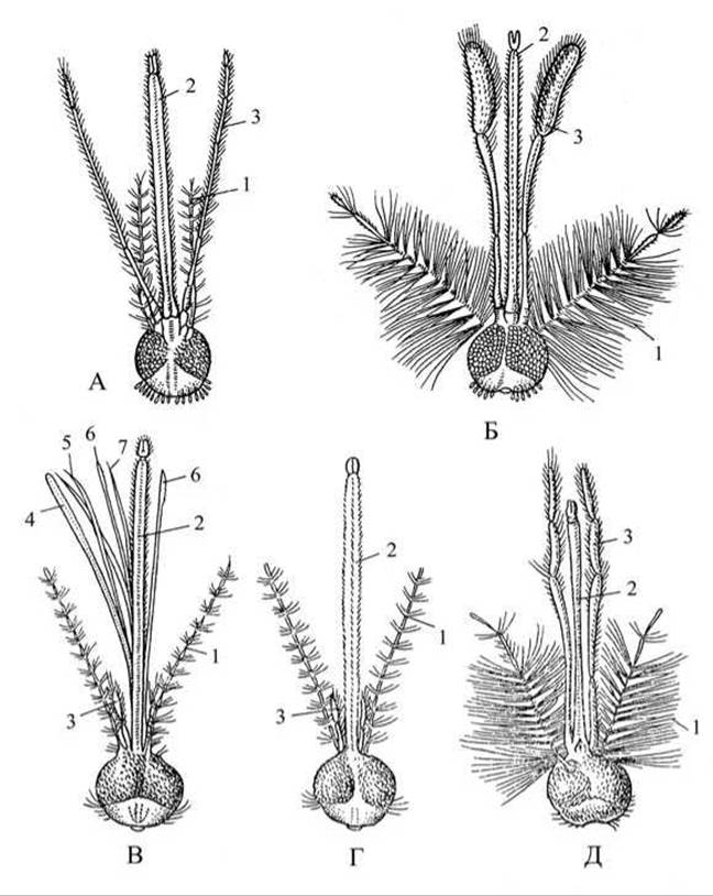
Fig. 108. Heads and mouthparts of Anopheles and Culex mosquitoes:
A — Anopheles female; B — Anopheles male; C — Culex female with split mouthparts; D — Culex female with unsplit mouthparts; E — Culex male; 1 — antennae; 2 — proboscis; 3 — palps; 4 — labrum; 5 — mandibles; 6 — maxillae; 7 — tongue (hypopharynx).
Study of whole mount preparations of mosquito larvae and pupae. Examine prepared slides of Anopheles and Culex mosquito larvae and pupae using a dissecting loupe and low microscope magnification. Note that the larva's body is divided into the head, thorax, and abdomen. Locate the eyes, a pair of antennae, and mouth appendages on the head. Examine the thorax and abdomen: the thorax consists of three segments with long, feathery setae protruding from their sides, and the abdomen consists of segments bearing tufts of hairs. On the ventral side of the penultimate segment are the respiratory openings (spiracles) leading into the tracheal system. In Anopheles, these are located directly on the segment, whereas in Culex and Aedes they are elevated onto the end of a long siphon tube. Note the anal gills and tufts of setae on the terminal segment of the abdomen.
Mosquito larvae are easily distinguished. At rest, Anopheles larvae lie parallel to the water surface, exposing their respiratory openings directly to the air. Culex and Aedes larvae hang by their siphons so that their bodies remain at an angle to the water surface.
Sketch the Anopheles mosquito larva.
Examine preparations of Anopheles and Culex mosquito pupae and identify their distinguishing features. Observe the thickened anterior region (representing the head and thorax of the future mosquito) and the abdomen curled underneath it. The abdomen is mobile, and two transparent caudal fins can be seen at its tip. Locate the breathing trumpets at the apex of the convex anterior thickened region, by which the pupa hangs from the surface film of water. In Anopheles mosquitoes, the breathing trumpets are funnel-shaped with a wide opening, whereas in Culex mosquitoes they are long and narrow.
Sketch the Anopheles mosquito pupa.
Horseflies, botflies, stable flies, houseflies.
Familiarity with parasitic insects of the order Diptera (horseflies, botflies, stable flies, houseflies) that act as vectors of infectious and invasive diseases in animals and humans is of great importance for livestock specialists and veterinarians.

Fig. 109. Botflies and gadflies:
A — cattle botfly (Hypoderma bovis); B — sheep botfly (Oestrus ovis); C — large horse botfly (Gastrophilus intestinalis).
Horseflies appear in May and disappear in August-September. They inhabit trees and shrubs in marshy, damp, and shady areas. They are active during the day in sunny, calm weather. Periodically, they fly toward water bodies, swoop down to the surface, and scoop up water droplets, as they drink heavily during peak activity periods. Only females feed on the blood of animals and humans, whereas males feed on plant juices. Female eggs mature only after a blood meal. The most common species are the common horsefly (Tabanus), the deerfly (Chrysops), and the twin-lobed deerfly (Chrysozona).
Horseflies cause distress to animals through their bites and act as vectors for the causative agents of anthrax and tularemia (Fig. 109).
The large horse botfly (*Gastrophilus intestinalis*, see Fig. 110) and the cattle botfly (*Hypoderma bovis*) are widespread across Russia, while the sheep botfly (*Oestrus ovis*) is found primarily in southern and southeastern regions. Adult female botflies fly about and deposit eggs on the hair of animals (in Stomach and cattle botflies) or spray live larvae directly (in the sheep nasal botfly). This occurs on dry, calm days, predominantly during the hottest hours. On overcast, cold, and windy days, botflies remain inactive, resting on trees, fences, and farm building walls. Larvae of the horse botfly inhabit The Stomach and intestines, causing gastrophilosis. Larvae of the cattle botfly settle under the skin, causing hypodermosis. Larvae of the sheep nasal botfly parasitize the nasal and frontal sinuses, causing oestrosis.
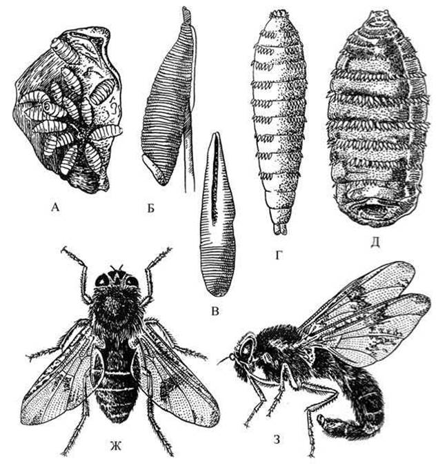
Fig. 110. Horse botfly (*Gastrophilus*):
A — larva on the wall of the horse's stomach; B — egg on a hair; C–E — larvae at various developmental stages; G — male; H — female.
The stable fly (*Stomoxys calcitrans*), also known as the biting house fly, appears toward the end of summer upon emerging from pupae. Both males and females feed on blood. While feeding, they transmit pathogens of anthrax, tularemia, septicemia, and other bacterial infections from sick animals and humans to healthy ones. Stable fly bites are painful and cause significant distress to livestock. The stable fly is frequently confused with the morphologically similar house fly.
The house fly (*Musca domestica*) carries the causative agents of intestinal diseases (typhoid fever, dysentery, cholera), glanders microbes, tuberculosis, and other infections, as well as helminth eggs and pathogenic protozoan cysts, on its tarsi, mouthparts, and body surface.
Required materials and equipment: microscope; hand magnifier; dry specimens (mounted on entomological pins) of the horsefly (*Tabanus*), deerfly (*Chrysops*), and twin-lobed deerfly (*Chrysozona*); botflies including stomach, cattle, and sheep botflies; house fly and stable fly; test tubes containing larvae of horseflies and botflies preserved in 70% alcohol; slide preparations of the mouthparts of the horsefly, house fly, and stable fly; forceps; Petri dishes.
Assignment: Study the external morphology of horseflies and botflies, and learn to distinguish between them. Examine the external Morphology of the stable fly and learn to differentiate it from the house fly.
Study of horsefly and botfly specimens and larvae preserved in 70% alcohol. Examine the dry specimens of horseflies and botflies using a hand magnifier. Pay attention to body size, hairiness, eyes, and the type of mouthparts.
Remove the horsefly and botfly larvae from the test tubes, place them in Petri dishes, and identify their distinctive features (see table).
Examination of the slide preparation of horsefly mouthparts.
Examine the mouthparts of a female horsefly under low magnification. They are of the cutting-sucking type, adapted for blood feeding and piercing the tough skin of humans and animals. Observe the maxillae and mandibles, which are shortened, shaped like sharp blades, and serve to slice the skin. Along with the jaws, the upper lip (labrum) and hypopharynx—also shortened and pointed—penetrate the wound. Note the lower lip (labium), which houses these cutting structures; it is soft and unsegmented at the tip. The lab lobes serve to sponge up liquids (blood, water). The upper jaws and labrum, closely apposed, form a channel through which blood is sucked.
Table
Distinctive features of botflies and horseflies
No. p/p |
Botflies |
Horseflies |
1 |
Botflies are medium-sized flies with a small head and small eyes. The body is densely covered with hair, giving the botfly a bumblebee-like appearance. |
Horseflies are predominantly large flies with a large head and large, brilliant eyes. The body is sparsely haired. Males and females differ: in the male, the eyes touch on the forehead, whereas in the female, they are separated by the frons. |
2 |
Mouthparts are reduced. Adults do not feed. |
Males feed on plant juices. Females are blood-sucking and deliver painful bites. Mouthparts are of the cutting-sucking type. |
3 |
Damage is caused by the larval stage. |
Damage is caused by the adult stage. |
4 |
Females approach animals to lay eggs (cattle and stomach botflies) or to spray live larvae (sheep botfly). |
Females approach animals to feed on blood. |
5 |
Botfly larvae that have parasitized under the skin (cattle botfly), in the intestine (horse stomach botfly), or in cavities (sheep nasal botfly) drop out of the host's body to pupate, forming cocoons (puparia). Botfly cocoons can be found in the soil. |
Blood-fed females undergo egg maturation. They lay batches of eggs (400–1000), gluing them to the undersides of leaves or stems of aquatic plants near water bodies. Larvae live at the bottom of water bodies, feeding on decaying organic matter, small worms, insects, and Mollusks. Large numbers of larvae can be found in marshy ditches near forest roads and in waterlogged pastures. In spring, before pupuration, larvae crawl onto plants on dry banks. Horsefly flight occurs near water at temperatures of 15–16°C. |
6 |
Botfly larvae are thick, firm, slightly tapered anteriorly, typically bearing spiny rings on the body. The posterior end bears a pair of spiracles with heavily chitinized margins (which are difficult to distinguish). |
Horsefly larvae are white, with a cylindrical, clearly segmented body tapering at both ends. The anterior end features a tiny head with barely perceptible jaws and antennae. |

Fig. 111. Flies:
A — house fly (*Musca domestica*); B — stable fly (*Stomoxys calcitrans*).
Examination of dry specimens of the house fly and stable fly. One should learn to distinguish the stable fly from the house fly (Fig. 111); pay attention to the body size and coloration of the integument.
Examine the flies under a magnifier. The body sizes of the stable fly and house fly are nearly identical (6–7 mm), but the stable fly is gray, whereas the house fly is grayish-brown.
The dorsal surface of the stable fly's abdomen features dark spots, and its thorax displays dark longitudinal stripes, whereas the house fly has four longitudinal stripes running down the DORSAL SIDE OF its thorax, and an indistinct dark pattern on its abdomen. The stable fly rests with its wings spread apart, and its abdomen is shorter and broader; unlike the house fly, it does not crawl on the skin or buzz, but rather flies up unnoticed, lands quietly, pierces the skin, and feeds on blood.

Fig. 112 Mouthparts of flies:
A — stable fly; B — house fly; 1 — labium; 2 — labellar lobes; 3 — oral orifice; 4 — labrum; 5 — hypopharynx; 6 — maxillary palps.
Examination of slide mounts of the mouthparts of the house fly and the stable fly. Examine the slide mounts of the mouthparts of the house fly and stable fly under low magnification and compare them. The house fly has sponging mouthparts; it lacks piercing structures (the maxillae and mandibles are reduced) and cannot bite. Locate the proboscis—a highly modified labium—and examine it (Fig. 112). It is elongated and terminates in cushion-like swellings, which represent heavily modified labial palps.
Examine these swellings, known as the labellar lobes. They are furrowed with chitinous Ribs and grooves through which food enters the mouth. Locate the oral opening between the lobes. Examine the labrum—it covers the proboscis groove from above. Within the channel formed by the proboscis and the labrum, locate the hypopharynx, or tongue. It is too soft to deliver a puncture. The hypopharynx is pierced by a salivary duct. Using saliva, the fly can dissolve food (such as sugar). Locate the palps of the reduced maxillae at the base of the proboscis. Flies lack antennae.
Examine the mouthparts of the stable fly, which are of the piercing-sucking type. The stable fly feeds on the blood of humans and animals. The long, sharp proboscis, containing the piercing apparatus within, projects forward and is clearly visible.
The proboscis of the stable fly develops similarly to that of the house fly, originating from the labium. Examine the pair of small lobes at the tip of the proboscis. Lying within the groove of the proboscis is a single piercing bristle—the hypopharynx. Locate the thin and sharp labrum covering the proboscis groove, and the maxillary palps at the base of the proboscis. The mandibles and maxillae in the stable fly are reduced.
Make a drawing of the stable fly's mouthparts.
Last update: 13/08/2026
Editorial and Educational Adaptation: This material has been compiled based on the primary/original source text. The project team performed an editorial review, corrected technical inaccuracies, structured sections, and adapted the content for an educational format.
What was processed:
- elimination of formatting defects (OCR errors, structural breaks, corrupted characters);
- editorial organization of content;
- standardization of terminology in accordance with academic sources;
- verification of factual statements against the original source text.
All mentions of the author, publication year, and origin of the primary text have been preserved in accordance with the source.