Practical Guide to Zoology: A Study Aid - T. A. Dauda 2014
Multicellular Animals
Arthropods
Subphylum Chelicerata – Class Arachnida
Arachnids are the most numerous (over 36,000 species) Class of terrestrial chelicerates. The class comprises more than 10 orders.
Arachnida are higher chelicerate Arthropods with 6 pairs of prosomal appendages. They breathe through Lungs or tracheae and, alongside coxal glands, possess an excretory system consisting of Malpighian tubules located in the abdomen.
The class Arachnida is divided into A number of orders, of which only three will be examined: the order Scorpions (Scorpiones), the order Spiders (Aranei), and the order Mites and Ticks (Acari).
Order Scorpions (Scorpiones)
Caucasian scorpion (Buthus eupeus).
Materials and equipment. The Caucasian scorpion is the most common scorpion species inhabiting the southeastern part of Russia, the Caucasus, and Central Asia. Scorpions are nocturnal, preying on insects and other small animals.
In localities where they occur, they can be collected by turning over piles of stones, individual rocks, or in crevices. Due to their venomous sting, long forceps should be used for capture. Live scorpions must be collected individually into separate containers, as they may cannibalize one another.
In captivity, the Caucasian scorpion, like other members of the family, lives for several months. Flies, mealworm larvae, and other insects, as well as spiders and small earthworms, are recommended as food for scorpions.
The Study of external Morphology is carried out on specimens killed with ether, chloroform, or alcohol. Dissections are best performed on freshly killed material, though fixed scorpions are also suitable.
Required tools and supplies: a hand magnifying Glass; forceps; dissecting needles; a dissecting tray; filter paper; scorpions fixed in 70% alcohol.
Assignment. Examine the external anatomy and draw the scorpion from the ventral side.
Study of external morphology. Take a scorpion with forceps, blot it dry with filter paper, and place it at the bottom of a dissecting tray with its dorsal side facing upward. Observe the body color—it is yellow, with noticeable brownish-black spots and stripes on the dorsal surface. The body length of the Caucasian scorpion is about 5 cm. The body is narrow and elongated dorsally, adapted to life in narrow crevices. It can be divided into a short, unsegmented prosoma (cephalothorax) bearing appendages, and a long, segmented, appendage-free opisthosoma (abdomen). The abdomen is subdivided into a broad mesosoma (pre-abdomen) and a narrow, elongated metasoma (post-abdomen) (Fig. 87).

Fig. 87 Scorpion (Buthus eupeus):
A — dorsal view; B — ventral view; 1 — prosoma; 2 — chelicerae; 3 — pedipalps; 4 — walking legs; 5 — terminal segment of the abdomen; 6 — sting; 7 — anterior region of the abdomen; 8 — posterior region of the abdomen; 9 — anus; 10 — pulmonary slit; 11 — pectines; 12 — genital operculum.
Note that the last segment of the metasoma is swollen. It is equipped with a pair of venom glands and terminates in a sharp spine—a stinger with two openings through which venomous fluid flows into the wound upon puncture. The metasoma can be curved upward.
The scorpion's body is covered on both the dorsal and ventral sides with hard chitinous plates, while the sides are equipped with chitinous membranes, except for the segments of the metasoma, which are covered by a continuous chitinous shell.
Using a magnifying glass, locate the two large median eyes in the anterior part of the prosoma and several smaller ocelli on the sides.
Turn the scorpion ventral side up. Draw it from the right or left side, indicating the appendages. Examine the prosoma: it consists of 6 segments. Locate 2 pairs of oral appendages and 4 pairs of walking legs on the prosoma. The first pair of oral appendages consists of the three-segmented chelicerae, or upper jaws, which are better visible from the dorsal side. These are followed by the second pair of oral appendages—the six-segmented, long pedipalps, or palps. The pedipalps terminate in powerful chelae (claws) used for capturing, holding, and crushing food, and they also function as tactile Organs. Between the basal segments of the pedipalps lies the preoral cavity with the Mouth opening.
Antennae are absent in scorpions, as in all arachnids. Examine the walking legs using a magnifying glass: scorpions have 4 pairs of them. Each leg consists of segments; the terminal segment ends in claws.
Using a magnifying glass, locate on the first segment of the pre-abdomen the small triangular plates—Valves that cover the genital opening. On the second segment, the comb-like pectines are clearly visible—these are vestigial abdominal limbs. On the next four segments, paired, obliquely positioned openings are visible—the spiracles or stigmata leading into the pulmonary sacs.
At the end of the metasoma, between the penultimate and ultimate segments, lies the anus.
Order Spiders (Aranei)
European garden spider (Araneus diadematus).
Materials and equipment. The garden spider is a common species in the European part of our country. Large sexually mature females are found in August and September in forests, parks, old buildings, and on fences, where they spin large orb webs and sit on them most of the time. Smaller males are usually found near female retreats under tree bark, in building crevices, etc. Eggs are laid by the female in autumn within silken egg cocoons, which are suspended in sheltered places. Juveniles hatch in spring, so only immature individuals are encountered in summer. Spiders intended for morphological studies are collected directly into alcohol. Live spiders are required for studying internal anatomy.
Materials and equipment required: dissecting magnifier; hand lens; dissecting tools (forceps, dish, needles); watch glasses; filter paper cut to fit the bottom of the dish; cross spiders (males and females) preserved in 96% alcohol.
Task. Examine and sketch the external Anatomy of the cross spider. Dissect and examine the chelicerae, pedipalps, and walking legs of the spider without sketching them.
Studying external anatomy. Place the spiders preserved in 96% alcohol ventral side down on the bottom of the dissecting dish, lined with a piece of filter paper to dry them. Examine the spiders from the dorsal side using a hand lens, assisted by forceps and dissecting needles.
Locate the body regions into which the spider's body is divided: the cephalothorax and the unsegmented abdomen, connected by a slender pedicel (see Fig. 88). Six pairs of appendages articulate with the cephalothorax. The abdomen lacks appendages.

Fig. 88 Cross spider (Aranea diadematus), female:
1 — cephalothorax; 2 — chelicerae; 3 — pedipalps; 4 — walking legs; 5 — abdomen.
The cephalothorax is covered by two chitinous shields. The dorsal shield covers it from above, in front, laterally, and behind. Ventrally, the cephalothorax is protected by a small, oval plate—the Sternum. The shields are unsegmented. The segments of the cephalothorax are thoroughly fused with one another. The fact that the cephalothorax is formed by the fusion of 6 segments can only be inferred from the presence of 6 pairs of appendages.
Find the cross-shaped pattern of white stripes and spots on the dorsal shield.
Learn to distinguish the male from the female: in the male, the abdomen is elongated and smaller than the cephalothorax, whereas in the female, it is rounded and larger than the cephalothorax.
Place the spider on a watch glass, dorsal side up, and set the watch glass on the stage of the dissecting magnifier. Locate the 8 simple eyes, arranged in two rows, on the anterior part of the dorsal shield. Examine the 2 pairs of oral appendages and the 4 pairs of walking legs extending from the cephalothorax.
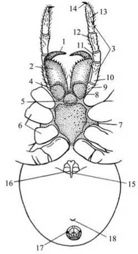
Fig. 89 Ventral view of the cross spider (female):
1 — claw-like segment of chelicera; 2 — basal segment of chelicera; 3 — pedipalp; 4 — gnathobase (maxillary lobe); 5 — "lower lip" plate; 6 — coxae of legs; 7 — sternum; 8 — coxa of pedipalp; 9 — trochanter; 10 — Femur; 11 — Patella; 12 — Tibia; 13 — tarsus; 14 — claw; 15 — epigyne (genital plate); 16 — spiracle; 17 — spinnerets; 18 — tracheal opening.
Sketch the spider from the dorsal side.
Turn the spider ventral side up. Examine the two anterior pairs of oral appendages. The first pair is called the chelicerae, and the second, the pedipalps. The chelicerae are short, powerful appendages used for capturing and killing prey. The mouth opening is located at the base of these appendages. Carefully separate the bases of the chelicerae and pedipalps with dissecting needles and try to observe the mouth opening using a magnifier. Dorsally, it is bounded by a fleshy tubercle—the labrum (upper lip), and ventrally, by a single unpaired chitinous plate—the labium (lower lip) (Fig. 89).

Fig. 90 Silk-spinning apparatus of the cross spider:
1 — anterior spinnerets; 2 — posterior spinnerets; 3 — internal spinnerets; 4 — anal opening.
Tear off the chelicerae with forceps at their point of attachment to the cephalothorax and examine them under the dissecting magnifier. Note that each chelicera consists of two segments: a thick basal segment and a sharp, hook-like terminal claw. At the tip of the claw opens the duct of the venom gland, which is located inside the cephalothorax and the basal segment of the chelicera.
Tear off the pedipalps and examine them on the stage of the dissecting magnifier. Like the chelicerae, the pedipalps are used for capturing and crushing food, but they are longer than the chelicerae. Locate the basal segment of the pedipalps, covered with setae, which bears a gnathobase (maxillary lobe); the remaining 5 segments form a palp. Dissect and examine the remaining 4 pairs of appendages—the walking legs. They share a similar Structure, differing only in length. Each leg consists of 7 segments. The structural details of the oral and walking appendages can only be observed on a permanent slide.
On the ventral side of the spider's abdomen, near the front, lies the genital opening, flanked on either side by narrow slits—the spiracles, which lead to the respiratory organs, the book lungs. At the posterior end of the abdomen, the spinnerets are visible, behind which lies the anal tubercle with the anal opening at its tip (Fig. 90). On the surface of the spinnerets are numerous microscopic pores (not visible) through which the spider secretes a sticky fluid that hardens upon contact with the air to form silk. This secretion is produced by silk glands located inside the lower part of the abdomen.
Just anterior to the spinnerets lies the tracheal spiracle (not visible), leading into the tracheae, which, along with the book lungs, perform a respiratory function.
Tarantula (Lycosa singoriensis).
Materials and equipment. The tarantula belongs to wandering spiders, which do not build web snares, but instead capture prey either in active pursuit or in steep vertical burrows dug with the help of their chelicerae. It is one of the larger spiders, with a body length of up to 3.5 cm. In Russia, tarantulas are found in steppe, forest-steppe, and semidesert zones. Their bites are very painful, but not life-threatening to humans.
Materials needed: hand lens; tweezers; dissecting needles; dissecting tray; filter paper; tarantulas preserved in 70% alcohol.
Task. Examine the External structure of the tarantula.
Study of external structure. Using tweezers, place the tarantula ventral side down on a piece of filter paper placed at the bottom of a dissecting tray, and observe it from the dorsal side.
Note that body segmentation is not pronounced: the body consists of an unsegmented cephalothorax and abdomen.

Fig. 91 Tarantula (Lycosa singoriensis)
The abdomen is connected to the cephalothorax by a narrow pedicel. Using a hand lens, locate the 4 pairs of eyes on the anterior part of the cephalothorax and observe their arrangement. Note the absence of antennae. Observe that the entire tarantula's body is densely covered with setae and appears hairy (hence the "hairy spider"), with a yellowish-brown color and a black pattern.
On the ventral side of the cephalothorax, there are 6 pairs of appendages: the upper jaws (chelicerae), pedipalps, and 4 pairs of walking legs. Poison glands are located at the Base of the chelicerae. Examine the chelicerae, which appear as strongly developed claws. The tarantula uses its long pedipalps to turn its prey while feeding. The tarantula's legs are built for running; they are transversely striated, long, and elevate the body high above the ground (Fig. 91).
Note that the abdomen of the tarantula, as in all spiders, bears no appendages. Compare it with the abdomen of the European garden spider. In the tarantula, the abdomen is lighter and more slender because its silk glands are less developed than those of the garden spider.
Using a hand lens, locate the genital opening covered by a plate on the anterior part of the female's abdomen. A pair of stigmata leading into book lungs is located on either side of it. Examine the 3 pairs of distinct spinnerets (rudiments of abdominal limbs) at the posterior end of the abdomen. Using a hand lens and dissecting needles, find the transverse slit lying anterior to them, which is the spiracle leading into the tracheae. At the posterior end of the abdomen, behind the spinnerets, lies the anus.
Steppe widow / Mediterranean black widow (Lathrodectus tredecimguttatus).
Material and equipment. The black widow spider is found in clay-sandy or clay-solonetzic steppes and deserts overgrown with wormwood, intersected by ravines and hollows.
Female spiders inhabit specially spun webs. Very often they build nests in the burrows of tortoises and rodents. Along the edges of this nest on The surface of the ground, webs are stretched in the form of randomly interwoven catching threads. Females feed on insects trapped in them. Males do not feed at all. Mating of the females occurs in June and early July. Many males are eaten by the females after mating, while the rest die later.
In the second half of summer (July–September), fertilized females migrate to other locations, build new nests, and lay numerous eggs. Each female lays from 100 to 700 eggs in a dense, specially spun cocoon. She weaves from 2 to 14 such cocoons. During egg-laying, the females feed intensively. After 5–7 days, spiderlings hatch and remain in the cocoon for the winter. In early spring, the spiderlings leave the cocoons and stay together for some time, spinning a common web near the maternal one; then each of them releases a fine thread, and the wind carries them away, sometimes over long distances from their hatching site.
Young black widows sequentially go through 7–8 instars. Females of the final instar (sexually mature) are the most venomous. Males are of little danger because their venom is weaker. Cases of black widow bites coincide with the migration seasons of the females (May–June, mid-July). Black widow bites are dangerous to humans and animals, and sometimes fatal.
Materials needed: hand lens; dissecting Microscope; tweezers; dissecting needles; watch glasses; filter paper; black widows preserved in 70% alcohol.
Task. Examine the external structure of spiders and draw the female and male.
Study of external structure. Using tweezers, take the black widows, dry them with filter paper, place them on a watch glass on the stage of a dissecting microscope, and turn them dorsal side up using dissecting needles.
Learn to distinguish the male from the female. The body length of the female is about 1.5–2 cm. The male's body is 3–4 times shorter. The body of the female black widow consists of a cephalothorax and a spherical, velvety-black abdomen with reddish or whitish spots. The male's abdomen is more elongated and features rows of red spots with a white border. The male's legs are longer than those of the female. The jaw feelers, or pedipalps, have a thickening — a complex copulatory organ (Fig. 92).
Locate the eyes of the black widows, situated on the upper side of the cephalothorax (two rows of 4 eyes each).
Turn the spiders ventral side up and examine The structure of their appendages. 4 pairs of walking legs and a pair of pedipalps are clearly visible. Pay attention to the chelicerae: they are located at the anterior end of the cephalothorax, between the pedipalps, with their sharp claws pointing downward. At the base of each chelicera is a venom gland, the ducts of which open at the tip of the claw. Having captured prey, the black widow spreads its chelicerae wide, then, pressing against the prey, brings them together, driving the claws into the prey's body. At this moment, venom pours from the strongly contracted venom gland into the wound.

Fig. 92 Black widow (Lathrodectus tredecimguttatus):
A — female; B — male.
Locate and examine the spinnerets at the end of the abdomen. The hind legs play a major role in web spinning. Using the 2nd and 3rd pairs of walking legs, the spider easily holds onto and moves along the web. The first pair of legs helps the black widow determine where prey has landed in the web and find the most vulnerable spots in its body.
Order Ticks and Mites (Acari)
Castor bean tick (Ixodes ricinus).
Materials and equipment. Ticks and mites (Acari) are a numerous group of arachnids, many of which are parasites of animals and humans. Among external parasites, some attack the host for a short period to obtain food, while others remain on the host's body throughout their entire lives. Examples include cheese, flour, and grain mites, as well as species that cause bee diseases (acariosis) and plant-parasitic mites (such as the red spider mite).
Blood-sucking ticks act as vectors for various infectious pathogens (transmissible diseases). Representatives of the family Ixodidae (hard ticks) are of particular importance as vectors: the castor bean tick or sheep tick (Ixodes ricinus) transmits the causative agents of bovine piroplasmosis, tularemia, etc.; the taiga tick (I. persulcatus) transmits the virus of Russian spring-summer encephalitis (tick-borne encephalitis), which affects various mammals and humans; ticks of the genus Dermacentor transmit brucellosis pathogens; and the steppe tick Dermacentor nuttali transmits the tick-borne relapsing fever virus from ground squirrels and field mice to domestic large mammals and humans.
Ticks attach to the host animal's body under the fur in areas with the thinnest Skin (in the groin, at the base of the tail, on the Scrotum of males, on the ears) and embed themselves deeply and firmly using their hypostome armed with barbs. When removing a tick, the mouthparts frequently break off and remain in the skin; therefore, caution is required. It is best to first immobilize the tick by applying kerosene or oil to block its spiracles. Once the Muscles controlling the movement of the chelicerae relax, the tick is easier to remove intact. Ticks can be grasped either with forceps or simply with fingers.
To study external anatomy, ticks are fixed in 70% alcohol. Internal anatomy is best studied by dissecting live ticks that have been pre-anesthetized with ether or chloroform.
Required equipment: microscope; dissecting magnifier; hand lens; watch glasses; forceps; dissecting needles; filter paper; test tubes containing live Ixodes ricinus ticks (unfed males and females; blood-gorged female; egg-laying female; larvae, nymphs); I. ricinus, I. persulcatus, Dermacentor species, and itch mites fixed in 70% alcohol; and a slide mount of the mouthparts of I. ricinus.
Assignment. Study the external anatomy of the castor bean tick and sketch the male and female in dorsal and ventral views. Examine the external anatomy of other ixodid ticks—Ixodes persulcatus and Dermacentor nuttali. Examine the mouthparts of the I. ricinus tick and sketch them. Examine its larvae and nymphs. Sketch the larva and nymph in ventral view. Study the external anatomy of the itch mite (see Fig. 97) and sketch it. Review the biological features and Prevention of itch mite infestation.
Examination of live adult ticks and specimens fixed in 70% alcohol. Using forceps, remove castor bean ticks (Ixodes ricinus) from the test tube with 70% alcohol, blot them dry with filter paper, and place them on a watch glass, dorsal side up. Place the watch glass on the stage of the dissecting magnifier. Examine the ticks under incident light, manipulating them with a dissecting needle. Pay attention to the body shape of the ticks: it is flattened, tapered toward the anterior end, and lacks segmentation—the cephalothorax and abdomen are fused into a single tagma. The body is covered by a dark brown chitinous cuticle that forms a scutum (shield) of varying sizes in males and females. The chitinous integument is perforated with numerous pores bearing setae, which function as Sensory Organs.

Fig. 93 Castor bean tick (Ixodes ricinus):
A — larva in dorsal and ventral views; B — nymph in dorsal and ventral views; C — unfed female (ventral view); D — blood-gorged female (dorsal view): 1 — mouthparts; 2 — genital opening; 3 — spiracle; 4 — genital groove; 5 — anus; 6 — anal groove.
Note the absence of eyes. Locate the capitulum (gnathosoma), which is clearly visible from the dorsal side at the anterior end of the body. What is commonly referred to as their "HEAD" is actually the set of mouthparts. An emargination is visible anteriorly on the scutum where the capitulum articulates; the lateral edges of this emargination project forward in the castor bean tick and are termed shoulders. Learn to distinguish between the male and female. The female is larger than the male (length 4 mm, width 3 mm, whereas the male is about 2.5 mm long and 1.5 mm wide). The male's dorsum is covered by an almost continuous chitinous shield, whereas in the female only a portion of the dorsum is covered by a chitinous shield (approximately 1/3 of the length of her oval body), with the rest consisting of soft, distensible Chitin. Due to this incomplete shield coverage, the female's body stretches significantly during blood feeding (reaching up to 11 mm in length and 7 mm in width), while blood feeding alters the male's body shape only negligibly. Separate the males from the females on the watch glass.
Take the test tubes containing live ticks and use a hand lens to examine an unfed male and female in the first tube, and a blood-gorged male and female in the second tube. Compare the size and coloration of the unfed male and female with those of the blood-gorged specimens (Fig. 93). Find a female laying eggs.

Fig. 94 Taiga tick (Ixodes persulcatus)
Return to examining the fixed ticks on the watch glass. Using dissecting needles, turn the male and female over to their ventral sides. Using a dissecting magnifier, locate the 4 pairs of six-segmented legs. With the help of a binocular microscope or low-power Cell/15.html">Microscopy, it is clearly visible that the tarsus terminates in two curved, sharp claws with a pulvillus (pad-like sucker) between them. The presence of claws and suckers allows the tick to move freely across any surface, even a smooth one (glass).
Using forceps, take taiga ticks from the test tube
(I. persulcatus) fixed in 70% alcohol, blot them dry with filter paper, place them on a watch glass, and examine them in dorsal and ventral views using a dissecting magnifier under incident light (Fig. 94). Pay attention to body shape and coloration, the scutum, and whether the capitulum is visible from above. Determine the sex and separate the males from the females. Examine the STRUCTURE OF THE male and female in dorsal and ventral views, as well as their legs.
It is advisable to also examine the steppe tick (Dermacentor nuttali) using the same Procedures as for the previous ticks. Note the distinctive Features of the steppe tick. The posterior margin of the capitulum base is rectangular (whereas in the castor bean tick it is slightly concave). The capitulum is wide and short. The scutum, which covers the entire dorsum in the male and only the anterior part of the dorsum in the female, is adorned with a white pattern (see Fig. 95), and eyes are present—located on the lateral margins of the dorsum. Festoons (rounded notches) can be seen along the posterior margin of the body.
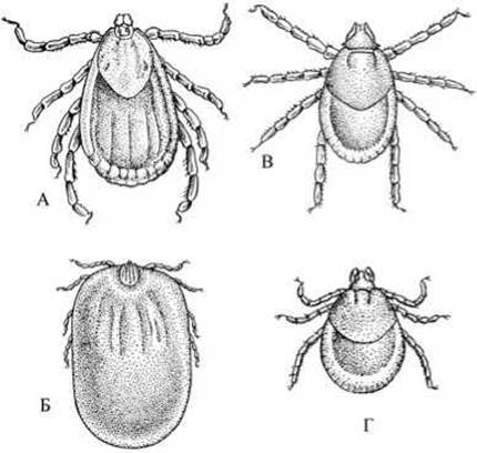
Fig. 95 Dermacentor tick (Dermacentor nuttali):
A — unfed female; B — heavily blood-gorged female; C — nymph; D — larva.
Examination of slide mounts of the mouthparts of Ixodes ricinus. Examine the prepared microscope slide of the castor bean tick's mouthparts under low magnification or using a dissecting magnifier, and sketch them in dorsal and ventral views. Dorsally, a pair of upper jaws, or chelicerae, covered by a sheath is visible (Fig. 96). The chelicerae are protrusible. Ventrally, examine the hypostome—a broad plate covered with denticles, among which the lateral ones are particularly well developed. The hypostome is formed from fused pedipalps. Locate the four-segmented palps on either side of the hypostome.
The tick's mouthparts are of the piercing-sucking type. Upon attaching to the host's skin, the tick uses the Teeth of its chelicerae to tear the skin and inserts the hypostome and cheliceral sheath into the wound. The denticles of the hypostome are oriented so as not to hinder its advancement into the wound, while preventing it from being pulled out. The ducts of two Salivary Glands (not visible) empty into the Oral Cavity. Much like in leeches, the salivary glands of ticks secrete a substance that inhibits blood clotting.
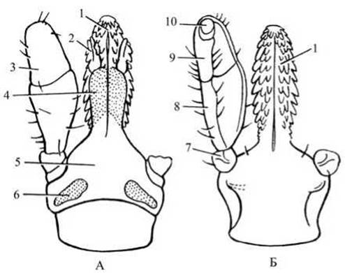
Fig. 96. Mouthparts of the female (Ixodes ricinus):
A — dorsal view; B — ventral view; 1 — hypostome; 2 — chelicerae; 3 — palps; 4 — cheliceral sheath; 5 — basis capituli; 6 — porose areas; 7–10 — palp segments.
The Development of the castor bean tick (*Ixodes ricinus*) proceeds with incomplete metamorphosis involving three hosts. After feeding to repletion and mating, the engorged female begins laying eggs. Throughout her lifetime, she deposits eggs onto the ground only once, sometimes numbering up to 12,000. Larvae hatch from the eggs and attack a host (rodents, birds, small animals). Having fed on blood, the larvae drop to the ground and molt. Following the molt, the larva transforms into a nymph. Nymphs attach to a new host, engorge with blood, drop to the ground, molt, and develop into adult ticks (imagos). The adult ticks attack a new (third) host, feed on blood, mate, and the females begin to lay eggs. Thus, the castor bean tick is a three-host tick. At all Selection/3.html">Stages of development, it parasitizes both domestic animals (horses, cattle, small ruminants, dogs, cats, etc.) and wild animals (lizards, hedgehogs, rodents, birds), as well as humans.
The ticks *I. persulcatus* and *Dermacentor nuttali* are also three-host species.

Fig. 97. Itch mite (*Sarcoptes scabiei*), female
The itch mite (*Sarcoptes scabiei*).
The itch mite is the CAUSATIVE AGENT OF Scabies in humans as well as in many domestic and wild mammals (Fig. 97). These mites spend their entire lives within the stratum corneum of the epidermis. Females measure 0.4 mm in length, while males are 0.15–0.20 mm long.
Females typically excavate long burrows (up to 2–3 mm per day) at night, specifically in areas with thinner and more delicate skin. Males do not dig burrows, but instead roam through existing ones. Itch mites cause agonizing itching, which leads to scratching, skin irritation, and even crust formation.
The female lays about 20 eggs inside the burrows she creates. Within 2–3 days, six-legged larvae hatch from the eggs. After only 1.5–3 days, the larvae transition
into the nymphal stage, possessing four pairs of legs but lacking a genital opening. After 2–3 days, the first-stage nymph transforms into a second-stage nymph, which subsequently molts into a sexually mature tick. Male second-stage nymphs do not undergo this stage. The lifespan of these mites is 40–50 days.
Prevention: personal hygiene (cleanliness of hands, body, clothing, and living quarters); Treatment of infected animals; veterinary supervision and sanitary treatment of animals introduced into farms; periodic veterinary inspection of animals to detect and eradicate scabies agents within agricultural premises.
Last update: 13/08/2026
Editorial and Educational Adaptation: This material has been compiled based on the primary/original source text. The project team performed an editorial review, corrected technical inaccuracies, structured sections, and adapted the content for an educational format.
What was processed:
- elimination of formatting defects (OCR errors, structural breaks, corrupted characters);
- editorial organization of content;
- standardization of terminology in accordance with academic sources;
- verification of factual statements against the original source text.
All mentions of the author, publication year, and origin of the primary text have been preserved in accordance with the source.