Human Anatomy - Kotsan I. Ya. 2009
Urogenital Apparatus
Genitalia - External Male Genitalia
Scrotum
The scrotum (scrotum) is a cutaneous-fascial pouch-like Structure located below the Pubic Symphysis, inferior to the ROOT of the Penis. The scrotal cavity is divided by the scrotal septum (septum scroti) into two compartments. Each of these compartments contains a Testis with its Epididymis and the scrotal portion of the Spermatic Cord. The scrotum acts as a "physiological thermostat," maintaining the Temperature of the Testes slightly below core body temperature, which is a necessary condition for normal Spermatogenesis.
The wall of the scrotum consists of 7 layers (tunics), also known as the Tunics of the testis (Fig. 179). In terms of structure and topography, the scrotal (testicular) tunics are similar to the layers of the anterior abdominal wall because the scrotum is formed by an outpouching of the abdominal wall. The following layers of the scrotum (tunics of the testis) are distinguished: 1) Skin (cutis); 2) dartos tunic (tunica dartos); 3) external spermatic fascia (fascia spermatica externa); 4) cremasteric fascia (fascia cremasterica); 5) cremaster Muscle (musculus cremaster); 6) internal spermatic fascia (fascia spermatica interna); 7) tunica vaginalis of the testis (tunica vaginalis testis). The tunica vaginalis features two layers (laminae): the parietal layer (lamina parietalis) and the visceral layer (lamina visceralis), which are continuous with each other and form a closed serous sac around the testis and epididymis, filled with a small amount of serous fluid.
The skin of the scrotum is thin, delicate, pigmented, covered with sparse and coarse Hair, and contains numerous sweat and Sebaceous Glands, the secretions of which have a distinct individual odor. Along the midline of the scrotal skin in the sagittal plane runs the scrotal raphe (raphe scroti), which extends anteriorly onto the Inferior surface of the penis and posteriorly reaches the anus.
Beneath the skin lies the dartos tunic, which is formed from the subcutaneous Connective Tissue of the inguinal region and Perineum, replacing the subcutaneous adipose layer. The dartos tunic contains bundles of muscle Cells and elastic fibers. The medial sections of the dartos tunic from both sides fuse to form the scrotal septum, which separates the right testis from the left. The dartos tunic is fused with the scrotal skin. Contraction of the muscular fibers of the dartos tunic gives the scrotal skin a wrinkled appearance.
Deeper beneath the dartos tunic lies the external spermatic fascia, which is a derivative of the superficial Fascia of the abdomen. Beneath it is the cremasteric fascia, formed from the deep fascia of the external oblique abdominal muscle and partially from the fibrous fibers of its aponeurosis. This fascia belongs to the cremaster muscle, which developed from muscle bundles branching off from the transversus abdominis and internal oblique Abdominal Muscles. Medial to the cremaster muscle is the internal spermatic fascia, originating from the transversalis fascia of the abdomen. Beneath the internal spermatic fascia lies the tunica vaginalis of the testis, derived from the vaginal process of the Peritoneum. The parietal lamina of the tunica vaginalis is fused with the internal spermatic fascia, while the visceral lamina is fused with the tunica albuginea of the testis. The closed serous cavity located between the two laminae of the tunica vaginalis is a derivative of the peritoneal cavity.
Class="center">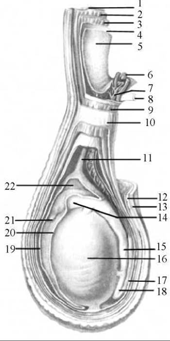
Fig. 179. Tunics of the testis and spermatic cord (diagram)
1 — external oblique abdominal muscle; 2 — internal oblique abdominal muscle; 3 — transversus abdominis muscle; 4 — transversalis fascia; 5 — peritoneum; 6 — testicular artery; 7 — pampiniform venous plexus; 8 — Ductus deferens; 9 — cremaster muscle; 10 — external spermatic fascia; 11 — remnant of the vaginal process; 12 — dartos tunic; 13 — skin; 14 — HEAD of the epididymis; 15 — body of the epididymis; 16 — testis; 17 — ductus deferens; 18 — tail of the epididymis; 19 — tunica vaginalis (parietal lamina); 20 — tunica vaginalis (visceral lamina); 21 — Appendix of the epididymis; 22 — serous cavity of the testis
The tunics of the testis are formed during the descent of the testis. In the embryo, the testis is located in the posterior part of the Abdominal cavity at the level of the lower lumbar vertebrae. An important role in the descent of the testis is played by the gubernaculum testis — a guiding ligament that forms during early developmental stages in a retroperitoneal position and extends from the caudal end of the testicular primordium to the anterior abdominal wall, where the scrotum subsequently begins to form. In the third month of intrauterine development, a blind peritoneal outpouching appears at the site of the future deep inguinal ring, forming its vaginal process. As the body of the embryo grows in length, the testis assumes a progressively lower position, shifting retroperitoneally into the pelvic cavity, and then, alongside the vaginal process, descends into the scrotum, where, enveloped in its serous covering, it reaches its final position. Along with the vaginal process, other layers of the anterior abdominal wall also pouch outward, forming the receptacle for the testis — the scrotum.
Blood supply to the scrotum is provided by the anterior scrotal Arteries (Branches of the external pudendal artery), posterior scrotal arteries (branches of the perineal artery), and the cremasteric artery (a branch of the inferior epigastric artery). The Veins OF THE scrotum accompany the corresponding arteries and drain into the venous System of the spermatic cords and the inferior rectal veins.
The scrotum receives its innervation from the sacral and lumbar plexuses.
Last update: 08/08/2026
Editorial and Educational Adaptation: This material has been compiled based on the primary/original source text. The project team performed an editorial review, corrected technical inaccuracies, structured sections, and adapted the content for an educational format.
What was processed:
- elimination of formatting defects (OCR errors, structural breaks, corrupted characters);
- editorial organization of content;
- standardization of terminology in accordance with academic sources;
- verification of factual statements against the original source text.
All mentions of the author, publication year, and origin of the primary text have been preserved in accordance with the source.