Human Anatomy - Kotsan I. Y. 2009
Urogenital System
Genitals - External Female Genitalia
Perineum
The perineum is a complex of soft Tissues (Skin, Muscles, fascia) that seal the pelvic outlet. In a narrow sense, the perineum refers to the region located between the external genitalia and the anus. In women, this area extends from the posterior margin of the pudendal cleft to the anterior margin of the anus (Fig. 185), whereas in men it extends from the posterior margin of the Scrotum to the anterior margin of the anus (Fig. 186). In shape, the perineum resembles a rhombus bounded posteriorly by the tip of the coccyx, anteriorly by the inferior point of the Pubic Symphysis, and laterally by the ischial tuberosities. An imaginary transverse line connecting the ischial tuberosities divides the perineum into two triangular regions: the urogenital region (anterosuperior) and the anal region (posteroinferior). The urogenital Diaphragm lies within the urogenital region, while the pelvic diaphragm occupies the anal region. Both diaphragms adjoin each other at their bases, with their apices directed toward the pubic symphysis and the coccyx, respectively. The urogenital diaphragm and the pelvic diaphragm form a musculo-fascial plate consisting of two Muscle layers (superficial and deep) and fasciae. This entire musculo-fascial plate is covered on the pelvic cavity side by the parietal layer of the pelvic fascia, and externally (inferiorly) by the superficial fascia.
The urogenital diaphragm (diaphragma urogenitale) occupies the anterior part of the perineum and is triangular in shape, with its apex directed toward the pubic symphysis and its base corresponding to the line connecting the ischial tuberosities. The Urethra passes through the urogenital diaphragm in men, while both the urethra and the Vagina pass through it in women. The urogenital diaphragm comprises two muscle layers: superficial and deep.
The SUPERFICIAL MUSCLES OF the urogenital diaphragm include three muscles: the superficial transverse perineal muscle, the ischiocavernosus, and the bulbospongiosus.
The superficial transverse perineal muscle (m. transversus perinei superficialis) is a paired muscle located in the Deep Layer of the subcutaneous adipose tissue. It originates from the ramus of the ischium near the ischial tuberosity, runs transversely to meet its counterpart from the opposite side, and inserts into the perineal body (central tendon of the perineum). Some fibers of this muscle blend into the external anal sphincter and the bulbospongiosus muscle. Function: the muscle participates in strengthening the perineal body.
The ischiocavernosus muscle (m. ischiocavernosus) is a paired muscle originating from the ischial ramus, curving from medial to lateral around the crus of the corpus cavernosum of the Penis (in men) and inserting into the tunica albuginea of the penis or Clitoris (in women). Function: it compresses the corpora cavernosa against the bone, thereby reducing venous outflow from the erectile tissue and inducing erection of the penis or clitoris.
Class="center">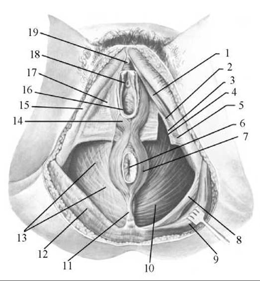
Fig. 185. MUSCLES AND FASCIAE of the female perineum
1 — ischiocavernosus muscle; 2 — inferior Fascia of the urogenital diaphragm; 3 — deep transverse perineal muscle; 4 — superior fascia of the urogenital diaphragm; 5 — superficial transverse perineal muscle; 6 — anus; 7 — external anal sphincter; 8 — sacrotuberous ligament; 9 — gluteus maximus muscle; 10 — levator ani muscle; 11 — anococcygeal ligament; 12 — ischial fascia; 13 — inferior fascia of the pelvic diaphragm; 14 — bulbospongiosus muscle; 15 — fascia lata; 16 — vaginal orifice; 17 — superficial perineal fascia; 18 — external urethral orifice; 19 — glans clitoridis
The bulbospongiosus muscle (m. bulbospongiosus) consists of two halves originating from the raphe on the Inferior surface of the bulb of the penis, enclosing the bulb and corpus spongiosum of the penis on the right and left sides, and inserting into its tunica albuginea and the superficial fascia on the dorsum of the penis. Function: upon contraction, the muscle compresses the bulb, corpora cavernosa, and dorsal vein of the penis, as well as the Bulbourethral Glands, facilitating erection. In women, the bulbospongiosus is a paired organ encircling the vagina around its orifice. This muscle originates from the perineal body and the external anal sphincter, inserting into the dorsal surface of the clitoris by blending into its tunica albuginea. Upon contraction, the muscle narrows the vaginal introitus and compresses the greater vestibular gland and the vestibular bulb.
The Deep Muscles of the urogenital diaphragm include the deep transverse perineal muscle and the urethral sphincter.
The deep transverse perineal muscle (m. transversus perinei profundus) is a paired muscle originating from the rami of the ischium and pubis. Along the midline of the perineum, this muscle connects with the tendon of the corresponding muscle of the opposite side and contributes to The formation of the perineal body. Both of these muscles reinforce the urogenital diaphragm.
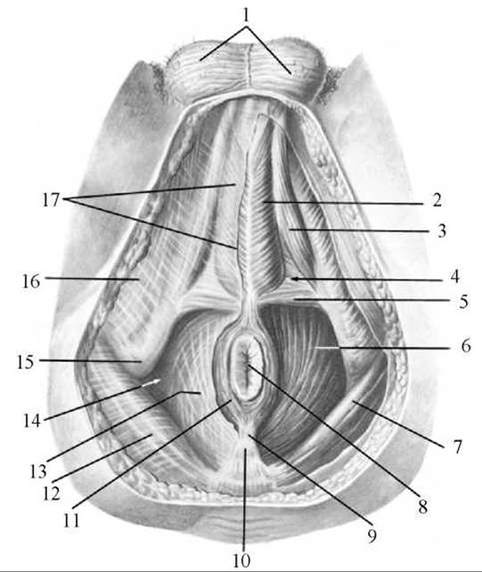
Fig. 186. Muscles and Fasciae of the male perineum
1 — scrotum; 2 — bulbospongiosus muscle; 3 — ischiocavernosus muscle; 4 — urogenital diaphragm; 5 — superficial transverse perineal muscle; 6 — levator ani muscle; 7 — gluteus maximus muscle; 8 — anus; 9 — anococcygeal ligament; 10 — coccyx; 11 — external anal sphincter; 12 — ischial fascia; 13 — inferior fascia of the pelvic diaphragm; 14 — ischioanal fossa; 15 — ischial tuberosity; 16 — fascia lata; 17 — superficial perineal fascia.
The urethral sphincter (m. sphincter urethrae) is an unpaired muscle with predominantly circular fibers that surrounds the membranous urethra in men and the urethra in women, originating in part from the inferior rami of the pubic bones. In men, muscle fiber bundles attach to the Prostate Gland, whereas in women, they blend into the vaginal wall. This muscle acts as a voluntary constrictor of the urethra.
The pelvic diaphragm (diaphragma pelvis) occupies the posterior part of the perineum and is shaped like a triangle, with its apex directed toward the coccyx and its other two angles directed toward the ischial tuberosities. The terminal part of the rectum passes through the pelvic diaphragm in both men and women. The pelvic diaphragm also features two muscle layers: superficial and deep.
The superficial Muscles of the pelvic diaphragm include the unpaired external anal sphincter.
The external anal sphincter (m. sphincter ani externus) is a muscular ring surrounding the anus and the adjacent part of the rectum. The muscle comprises three parts: the subcutaneous part (pars cutanea), whose muscle bundles originate and insert beneath the skin; the superficial part (pars superficialis), which terminates as a pair of bundles on the anococcygeal ligament; and the deep part (pars profunda), which is the most prominent portion, surrounding the lower rectum and adjoining the levator ani muscle. Upon contraction, all bundles of the external anal sphincter compress (close) the anus.
The deep muscles of the pelvic diaphragm include two muscles: the levator ani and the coccygeus.
The levator ani muscle (m. levator ani) is paired and forms, together with its counterpart from the opposite side, a funnel with its wide end directed upward. The levator ani is the largest muscle of the pelvic diaphragm. It originates from the lateral wall of the lesser pelvis via several bundles. The anterior bundles arise from the inner surface of the inferior pubic ramus, while the lateral bundles arise from the tendinous arch of the levator ani (arcus tendineus musculi levatoris ani), which is a tendinous thickening of the pelvic fascia where it covers the obturator fascia. The bundles of the right and left levator ani muscles run downward and backward, unite with each other, encircle the rectum, and then course downward and medially. Some fibers of the levator ani blend into the prostate gland (in men), the vaginal wall (in women), as well as the Urinary Bladder and rectum, while the muscles themselves terminate near the tip of the coccyx via the anococcygeal ligament (lig. anococcygeum). Contraction of the levator ani strengthens and elevates the pelvic floor, pulls the lower rectum upward and forward while compressing it. In women, this muscle also narrows the vaginal introitus and approximates the posterior vaginal wall to the anterior one.
The coccygeus muscle (m. coccygeus) is paired, originating from the ischial spine and the sacrospinous ligament, runs medially and posteriorly, and inserts into the lateral margin of the coccyx and the apex of the sacrum. The bundles of this muscle lie medial to the sacrospinous ligament, partially blending into it. The muscle reinforces the posterior part of the pelvic diaphragm.
Last update: 08/08/2026
Editorial and Educational Adaptation: This material has been compiled based on the primary/original source text. The project team performed an editorial review, corrected technical inaccuracies, structured sections, and adapted the content for an educational format.
What was processed:
- elimination of formatting defects (OCR errors, structural breaks, corrupted characters);
- editorial organization of content;
- standardization of terminology in accordance with academic sources;
- verification of factual statements against the original source text.
All mentions of the author, publication year, and origin of the primary text have been preserved in accordance with the source.