Human Anatomy - Kotsan I. Y. 2009
Urogenital System
Genitals - Female Genitals
Ovary
The ovary (ovarium) is a paired female reproductive gland which, much like the Testis in males, performs two main Functions: exocrine (the development and maturation of Female Germ Cells, or ova) and endocrine (The production of Female Sex Hormones, which enter the Blood AND Lymph) (Fig. 181).
The ovary is situated against the lateral walls of the lesser pelvis, on either side of the uterine fundus. It is held in place by two primary structures: the proper ovarian ligament (lig. ovarii proprium) and the suspensory ligament of the ovary (lig. suspensorium ovarii). The proper ovarian ligament is a rounded cord extending from the uterine end of the ovary to the uterine wall, running between the two layers of the broad ligament of the Uterus. The suspensory ligament of the ovary is a peritoneal fold extending from the pelvic wall to the ovary, enclosing ovarian Blood Vessels and bundles of fibrous fibers. Additionally, the ovary is anchored by a short mesovarium, which is a peritoneal fold extending from the posterior layer of the broad ligament of the uterus to the mesovarian margin of the ovary. The Ovaries themselves are not covered by Peritoneum. The Topography of the ovaries is largely influenced by THE POSITION OF the uterus and its enlargement during Pregnancy. The ovaries are classified as quite mobile Organs within the lesser pelvis.
Class="center">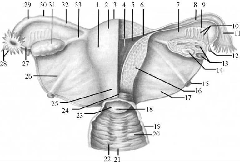
Fig. 181. Internal female reproductive organs (posterior view)
1 — body of uterus; 2 — serous coat (perimetrium); 3 — fundus of uterus; 4 — uterine cavity; 5 — mucous membrane (endometrium); 6 — uterine opening of tube; 7 — mesosalpinx; 8 — mucosal folds of tube; 9 — longitudinal duct of epoophoron; 10 — transverse ductules; 11 — infundibulum of Uterine tube; 12 — vesicular Appendix; 13 — vesicular ovarian follicles; 14 — corpus luteum; 15 — round ligament of uterus; 16 — muscular coat (myometrium); 17 — broad ligament of uterus (posterior layer removed); 18 — external orifice of uterus (ostium uteri); 19 — muscular coat of Vagina; 20 — mucous membrane of vagina; 21 — anterior vaginal Column; 22 — vaginal rugae; 23 — vaginal part of cervix; 24 — cervical canal; 25 — cervix of uterus (supravaginal part); 26 — broad ligament of uterus (posterior layer); 27 — ovarian fimbria; 28 — fimbriae of uterine tube; 29 — ampulla of uterine tube; 30 — uterine tube; 31 — ovary; 32 — isthmus of uterine tube; 33 — proper ovarian ligament
In an adult woman, the ovary is egg-shaped and slightly compressed in the anteroposterior direction. The mass of the ovary is 5–8 g, length 2.5–5 cm, width 1.5–3 cm, and thickness 0.5–1.0 cm.
The ovary has two extremities: the upper tubal extremity (extremitas tubaria), directed toward the uterine tube, and the lower uterine extremity (extremitas uterina), directed toward the uterus; it also has two surfaces: the lateral surface (facies lateralis), adjacent to the pelvic wall, and the medial surface (facies medialis), facing the pelvic cavity. The surfaces of the ovary transition anteriorly into the straight mesovarian margin (margo mesovaricus), to which the mesovarium attaches, and posteriorly into the convex free margin (margo liber), facing the Abdominal cavity. The mesovarian margin features a small depression known as the hilum of the ovary (hilum ovarii), which serves as the entry and exit point for blood Vessels and nerves.
Structure OF THE ovary. Externally, the ovary is covered by a single layer of cuboidal (germinal) epithelium, beneath which lies the Connective Tissue tunica albuginea. The connective tissue stroma of the ovary (stroma ovarii) is rich in elastic fibers and cellular elements. The Internal Structure of the ovary varies depending on the age and functional state of the body. In the adult woman's ovary, two layers are distinguished: an outer cortex and an inner medulla.
The medulla of the ovary (medulla ovarii) is located in the center of the organ, closer to its hilum. This substance is formed of loose connective tissue containing A large number of elastic fibers, smooth Muscle cells, nerves, blood vessels, and Lymphatic vessels.
The cortex of the ovary (cortex ovarii) lies beneath the tunica albuginea and constitutes the primary functional part of the organ. It consists of dense Fibrous connective tissue housing numerous ovarian follicles: primary (growing and maturing), mature vesicular follicles (Graafian follicles), atretic follicles (undergoing regression), as well as corpora lutea and scars (Fig. 182).
Unlike Male Germ Cells, the proliferation of female germ cells occurs during the prenatal period. Primary ovarian follicles (folliculi ovarici primarii) are formed, each containing a female germ Cell (oocyte) surrounded by a single layer of follicular epithelial cells. In a newborn girl's ovary, there are about 400,000 primary follicles. After birth, the number of follicles no longer increases, but instead decreases through resorption. By the onset of Puberty, none remain in the medullary substance of the ovary, while about 15,000 remain in the cortical substance. Primary follicles mature only during puberty. Over a woman's lifetime, about 400–500 follicles mature, gradually transforming into mature vesicular follicles (folliculi ovarici vesiculosi), while the rest degenerate. The death (atresia) of follicles is accompanied by The breakdown of the oocyte and follicular epithelium, their replacement by connective tissue, and The formation of an atretic body (corpus atreticum).
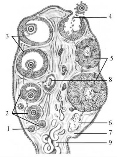
Fig. 182. Diagram of ovarian follicle development, ovulation, and corpus luteum formation
1 — primordial follicle; 2 — primary (growing, maturing) follicle; 3 — vesicular follicle (Graafian follicle); 4 — ovulation; 5 — corpus luteum; 6 — scar of corpus luteum; 7 — ovarian stroma; 8 — atretic body; 9 — blood vessel
A mature follicle (Graafian follicle) reaches about 1 cm in diameter and is externally covered by a connective tissue sheath known as the theca folliculi, which consists of two layers: external and internal. The outer layer of the theca is built of Cytology/practical/45.html">Dense connective tissue poor in cellular elements and blood vessels. The connective tissue of the internal theca layer contains numerous blood vessels and lymphatic capillaries, as well as interstitial cells that produce the hormone estradiol, which is responsible for oocyte maturation and The Development of secondary female sex characteristics. Adjacent to the internal theca layer is the stratum granulosum, which thickens in one area to form the cumulus oophorus. The cumulus oophorus contains the oocyte (ovocytus). Inside the mature follicle is a cavity filled with follicular fluid (liquor follicularis), which exhibits hormonal activity. The oocyte located within the cumulus oophorus is surrounded by the zona pellucida and the corona radiata, both formed by follicular cells. As it matures, the ovarian follicle grows, increases in size, and gradually reaches the Superficial layer of the organ, projecting slightly above its surface. During ovulation, the wall of such a follicle ruptures, and the oocyte, along with the follicular fluid, is released into the peritoneal cavity, where it is caught by the fimbriae of the uterine tube and enters the abdominal ostium of the uterine tube.
In humans, ovulation normally occurs regularly every 28–30 days. Follicles rupture under the Influence of the pituitary follicle-stimulating hormone (FSH), or follitropin, while ovulation itself is triggered by the pituitary luteinizing hormone (LH), or lutropin. The cavity of the ruptured and emptied follicle fills with blood, and the blood clot is rapidly replaced by connective tissue. Under the action of lutropin, a corpus luteum develops here. The follicular epithelial cells proliferate, accumulate a yellow pigment, and transform into luteocytes, which produce the hormone progesterone. If Fertilization of the oocyte does not occur, the corpus luteum remains small (up to 1.0–1.5 cm), functions briefly (12–14 days), and is called the cyclic (menstrual) corpus luteum (corpus luteum cyclicum [menstruationis]). Later, it is invaded by connective tissue and transforms into a corpus albicans, which is resorbed after several years. If the oocyte is fertilized and pregnancy ensues, the corpus luteum enlarges significantly, reaching 1.5–2 cm in diameter, and persists throughout the entire period of pregnancy. This is known as the true corpus luteum of pregnancy (corpus luteum graviditatis). It performs an endocrine function by producing progesterone, which acts on the uterine mucosa, preparing it for the implantation of the fertilized ovum, fetal GROWTH AND DEVELOPMENT, and inhibiting the growth of new follicles. Following childbirth, the corpus luteum of pregnancy is also replaced by connective tissue and turns into a corpus albicans. Small scars remain On the surface of the ovary at the sites of ruptured vesicular follicles. In an adult woman, such scars cover the entire surface of the ovary.
In older women (aged 45 and over), follicle maturation and ovulation become irregular and eventually cease altogether, marking the onset of the Climacteric period (menopause).
Located near each ovary are rudimentary structures—the epoophoron, paroophoron, and vesicular Appendices—which represent remnants of the tubules of the pronephros/mesonephros and their ducts.
The epoophoron is situated posterior and lateral to the ovary between the layers of the mesosalpinx. It consists of the longitudinal duct of the epoophoron (ductus epoophori longitudinalis) and a delicate network of convoluted transverse ductules (ductuli transversi) that drain into the longitudinal duct (Fig. 181).
The paroophoron is a small structure also located in the mesosalpinx near the tubal end of the ovary. It consists of a few scattered blind ductules, which are remnants of the caudal part of the mesonephric duct.
The vesicular appendices (appendices vesiculosae) are one or more small cysts suspended on long stalks from the mesosalpinx or one of its fimbriae.
The ovary is supplied with blood by Branches of the ovarian artery, which originates from the Abdominal Aorta, and by ovarian branches arising from the uterine artery. Venous blood drains via corresponding Veins into the INFERIOR VENA CAVA (from the right ovary) and the left renal vein (from the left ovary). Lymphatic vessels carry lymph to the lumbar and internal iliac Lymph Nodes.
The ovary is innervated by branches of the abdominal aortic and inferior hypogastric plexuses (sympathetic innervation) and the pelvic splanchnic nerves (parasympathetic innervation).
Last update: 08/08/2026
Editorial and Educational Adaptation: This material has been compiled based on the primary/original source text. The project team performed an editorial review, corrected technical inaccuracies, structured sections, and adapted the content for an educational format.
What was processed:
- elimination of formatting defects (OCR errors, structural breaks, corrupted characters);
- editorial organization of content;
- standardization of terminology in accordance with academic sources;
- verification of factual statements against the original source text.
All mentions of the author, publication year, and origin of the primary text have been preserved in accordance with the source.