Human Anatomy, Part 1 - K. A. Dyubenko, A. K. Kolomiyytsev, Yu. B. Chaikovsky 2002
Special Part
Myology, myologia [the study of muscles] – Muscles and fascia of the trunk
Muscles and fascia of the abdomen – Muscles of the abdomen, musculi abdominis – Fascia of the abdomen
1. The superficial Fascia of the abdomen, fascia subcutanea abdominis, is thin in its upper part, but thickens as it descends, acquiring elastic fibers. Along the midline, this fascia fuses with the linea alba, and inferiorly with the inguinal ligament. In the lower region above the Pubic Symphysis, it forms specific strands:
- the fundiform ligament, lig. fundiforme Penis, which originates from the pubic symphysis and divides into two crura that embrace the penis laterally;
- the suspensory ligament of the penis, lig. suspensorium penis, and in females, the suspensory ligament of the Clitoris, lig. suspensorium clitoridis. It extends from the pubic symphysis to the dorsal surface of the penis (clitoris).
2. The transversalis fascia, fascia transversalis, covers the internal surface of the transversus abdominis Muscle and the inner aspect of the posterior layer of the rectus sheath, and below the arcuate line, it covers the inner posterior surface of the rectus abdominis muscle. Inferiorly, the fascia fuses with the edge of the inguinal ligament. In the umbilical region, it is denser, whereas in the inguinal region, it continues along the Spermatic Cord into the Scrotum as the internal spermatic fascia, fascia spermatica interna. An expanded oval depression On the surface of the fascia transversalis represents the deep inguinal ring, anulus inguinalis profundus.
The inguinal region, regio inguinalis (see Fig. 14A), is of paramount clinical importance because surgical interventions for inguinal hernias are performed within its boundaries. Situated within the inguinal region are the inguinal triangle, trigonum inguinale, and the inguinal interstice. The BOUNDARIES OF THE inguinal triangle are: inferiorly, the inguinal ligament; medially, the lateral margin of the rectus abdominis muscle; superiorly, an imaginary perpendicular dropped from the middle third of the inguinal ligament to the rectus abdominis muscle.
The inguinal triangle is a static concept (its boundaries are constant and unchanging), whereas the inguinal interstice is a dynamic concept (it can vary in shape and size) (Fig. 137).
Class="center">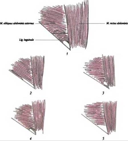
Fig. 137. The inguinal triangle and variations of the inguinal interstices (after B. V. Ognev, Z. Kh. Frauchie):
1 - inguinal triangle; 2 - slit-like inguinal interstice; 3 - circular inguinal interstice; 4 - oval inguinal interstice; 5 - triangular inguinal interstice
Four shapes of the inguinal interstice are distinguished: slit-like, triangular, circular, and oval (B. V. Ognev,1 V. Kh. Frauchie, 1960).
The Inguinal Canal, canalis inguinalis, is located in the inguinal region, reg. inguinalis, superior to the inguinal ligament, lig. inguinale (Figs. 138, 139). The formation of the inguinal canal is associated with the descent of the Testis into the scrotum. The canal appears as an oblique slit-like passage in the lower part of the abdominal wall, measuring 4-5 cm in length. In males, the canal transmits the spermatic cord, funiculus spermaticus; in females, it transmits the round ligament of the Uterus, lig. teres uteri. The inguinal canal has four walls:
- anterior wall - the aponeurosis of the external oblique abdominal muscle;
- posterior wall - the transversalis fascia;
- inferior wall - the groove of the inguinal ligament;
- superior wall - the lower margins of the internal oblique and transversus abdominis Muscles.
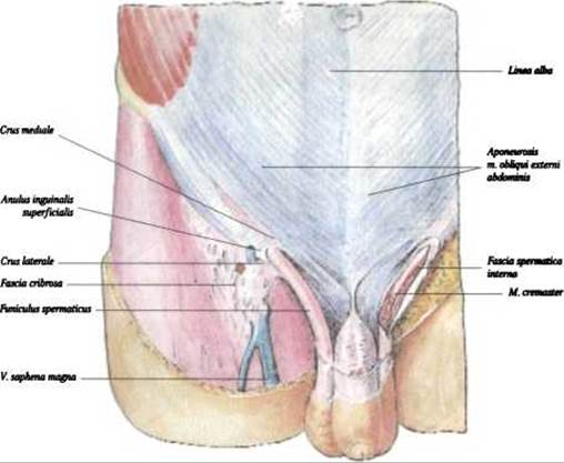
Fig. 138. Inguinal canal, canalis inguinalis - anterior view (after R. D. Sinelnikov)
1 Ognev, Boris Vladimirovich (1901-1978) - a renowned Russian topographic anatomist and surgeon.
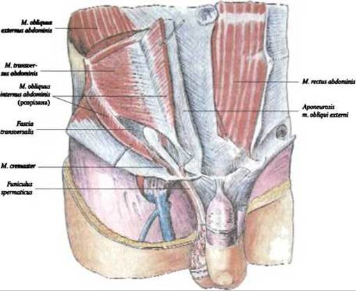
Fig. 139. Inguinal canal, canalis inguinalis - anterior view in dissection (after R. D. Sinelnikov)
The inguinal canal features superficial and deep rings.
The superficial inguinal ring, anulus inguinalis superficialis, is located above the pubic bone (see Fig. 138). Superiorly and inferiorly, it is bounded by the medial and lateral crura of the external oblique aponeurosis; laterally, by the intercrural fibers, fibrae intercrurales; medially, by the reflected inguinal ligament, lig. reflexum.
The deep inguinal ring, anulus inguinalis profundus, is bounded medially by the intervolar ligament, lig. interfoveolare (see Fig. 139). The posterior wall of the inguinal canal is reinforced by the fibers of the lower edge of the aponeurosis of the m. transversus abdominis, which curve and attach to the pubic tubercle and pubic crest, forming the conjoint tendon (falx inguinalis).
The deep inguinal ring corresponds to the Location OF THE lateral inguinal fossa, which resembles a funnel-shaped depression.
The inguinal canal is a potential site for the formation and passage of inguinal hernias, a factor that must be considered in clinical practice.
Last update: 08/08/2026
Editorial and Educational Adaptation: This material has been compiled based on the primary/original source text. The project team performed an editorial review, corrected technical inaccuracies, structured sections, and adapted the content for an educational format.
What was processed:
- elimination of formatting defects (OCR errors, structural breaks, corrupted characters);
- editorial organization of content;
- standardization of terminology in accordance with academic sources;
- verification of factual statements against the original source text.
All mentions of the author, publication year, and origin of the primary text have been preserved in accordance with the source.