Review of Medical Physiology - William F. Ganong 2002
Endocrine System, Metabolism, and Reproduction
Hormonal Regulation of Calcium Metabolism and Bone Physiology
Calcitonin
Origin
In dogs, perfusion of the thyroid-parathyroid region with solutions containing high concentrations of Ca2+ leads to a decrease in peripheral plasma Ca2+ levels. Conversely, following damage to this region, Ca2+ infusions cause an increase in plasma Ca2+ compared to control animals. These and other observations led to the discovery that Ca2+-lowering and Ca2+-raising Hormones are secreted by structures in the neck; the Ca2+-lowering hormone was named calcitonin. In non-mammalian vertebrates, the source of calcitonin is the ultimobranchial bodies—a pair of glands embryologically derived from the fifth pharyngeal pouches. In mammals, these bodies are mostly located within The Thyroid Gland, where ultimobranchial tissue spreads around the follicles as parafollicular Cells, also known as light cells or C cells (Figs. 18-2 and 21-15).
Human calcitonin has a Molecular Weight of 3,500 and consists of 32 amino acid residues (Fig. 21-16). A large amount of mRNA transcribed from the calcitonin Gene undergoes alternative Processing into other mRNAs in The Nervous system, producing calcitonin gene-related peptide (CGRP) rather than calcitonin (see Chapter 4). Calcitonins from other studied species also contain 32 amino acid residues, although the Amino Acid Composition varies significantly. Salmon calcitonin is of particular interest because its activity is 20 times that of human calcitonin.
Class="center">
Fig. 21-15. Parafollicular cells in the thyroid gland (modified from Poirier J, Dumas JLR: Review of Medical Histology. Saunders, 1977).
Secretion and METABOLISM
Calcitonin secretion increases when the thyroid gland is perfused with solutions containing high concentrations of Ca2+. Radioimmunoassay measurements of circulating calcitonin indicate that it is not secreted until plasma calcium reaches approximately 9.5 mg/dL; above this level, plasma calcitonin is directly proportional to calcium. Beta-adrenergic agonists, dopamine, and estrogens also stimulate calcitonin secretion, as do gastrin, CCK, Glucagon, and secretin, with gastrin having the strongest effect (see Chapter 26). Plasma calcitonin levels are elevated in Zollinger-Ellison syndrome (see Chapter 26) and pernicious anemia, conditions that are also associated with elevated plasma gastrin levels. However, the dose of gastrin required to stimulate calcitonin secretion results in a much greater increase in plasma gastrin concentration than does a meal, so it is premature to conclude that calcium in the gut initiates the secretion of the calcium-lowering hormone before its absorption.
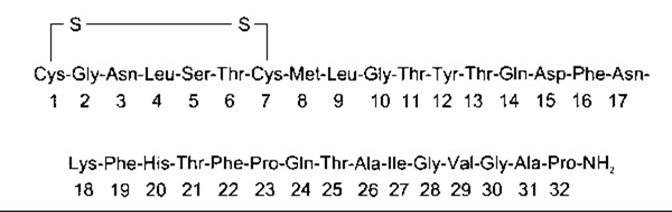
Fig. 21-16. Human calcitonin.
The half-life of human calcitonin is less than 10 min.
Actions
Serpentine receptors for calcitonin have been identified in bones and Kidneys. Calcitonin decreases circulating levels of calcium and phosphate. It exerts its calcium-lowering effect by inhibiting bone resorption. This action is direct: calcitonin inhibits osteoclast activity in vitro and also enhances the urinary excretion of Ca2+.
The exact Physiological Role of calcitonin remains uncertain. Human thyroid stores of calcitonin are low, so following thyroidectomy, bone density and plasma calcium levels remain normal provided the Parathyroid glands are intact. Furthermore, only transient abnormalities in calcium metabolism occur when calcitonin is administered after thyroid removal. This can be partly explained by the secretion of calcitonin from Tissues other than the thyroid. Nevertheless, the prevailing view is that the hormone has little long-term effect on plasma calcium levels in adult animals and humans. In addition, patients with medullary thyroid carcinomas have very high circulating levels of calcitonin yet exhibit no symptoms directly attributable to the hormone, and their bones are essentially normal. No deficiency syndrome has been described for calcitonin. The hormone is secreted in larger amounts in young individuals, where it may play a role in skeletal development and protect against postprandial hypercalcemia. Calcitonin protects maternal bones from excessive calcium loss during Pregnancy. Fetal Bone Formation and Lactation are major drains on calcium reserves, and 1,25-dihydroxycholecalciferol levels are elevated during pregnancy. These would cause maternal bone loss were it not for the simultaneous suppression of bone resorption by increased plasma calcitonin levels.
Clinical Parallels
Calcitonin is useful in the Treatment of Paget's Disease, a condition in which increased osteoclastic activity leads to the compensatory formation of disorganized new bone. It is also therapeutically effective in severe hypercalcemia, although the hormone must be administered by injection and its efficacy typically wanes over time.
Summary
The actions of the three main hormones regulating plasma Ca2+ concentration can now be summarized. Parathyroid hormone increases plasma calcium concentration by mobilizing this ion from bone. It increases renal reabsorption of Ca2+, though this may be a consequence of increased filtered Ca2+, and it enhances The formation of 1,25-dihydroxycholecalciferol, which promotes intestinal absorption of Ca2+, mobilizes the ion from bone, and increases renal calcium reabsorption. Calcitonin inhibits bone resorption and increases urinary Ca2+ excretion.
Last update: 10/08/2026
Editorial and Educational Adaptation: This material has been compiled based on the primary/original source text. The project team performed an editorial review, corrected technical inaccuracies, structured sections, and adapted the content for an educational format.
What was processed:
- elimination of formatting defects (OCR errors, structural breaks, corrupted characters);
- editorial organization of content;
- standardization of terminology in accordance with academic sources;
- verification of factual statements against the original source text.
All mentions of the author, publication year, and origin of the primary text have been preserved in accordance with the source.