Review of Medical Physiology - William F. Ganong 2002
Endocrine System, Metabolism, and Reproduction
Hormonal Regulation of Calcium Metabolism and Bone Physiology
Parathyroid Glands
Anatomy
Humans normally have four Parathyroid glands: two embedded in the upper poles of The Thyroid Gland and two in the lower poles (Fig. 21-9). However, the exact Location and number of parathyroid glands can vary significantly among individuals. Parathyroid tissue is occasionally found in the Mediastinum. Each parathyroid gland is highly vascularized, measures approximately 3x6x2 mm, and contains two Cell types. Numerous chief Cells, which feature prominent Golgi complexes, Endoplasmic reticulum, and secretory granules (Fig. 21-10), synthesize and secrete parathyroid hormone. Less numerous and larger oxyphil cells contain oxyphil granules and abundant Mitochondria in their Cytoplasm. In humans, only a few are present before Puberty, but their number increases with age. Their exact function remains unknown, although some consider them to be degenerated chief cells.
Class="center">
Fig. 21-9. Human parathyroid glands, posterior view.
Synthesis and METABOLISM of Parathyroid Hormone
Human parathyroid hormone is a linear polypeptide with a Molecular Weight of 9,500, consisting of 84 amino acid residues (Fig. 21-11). Its Structure is very similar to that of bovine and porcine parathyroid Hormones. It is synthesized as part of a larger precursor molecule containing 115 amino acid residues (preproparathyroid hormone). Upon entering The endoplasmic reticulum, the leader sequence is cleaved from the amino terminus, yielding the 90-amino-acid polypeptide proparathyroid hormone. Six additional amino acid residues are removed from the amino terminus of proparathyroid hormone in the Golgi apparatus, and the 84-amino-acid polypeptide parathyroid hormone enters secretory granules to be released as the primary secretory product of the chief cells.

Fig. 21-10. Cross-section of a human parathyroid gland (reduced 50% from x960). Small cells are chief cells; large, intensely stained cells (especially prominent in the lower left corner) are oxyphil cells (reproduced with permission from Fawcett DW: Bloom and Fawcett, A Textbook of Histology, 11th ed. Saunders, 1986).
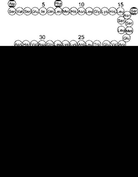
Fig. 21-11. Parathyroid hormone. Symbols below and above the human hormone structure indicate differing amino acid residues in bovine and porcine parathyroid hormones (reproduced with permission from Keutmann HT et al: Complete Amino Acid Sequence of human parathyroid hormone. Biochemistry 1978; 17-5723).
The normal plasma level of intact parathyroid hormone ranges from 10 to 55 pg/mL; its half-life is approximately 10 minutes. The secreted polypeptide is rapidly cleaved by Kupffer cells in the Liver into carboxy-terminal and mid-region fragments, which appear to be biologically inactive. Parathyroid hormone and these fragments are subsequently cleared via the Kidneys. Interestingly, a synthetic polypeptide containing the 34 amino acid residues of the amino terminus of parathyroid hormone exhibits all the known BIOLOGICAL EFFECTS OF the intact molecule.
Because many older radioimmunoassays for parathyroid hormone utilized Antibodies directed against the mid-region of the molecule, they detected both the intact hormone and its fragments, leading to falsely elevated values. This was especially problematic in patients with renal failure, as these fragments are not cleared efficiently in such conditions. To overcome this issue, researchers developed two-site immunoassays utilizing one antibody directed against the amino terminus and another against the carboxy terminus of the molecule. Plasma first reacts with radioactively labeled amino-terminal antibodies, and is then exposed to beads bound with unlabeled carboxy-terminal antibodies. The beads are washed, and their bound radioactivity is measured. Only the intact parathyroid hormone molecule is recognized by both antibodies, thus providing an accurate measurement of circulating parathyroid hormone (1-84).
Actions
Parathyroid hormone acts directly on bone, enhancing its resorption and mobilizing Ca2+. In addition to elevating plasma Ca2+ and lowering plasma phosphate levels, parathyroid hormone increases urinary phosphate excretion. This phosphaturic effect results from decreased phosphate reabsorption in the proximal tubules. Parathyroid hormone also enhances Ca2+ reabsorption in the distal tubules, although urinary Ca2+ excretion is often elevated in hyperparathyroidism because the filtered load exceeds the reabsorptive capacity. Furthermore, parathyroid hormone stimulates the synthesis of 1,25-dihydroxycholecalciferol, which in turn promotes intestinal Ca2+ absorption.
Over a longer time scale, parathyroid hormone stimulates both osteoclasts and osteoblasts (see Table 21-2), with the effect on osteoclasts predominating, thereby mobilizing greater amounts of Ca2+ from Bone tissue.
It is now believed that at least three distinct parathyroid Hormone Receptors exist. One of these also binds parathyroid hormone-related peptide (PTHrP) (see below) and is known as the hPTH/PTHrP receptor. Another receptor, PTH2 (hPTH2-R), does not bind PTHrP and has been identified in the Brain, Placenta, and Pancreas. Additionally, evidence points to the existence of a third receptor, CPTH, which interacts with the carboxy terminus rather than the amino terminus of parathyroid hormone. The first two receptors are serpentine, Gs-protein-coupled receptors that activate adenylate cyclase via this heterotrimeric G protein, thereby increasing intracellular cAMP levels. The hPTH/PTHrP receptor also activates PLC via Gq, leading to elevated intracellular Ca2+ levels and activation of protein kinase C (Fig. 21-12). However, the exact Intracellular Signaling pathways mediating the effects of these second messengers on bone Calcium Homeostasis are not yet fully elucidated.
In a disorder known as pseudohypoparathyroidism, the clinical signs and symptoms of hypoparathyroidism develop, yet circulating parathyroid hormone levels are normal or elevated. Because the target Tissues fail to respond to the hormone, the condition is receptor-mediated. There are two main forms of this disorder. The more common form involves a hereditary 50% reduction in Gs activity, preventing parathyroid hormone from eliciting a sufficient increase in cAMP concentration. In the less common form, the cAMP response is normal, but the phosphaturic action of the hormone is defective.
Regulation of Secretion
Circulating ionized calcium acts directly on the parathyroid glands via a negative feedback loop to regulate parathyroid hormone secretion (Fig. 21-13). The key mediator of this regulation is The Cell-surface Ca2+-sensing receptor. This serpentine, G-protein-coupled receptor is linked to phosphoinositide metabolism and is expressed in numerous tissues. In the parathyroid glands, its activation suppresses parathyroid hormone secretion. When plasma Ca2+ levels are high, parathyroid hormone secretion is inhibited and calcium is deposited in bones; conversely, when Ca2+ concentrations are low, secretion is stimulated and Ca2+ is mobilized from the Skeleton.
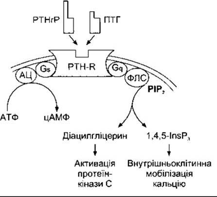
Fig. 21-12. Signal Transduction pathways activated by the binding of PTH or PTHrP to the hPTH/hPTHrP receptor. Intracellular cAMP is increased via Gs and adenylate cyclase (AC). Diacylglycerol and IP3 (1,4,5-InsP3) are elevated through the action of Gq and phospholipase C (PLC) (modified with permission from Shoback DM, Strewler GJ: Disorders of the parathyroids and calcium metabolism. In McPhee SH et al. Pathophysiology of Disease, 3rd ed. McGraw-Hill, 2000).
1,25-Dihydroxycholecalciferol acts directly on the parathyroid glands to suppress preproparathyroid hormone mRNA expression. Elevated plasma phosphate levels stimulate parathyroid hormone secretion indirectly by lowering plasma Ca2+ concentrations and inhibiting The production of 1,25-dihydroxycholecalciferol. Magnesium is required to maintain normal secretory responsiveness of the parathyroid glands. Impaired parathyroid hormone release, combined with a blunted target-organ response to the hormone, leads to the hypocalcemia sometimes observed in magnesium deficiency.
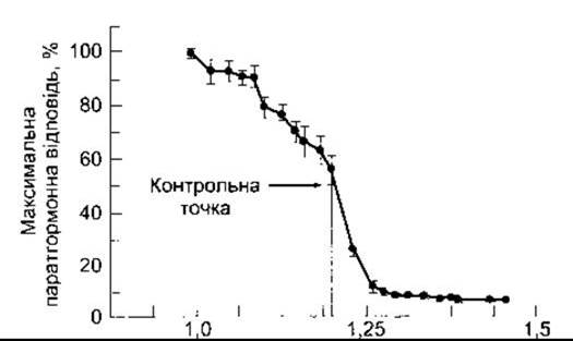
Fig. 21-13. Relationship between plasma Ca2+ concentration and the parathyroid hormone response in humans. The set point corresponds to the plasma Ca2+ concentration that produces half-maximal response (modified with permission from Brown E: Extracellular Ca2+ sensing, regulation of parathyroid cell Functions, and role of Ca2+ and other ions as extracellular (first) messengers. Physiol Rev 1991;71:371).
Effects of Parathyroidectomy
Parathyroid hormone is absolutely essential for life. Following parathyroidectomy, plasma Ca2+ levels drop persistently. Signs of neuromuscular hyperexcitability appear, accompanied by full hypocalcemic tetany (see above). Typically, the drop in calcium levels after parathyroidectomy is accompanied by an elevation in plasma phosphate, though not always.
In humans, tetany is most commonly the result of inadvertent parathyroidectomy during thyroid surgery. Symptoms usually manifest two to three days post-operatively, but may take several weeks or longer to appear. In rats fed a low-calcium diet, tetany develops much more rapidly, within 6–10 hours following parathyroidectomy. Administration of parathyroid hormone reverses these chemical abnormalities, causing the symptoms to subside. Administration of calcium salts provides temporary relief.
Signs of tetany in humans include Chvostek's sign—a brisk contraction of the ipsilateral facial Muscles triggered by tapping the Facial Nerve just anterior to the earlobe at the angle of the jaw; and Trousseau's sign—carpopedal spasm causing flexion of the wrist and thumb with extension of the other fingers (Fig. 21-14). In moderately tetanic individuals who do not exhibit spontaneous spasms, Trousseau's sign can sometimes be elicited by interrupting Blood flow using a blood pressure cuff.
Excess Parathyroid Hormone
Hyperparathyroidism resulting from the administration of large doses of parathyroid extract in animals or the hypersecretion of a functioning parathyroid tumor in humans causes hypercalcemia, hypophosphatemia, bone demineralization, hypercalciuria, and The formation of calcium-containing Kidney stones.
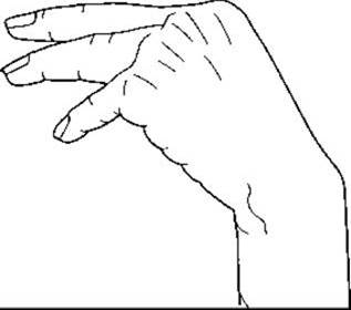
Fig. 21-14. Hand posture in hypocalcemic tetany (Trousseau's sign).
The bone disease caused by prolonged hyperparathyroidism is osteitis fibrosa cystica, characterized by Bone Marrow fibrosis and increased bone remodeling with resorption outpacing Bone Formation.
Secondary Hyperparathyroidism
In conditions such as Chronic Kidney Disease and Rickets, where plasma Ca2+ levels are chronically low, stimulation of the parathyroid glands leads to compensatory parathyroid hypertrophy and secondary hyperparathyroidism. Plasma Ca2+ levels are low in chronic kidney disease because diseased kidneys lose their ability to produce 1,25-dihydroxycholecalciferol. However, phosphate retention with resultant hyperphosphatemia also contributes to the lowering of plasma Ca2+.
Familial Hypercalcemia and Hypocalcemia
Mutations in the Ca2+-sensing receptor Gene lead to predictable, long-term alterations in plasma Ca2+ levels. Individuals heterozygous for an inactivating mutation have familial hypocalciuric hypercalcemia, a condition characterized by a chronic, mild elevation in plasma Ca2+ because the negative feedback inhibition of parathyroid hormone secretion by calcium is blunted. Plasma parathyroid hormone levels are normal to modestly elevated. In contrast, individuals homozygous for an inactivating mutation develop neonatal severe primary hyperparathyroidism. Conversely, individuals with activating mutations in the Ca2+-sensing receptor gene exhibit familial hypocalciuric hypocalcemia due to an increased sensitivity of the parathyroid glands to plasma Ca2+.
Parathyroid Hormone-Related Peptide
A protein with parathyroid hormone-like activity is produced by many different tissues throughout the body. It consists of 140 amino acid residues, compared to 84 in parathyroid hormone, and is encoded by a gene on human chromosome 12, whereas parathyroid hormone is encoded by a gene on chromosome 11. Despite this, parathyroid hormone and parathyroid hormone-related peptide share marked amino-terminal Homology. Specifically, 8 of the first 13 Amino Acids occupy identical positions, and both bind to the hPTH/PTHrP receptor. In developing Cartilage, PTHrP stimulates chondrocyte proliferation and inhibits their mineralization. It also appears to act as a growth factor in The Development of Skin, Hair follicles, and Mammary Glands. Mice with a knockout of both copies of the PTHrP gene die shortly after birth and exhibit severe skeletal abnormalities. A mutation rendering the hPTH/PTHrP receptor constitutively active is associated with a form of dwarfism.
In adults, PTHrP is produced in large quantities by the mammary glands and is found in high concentrations in milk. However, its plasma levels do not rise during Lactation, and the physiological function of PTHrP in milk remains unknown. PTHrP is also detected in the renal glomeruli and tubules, as well as in the brain, where it is localized to the Cerebral Cortex, hippocampus, and the granular layer of the cerebellar cortex.
Humoral Hypercalcemia of Malignancy
Hypercalcemia is a common metabolic complication of Cancer. Approximately 20% of hypercalcemic patients have bone metastases driven by osteolytic bone destruction (local osteolytic hypercalcemia). Evidence suggests this process is mediated by tumor-derived Prostaglandins, such as PGE. While a few lymphomas have been shown to secrete 1,25-dihydroxycholecalciferol, this is rare. In the remaining 80% of patients, hypercalcemia appears to result from elevated levels of circulating PTHrP (humoral hypercalcemia of malignancy). The tumors most commonly responsible for this hypersecretion include breast, renal, ovarian, and squamous cell carcinomas.
Last update: 10/08/2026
Editorial and Educational Adaptation: This material has been compiled based on the primary/original source text. The project team performed an editorial review, corrected technical inaccuracies, structured sections, and adapted the content for an educational format.
What was processed:
- elimination of formatting defects (OCR errors, structural breaks, corrupted characters);
- editorial organization of content;
- standardization of terminology in accordance with academic sources;
- verification of factual statements against the original source text.
All mentions of the author, publication year, and origin of the primary text have been preserved in accordance with the source.