Human Anatomy - Kotsan I. Ya. 2009
Endocrine glands
Parathyroid glands
The Parathyroid Glands (glandulae parathyroideae) are oval or round epithelial bodies located on the posterior surface of the right and left lobes of The Thyroid Gland (Fig. 225). The number of these glands is variable, ranging from 2 to 12, though in most cases there are 4 (two behind each lobe of the thyroid: one superior and one inferior). Each parathyroid gland measures 4–8 mm in length, 3–4 mm in width, and 2–3 mm in thickness. The combined weight of these glands in an adult is 0.13–0.36 g.
The parathyroid glands differ from the thyroid gland by a somewhat lighter coloration (pale pink in children and yellowish-brown in adults). Sometimes their color makes them easy to confuse with Lymph Nodes, adipose lobules, or accessory thyroid tissue. Microscopic examination is the only definitive way to confirm that a given Structure is indeed a parathyroid gland.
Most commonly, the parathyroid glands are situated near the entry points of the inferior thyroid Arteries or their branches into the thyroid tissue. Each parathyroid gland is encased in a Connective Tissue capsule, from which connective tissue septa extend into the interior of the gland. These septa contain a rich network of Blood Vessels and divide the glandular Cells into distinct cords or clusters.
While the precise function of the parathyroid glands is not yet fully understood, it is well established that they secrete parathyroid hormone (parathormone), which regulates Calcium and phosphorus METABOLISM in the blood and thereby influences the excitability of the nervous and muscular systems. Removal of these glands leads to heightened Central Nervous system excitability, Muscle spasms, and death from tetany.
The parathyroid glands develop from the epithelium of the third and fourth pharyngeal pouches. By the 7th week of development, the gland primordia separate from the walls of the pharyngeal pouches and migrate caudally as they grow. As they develop, they gradually assume their permanent positions on the posterior surface of the right and left lobes of the thyroid gland.
In a newborn, the total mass of the parathyroid glands does not exceed 10 mg; by one year of age, it reaches 20–30 mg, doubles by age 5, triples by age 10, and reaches its final adult size by age 20, remaining unchanged throughout the rest of adult life.
Class="center">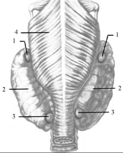
Fig. 225. Parathyroid glands (posterior view)
1 — superior parathyroid glands; 2 — thyroid gland; 3 — inferior parathyroid glands; 4 — Pharynx
Blood supply to the parathyroid glands is provided by Branches of the superior and inferior thyroid arteries, as well as from esophageal and tracheal branches. Venous blood drains via corresponding Veins.
The Innervation of the parathyroid glands is analogous to that of the thyroid gland.
Last update: 08/08/2026
Editorial and Educational Adaptation: This material has been compiled based on the primary/original source text. The project team performed an editorial review, corrected technical inaccuracies, structured sections, and adapted the content for an educational format.
What was processed:
- elimination of formatting defects (OCR errors, structural breaks, corrupted characters);
- editorial organization of content;
- standardization of terminology in accordance with academic sources;
- verification of factual statements against the original source text.
All mentions of the author, publication year, and origin of the primary text have been preserved in accordance with the source.