Human Anatomy - Kotsan I. Y. 2009
Endocrine glands
Thyroid gland
The Thyroid Gland (glandula thyroidea) is the largest endocrine gland in The Human Body. It is located in the neck region, anterior to the Trachea and along the lateral walls of the Larynx, partially adhering to the thyroid Cartilage, which gave the gland its name (Fig. 224).
The thyroid gland consists of right and left lobes (lobi dexter et sinister), which are connected inferiorly by a narrow isthmus. The ISTHMUS OF THE thyroid gland (isthmus glandulae thyroideae) is typically situated at the level of the II–III tracheal cartilages. In some cases, the isthmus lies at the level of the I tracheal cartilage or even the arch of the cricoid cartilage. Occasionally, the isthmus may be entirely absent, meaning the lobes are not connected at all. A pyramidal lobe (lobus pyramidalis) may extend upward from the isthmus or one of the lobes; this is a non-constant Structure found in approximately 30% of cases.
The upper pole of both the right and left thyroid lobes is located slightly below the upper margin of the corresponding plate of the thyroid cartilage of the larynx, while the lower pole reaches the level of the V–VI tracheal cartilages. The apex of the pyramidal lobe sometimes reaches the body of the Hyoid bone. The posterolateral surfaces of the right and left lobes are in contact with the laryngeal part of the Pharynx and the beginning of the Esophagus. The outer surface of the gland is convex, whereas the inner surface facing the trachea is concave. Anteriorly, the thyroid gland is covered by Skin, subcutaneous tissue, the Cervical fascia, and the Infrahyoid Muscles.
The transverse diameter of the adult thyroid gland reaches 50–60 mm. The longitudinal dimension of each lobe does not exceed 50 mm. The vertical height of the isthmus ranges from 5 to 15 mm, with a thickness of 6–8 mm. Maximum glandular mass (30–60 g) is observed in individuals aged 25–30 years. After 55–65 years, There is a slight decrease in both volume and mass. The mass and volume of the thyroid gland are generally greater in women than in men. In the fetus and early childhood, the thyroid gland is relatively larger than in adults.
Class="center">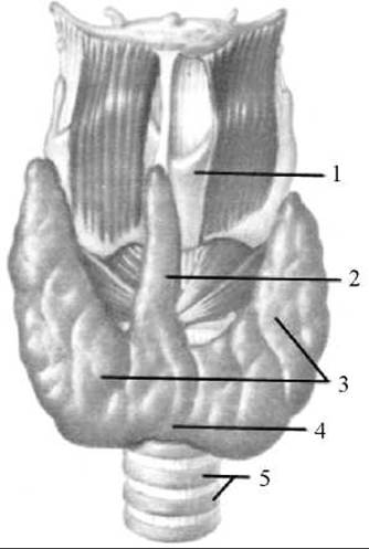
Fig. 224. Thyroid gland
1 — thyroid cartilage; 2 — pyramidal lobe; 3 — right and left lobes; 4 — isthmus of the thyroid gland; 5 — trachea
Externally, the thyroid gland is enclosed in a Connective Tissue fibrous capsule (capsula fibrosa) that fuses with the larynx and trachea, causing the thyroid gland to move during laryngeal movements. Connective tissue septa, or trabeculae, extend inward from the capsule to form the stromal framework (stroma) of the organ, dividing its parenchyma into lobules (lobuli). These lobules consist of follicles (folliculi). The walls of the follicles are lined internally with single-layered cuboidal follicular epithelial Cells. The follicular lumen contains a dense, viscous substance called colloid (colloidum), which stores THYROID Hormones. The glandular follicular epithelium of the thyroid exhibits a higher selective capacity for iodine accumulation than other Tissues. The concentration of iodine in thyroid tissue is 300 times greater than in Blood Plasma. Thyroid hormones (thyroxine and triiodothyronine), which are complex compounds of iodinated Amino Acids and Proteins, accumulate in the follicular colloid and are released into the bloodstream as needed, being delivered to Organs and tissues. Thyroxine and triiodothyronine regulate METABOLISM, increase heat production, enhance oxidative processes and the Breakdown of Proteins, fats, and CARBOHYDRATES, promote the Excretion of Water and potassium from the body, regulate GROWTH AND DEVELOPMENT, activate the adrenal, sex, and Mammary Glands, and stimulate the Central Nervous system. Another thyroid hormone, thyrocalcitonin, regulates Calcium and phosphorus metabolism, inhibits calcium resorption from bones, and lowers blood calcium levels.
The function of the thyroid gland is regulated by The Nervous System and the thyroid-stimulating hormone (TSH) secreted by the anterior Pituitary Gland.
The Importance of the thyroid gland to the Organism is profound. Congenital underdevelopment leads to cretinism (characterized by delayed growth, mental, and sexual development). In adults, thyroid hypofunction results in Myxedema (mucous edema). Hyperthyroidism leads to thyrotoxicosis, or Graves' disease, which is accompanied by enlargement of the thyroid gland (goiter), exophthalmos, tachycardia (accelerated Heart rate), elevated metabolism, increased irritability, and other symptoms.
The thyroid gland develops during early Embryogenesis from the foregut epithelium as an unpaired median diverticulum located between the 1st and 2nd visceral arches, just posterior to the Tongue anlage. Until the 4th week of embryonic development, this outgrowth contains a lumen, hence it is termed the thyroglossal duct (ductus thyreoglossus). By the end of the 4th week, the upper segment of the thyroglossal duct atrophies, while the lower segment divides into two processes—the future right and left lobes—which gradually shift caudally during gland formation to assume their definitive anatomical position. In adults, the Origin of the embryonic thyroglossal duct corresponds to the foramen cecum on the dorsum of the tongue. Ectopic patches of thyroid tissue in the form of accessory thyroid glands (glandulae thyroideae accessoriae) may occasionally be found along the course of this regressed duct in adults, most commonly at the ROOT of the tongue.
Blood supply to the thyroid gland is provided by the right and left superior thyroid Arteries (Branches of the external carotid arteries) and the right and left inferior thyroid arteries (branches of the thyrocervical trunks originating from the subclavian arteries).
Venous blood drains from the gland via the superior and middle thyroid Veins into the Internal jugular vein, and via the inferior thyroid vein into the brachiocephalic vein (or the lower part of the internal jugular vein). Lymphatic vessels form a rich plexus within the glandular tissue and transport hormone-rich Lymph to the thyroid, pretracheal, deep cervical, and mediastinal Lymph Nodes.
The thyroid gland is innervated by branches of the Vagus nerve and nerve fibers originating from the cervical Sympathetic trunk.
Last update: 08/08/2026
Editorial and Educational Adaptation: This material has been compiled based on the primary/original source text. The project team performed an editorial review, corrected technical inaccuracies, structured sections, and adapted the content for an educational format.
What was processed:
- elimination of formatting defects (OCR errors, structural breaks, corrupted characters);
- editorial organization of content;
- standardization of terminology in accordance with academic sources;
- verification of factual statements against the original source text.
All mentions of the author, publication year, and origin of the primary text have been preserved in accordance with the source.