Review of Medical Physiology - William F. Ganong 2002
Formation and Excretion of Urine
Renal Function and Urination
Excretion of Water
Normally, the glomeruli filter 180 L of fluid per day, while the average daily urine output is about 1 L. This same amount of solutes can be excreted in a daily urine volume of 500 mL at a concentration of 1400 mOsm/kg, or in a urine volume of 23.3 L at a concentration of 30 mOsm/kg (Table 38-7).
Class="center">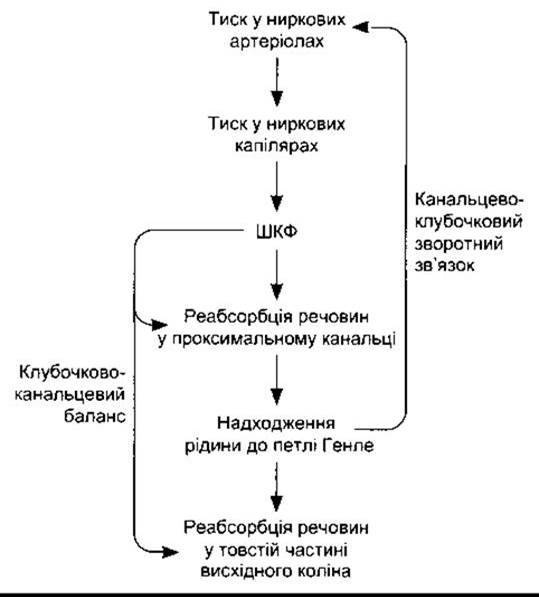
Fig. 38-14. Mechanisms of glomerulotubular balance and tubuloglomerular feedback.
Table 38-7. Effects of vasopressin on human Water METABOLISM. In all cases, the osmotic load of the excreted fluid is 700 mOsm/day
GFR, mL/min |
Water reabsorption percentage |
Urine output, L/day |
Urine concentration, mOsm/kg H2O |
Fluid loss or excess, L/day |
|
Isotonic plasma urine |
125 |
98.7 |
2.4 |
290 |
|
Vasopressin (maximum antidiuresis) |
125 |
99.7 |
0.5 |
1400 |
+1.9 |
Vasopressin absence (diabetes insipidus) |
125 |
87.1 |
23.3 |
30 |
-20.9 |
These figures highlight two crucial facts: first, at least 87% of water is reabsorbed even when daily urine output reaches 23 L; second, water reabsorption can vary widely without affecting solute excretion. Therefore, concentrated urine indicates water conservation by the body, whereas dilute urine signifies water loss. Both mechanisms are essential for the efficient utilization of water and the maintenance of osmotic Homeostasis in Body Fluids. Water balance is regulated primarily by vasopressin, which acts on the collecting ducts.
Proximal Tubule
Although numerous substances are transported in the proximal tubule, micropuncture studies show that the Osmotic Pressure of the fluid remains equal to that of Blood Plasma along its entire length (see Fig. 38-9). Consequently, water is reabsorbed passively in the proximal tubule along osmotic gradients established by The Active Transport of solutes, thereby maintaining fluid isotonicity. Water movement is facilitated by water channels in the apical membranes of proximal tubule epithelial Cells; HgCl2 reversibly inhibits these channels. In the proximal tubule, these channels consist of the aquaporin-1 protein. Four additional members of this protein family have been identified to date. They mediate water movement via simple diffusion. Aquaporin-2, which shares 42% Homology with aquaporin-1, is localized in the Cells of the collecting ducts. It acts as a mediator of these cells' response to vasopressin (see below and Chapter 14). Aquaporin-3 is located in the basolateral membrane of the collecting ducts and participates in The transport of urea, glycerol, and water. Aquaporin-4 is found in the Brain, whereas aquaporin-5 is present in the salivary and lacrimal glands, as well as the Respiratory system.
Although vasopressin does not affect aquaporin-1, this protein plays a vital role in water conservation. In knockout mouse models, proximal tubule water permeability drops by 80%, and the animals lose up to 35% of their body weight; upon dehydration, their plasma osmotic pressure rises to 500 mOsm/kg H2O, despite Aquaporins-2, -3, and -4 remaining intact.
The concentration of inulin (which is not reabsorbed) in the fluid at the end of the proximal tubule is 2.5 to 3.3 times higher than in blood plasma. This indicates that 60% to 70% of water and filtered solutes are reabsorbed during passage through the proximal tubule (Fig. 38-15).
As noted above, the loops of juxtamedullary nephrons penetrate deep into the medullary pyramids before ascending into the distal convoluted tubules in the cortex. All collecting ducts pass back through the renal pyramids, emptying through the papillary tips into the renal pelvis. The interstitial fluid of the pyramids exhibits a progressive increase in osmolality, reaching approximately 1200 mOsm/kg H2O at the papillary tips—nearly four times the plasma osmolality. The descending limb of the loop of Henle is permeable to water, whereas the ascending limb is not (Table 38-8). Na+, K+, and Cl- ions are reabsorbed from the thick ascending limb via cotransport (see below). Consequently, as water moves into the hypertonic medullary interstitium, the fluid within the descending limb of Henle's loop also becomes hypertonic. In the ascending limb, the fluid becomes progressively diluted, and by the time it reaches the apex, its osmotic pressure is lower than that of plasma due to the outward transport of Na+ and Cl- ions. Approximately another 15% of water is reabsorbed during passage through the loop of Henle, leaving only 20% of the initial water volume to enter the distal tubule, where the inulin concentration is already five times that of plasma.
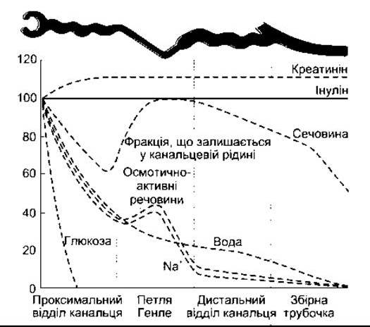
Fig. 38-15. Changes in the percentage of filtered substances remaining in the tubular fluid along the nephron (reprinted with permission from Sullivan LP, Grantham JJ: PHYSIOLOGY OF THE Kidney, 2nd ed. Lea&Febiger, 1982).
Table 38-8. Permeability and Transport in Various Nephron Segments1
Permeability |
Active Na+ transport |
|||
H2O |
Urea |
NaCl |
||
Loop of Henle |
||||
Thin descending limb |
4+ |
+ |
± |
0 |
Thin ascending limb |
0 |
+ |
4+ |
0 |
Thick ascending limb |
0 |
± |
± |
4+ |
|
Collecting duct |
± |
± |
± |
3+ |
Cortical portion |
3+* |
0 |
± |
2+ |
Outer medullary portion |
3+* |
0 |
± |
1 + |
Inner medullary portion |
3+* |
3+ |
± |
1 + |
1 Results obtained from rabbit and human kidney studies. Data marked with * were obtained under The Influence of vasopressin; +1 - in the absence of vasopressin (modified and reprinted with permission from Kokko JP: Renal concentrating and diluting mechanisms. Hosp Pract [Feb] 1979; 110:14).
The transport mechanism in the thick ascending limb of the loop of Henle relies on a carrier protein that transports one Na+ ion, one K+ ion, and two Cl- ions from the tubular lumen into The Cell (Fig. 38-16). In addition, Na+-K+-ATPase actively pumps Na+ out of the cells into the intercellular space; the concentration of this enzyme is highest in this segment of the renal tubule. Potassium (K+) moves back into the tubular lumen via simple diffusion. One Cl- ion diffuses passively into the intercellular space, while another moves together with K+.
The Na+-K+-2Cl- cotransporter in the thick ascending limb contains 12 transmembrane domains, with its amino and carboxyl terminals facing the cell interior. This protein belongs to a family of transporters found in many other Organs, including the Salivary Glands, gastrointestinal tract, and respiratory pathways.
Distal Tubule
The distal tubule, particularly its initial segment, is essentially a continuation of the thick ascending limb of the loop of Henle. It is relatively impermeable to water, and ongoing solute reabsorption without solvent results in further dilution of the tubular fluid. This segment accounts for only 5% of total water reabsorption.
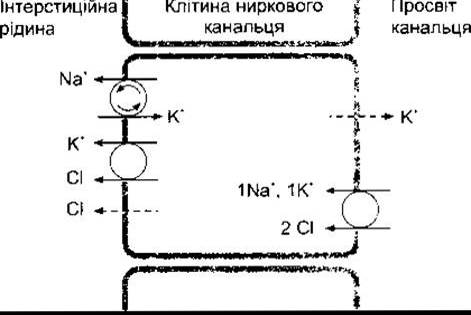
Fig. 38-16. Secondary active cotransport of Na+, K+, and Cl- in the cells of the thick ascending limb of the loop of Henle. Solid lines indicate active or secondary active transport, and dashed lines indicate diffusion. Active transport via Na+-K+-ATPase is denoted by arrows within the circle.
Collecting Ducts
The collecting ducts consist of two segments: cortical and medullary, through which the filtrate passes from the cortex into the renal pelvis. The osmotic pressure and volume of the fluid within the collecting ducts depend on the level of vasopressin acting upon them. This antidiuretic hormone, secreted by the posterior pituitary, increases the water permeability of the collecting ducts. This effect is achieved through the rapid appearance of aquaporin-2 water channels on the luminal membrane of principal cells (see Chapter 14). In the presence of vasopressin levels sufficient for maximum antidiuresis, water leaves the hypotonic fluid and enters the cortical interstitium via the cortical collecting ducts, rendering the tubular fluid isotonic. This mechanism accounts for the reabsorption of up to 10% of the filtered water. This isotonic fluid then enters the medullary collecting ducts, where its inulin concentration reaches 20 times that of plasma. An additional 4.7% of the filtrate is reabsorbed in the hypertonic medullary interstitium, producing concentrated urine in which the inulin concentration is up to 300 times that of plasma. Human urine osmotic pressure can reach 1400 mOsm/kg H2O—nearly five times that of blood plasma—resulting in the reabsorption of 99.7% of the filtered water, with only 0.3% excreted in the urine (see Table 38-7). Other species exhibit even greater urine-concentrating abilities. Maximum urine osmotic pressure is approximately 2500 mOsm/kg in dogs, 3200 mOsm/kg in laboratory rats, and up to 5000 mOsm/kg in certain desert reptiles.
In the absence of vasopressin, the epithelium of the collecting ducts is relatively impermeable to water. Consequently, the fluid remains hypotonic, and large quantities of it flow into the renal pelvis. In humans, urine osmolality can be as low as 30 mOsm/kg H2O. The impermeability of the distal nephron segments is not absolute; even without vasopressin, about 2% of the filtered fluid is excreted along with electrolytes. In any case, up to 13% of the filtered fluid is excreted, and urine flow can reach over 15 mL/min (21.6 L/day) under these conditions. Data on the effects of the presence and absence of vasopressin on water metabolism are presented in Table 38-7.
The causes of diabetes insipidus—a condition caused by vasopressin deficiency or insufficient target-organ sensitivity to this hormone—are described in Chapter 14. For example, in nephrogenic diabetes insipidus, the collecting ducts fail to respond to vasopressin. Two forms of this disorder have been described. The first is caused by a mutation in the V2 receptor Gene, rendering it unresponsive. The V2 receptor gene is located on the X chromosome, and the inheritance pattern is X-linked recessive. The second form is caused by a mutation in the autosomal aquaporin-2 gene.
Countercurrent Mechanism
The effective operation of the concentration mechanism depends on maintaining an increasing osmotic gradient along the renal medullary pyramids. This gradient is established by the loops of Henle, which act as a countercurrent multiplier, and the vasa recta, which function as countercurrent exchangers.
A countercurrent system is one in which the input flow runs parallel to and in close proximity to the output flow, but in the opposite direction.
This process takes place in both the loops of Henle and the vasa recta within the renal medulla (see Fig. 38-2).
The function of each loop of Henle as a countercurrent multiplier depends on the active transport of Na+ and Cl- ions out of the thick ascending limb (see above), the high water permeability of the thin descending limb (see Table 38-8), the influx of fluid from the proximal tubule, and its outflow into the distal tubule. This process is best understood through a hypothetical step-by-step model for achieving equilibrium. In vivo, however, these discrete steps do not occur sequentially; rather, a steady-state equilibrium is maintained at all times (unless the osmotic gradient is "washed out"). These steps are illustrated in Fig. 38-17 for a cortical nephron model lacking the thin ascending limb. First, let us imagine a situation where the osmotic pressure along the descending and ascending limbs, as well as in the interstitium, is 300 mOsm/kg H2O (see Fig. 38-17A). Now assume that pumps in the thick ascending limb can transport 100 mOsm/kg of Na+ and Cl- from the tubular fluid into the interstitium, increasing the interstitial osmotic pressure to 400 mOsm/kg H2O. In response, water moves out of the thin descending limb into the interstitium, equilibrating the osmotic pressure between the interstitium and the fluid in the thin descending limb (see Fig. 38-17B). However, fluid with an osmotic pressure of 300 mOsm/kg H2O continuously enters the thin descending limb from the proximal tubule (see Fig. 38-17C); consequently, the gradient against which Na+ and Cl- are pumped is reduced, and more of these ions are transported into the interstitium (see Fig. 38-17). At the same time, hypotonic fluid enters the distal tubule, while isotonic and subsequently hypertonic fluid enters the thick ascending limb. This process is repeated over and over, ultimately resulting in the generation of an osmotic gradient between the top and the bottom of the loop.
In juxtamedullary nephrons, which have long loops and long thin ascending limbs, the osmotic gradient extends over a greater distance, and the osmotic pressure reaches its maximum at the tip of the loop. This occurs because the thin ascending limb is relatively impermeable to water but permeable to Na+ and Cl- ions. As a result, Na+ and Cl- move down their concentration gradients into the interstitium, providing additional passive countercurrent multiplication. The longer the loop of Henle, the higher the osmotic pressure generated at the apex of the pyramid.
The osmotic gradient in the medullary pyramids would not persist for long if the Na+ and urea present in the interstitium were washed away into the bloodstream. These substances remain in the pyramids because the vasa recta function as countercurrent exchangers (Fig. 38-18). Solutes diffuse from the Blood Vessels ascending toward the cortex into the vessels descending into the pyramids. Conversely, water diffuses from the descending vessels into the fenestrated ascending vessels. Thus, electrolytes and other solutes tend to return to the cortical Circulation while water bypasses it, thereby maintaining the elevated osmotic pressure. Water exiting the collecting ducts is also picked up by the vasa recta and returned to the general circulation. Countercurrent exchange is a passive process driven by water movement; it would be unable to maintain the medullary osmotic gradient if the countercurrent multiplication process in the loops of Henle were to cease.
It is worth noting that a very steep osmotic gradient is established across the loop of Henle and the collecting ducts (in the presence of vasopressin). The countercurrent system makes this possible by distributing the gradient along a total tubular length of over 1 cm, rather than dropping it across a single cell layer only a few micrometers thick. Other Examples of countercurrent systems exist in animals. One of these is heat exchange between the arterial and venous Blood vessels of the extremities. This process is minimally present in humans but is most pronounced in marine mammals living in cold water. Heat is transferred from the arterial blood flowing toward the limbs to the venous blood returning to the trunk, keeping the distal PARTS OF THE extremities cool while conserving body heat overall.
Role of Urea
Urea also plays a major role in establishing the osmotic gradient in the medullary pyramids and in concentrating urine within the collecting ducts. It is transported by specific transporters, most likely via Facilitated Diffusion. There are at least four isoforms of this transport protein in the Kidneys: UT-A (UT-A1 through UT-A4), and UT-B, which is found in erythrocytes. Urea leaves the proximal tubule, but with the exception of the inner medullary collecting duct, the rest of the tubular epithelium is impermeable to it. Consequently, as water is removed along the loop of Henle and the distal tubule, the concentration of urea in the tubular fluid rises. However, at the level of the inner medullary collecting duct, urea diffuses into the medullary interstitium, raising its osmotic pressure. In this segment of the collecting duct, urea is transported by the UT-A1 transporter, the function of which is regulated by vasopressin. Another vasopressin-regulated urea transporter with 10 transmembrane domains has been isolated from rabbit kidney cells. When vasopressin is absent and the osmotic pressure of the tubular fluid in the inner medullary collecting duct falls, urea diffuses from the interstitium back into the tubular lumen, reducing the osmotic gradient. The amount of urea in the medullary interstitium and, consequently, in the urine depends on its delivery, which in turn is related to Dietary Protein Intake. Therefore, a high-protein diet enhances the urine-concentrating ability of the kidneys.
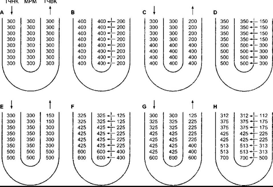
Fig. 38-17. Mechanism of the loop of Henle acting as a countercurrent multiplier that establishes an osmotic gradient in the renal medullary interstitium (RMI); TDLH, thin descending limb of the loop of Henle; TALH, thick ascending limb of the loop of Henle. The gradient-generating process is shown as a hypothetical sequence of steps, beginning with A, where the osmotic pressure in both limbs and the interstitium is 300 mOsm/kg. Pumps located in the thick ascending limb transport Na+ and Cl- into the interstitium, increasing the osmotic pressure to 400 mOsm/kg. Osmotic equilibrium is thus achieved with the fluid in the thin descending limb. Simultaneously, isotonic fluid continues to enter the thin descending limb while hypotonic fluid leaves the thick ascending limb. Through the cyclical repetition of this process, the fluid exiting the thick ascending limb becomes progressively more hypotonic, whereas fluid of extremely high osmotic pressure accumulates at the tip of the loop of Henle (modified and reproduced with permission from Johnson LR, Essential Medical Physiology, Raven Press, 1992).
Water Diuresis
The feedback mechanism regulating vasopressin secretion in response to plasma osmolality is discussed in Chapter 14. Following the ingestion of a large volume of hypotonic fluid, water excretion begins within 15 min and peaks within 40 min. The act of swallowing fluids alone causes a slight decrease in vasopressin secretion, but the primary stimulus for its reduction is the decrease in plasma osmolality resulting from water absorption.
Water Intoxication
The maximum urine excretion rate is 16 mL/min. If fluid absorption exceeds this rate, water shifts from the hypotonic extracellular fluid into cells, causing cellular Swelling and, in rare cases, clinically overt water intoxication. Brain cell swelling leads to seizures and coma, ultimately resulting in death. Water intoxication can occur if fluid intake is not appropriately restricted following the administration of vasopressin, or as a result of elevated vasopressin secretion triggered by non-osmotic stimuli, such as surgical trauma.
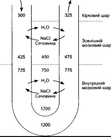
Fig. 38-18. Vasa recta and countercurrent multiplier in the kidney; NaCl and urea diffuse from the ascending limb into the descending limb, whereas water diffuses from the descending limb into the ascending limb.
Osmotic Diuresis
The presence of a large quantity of unabsorbed solutes in the renal tubules leads to an increase in urine volume known as osmotic diuresis. Substances that are not reabsorbed in the proximal tubules exert a significant osmotic effect because their concentration rises as fluid volume decreases. Consequently, they "hold water" in the tubules. It should also be noted that Na+ ions cannot be transported out of the tubule indefinitely; There is a limit to the concentration gradient they can establish. Under normal conditions, water reabsorption in the proximal tubule prevents any marked changes in concentration gradients, but when The rate of water reabsorption declines, the Na+ concentration also falls. This occurs due to the presence of various unabsorbed solutes in the tubular fluid. Once the limiting concentration gradient is reached, further Na+ reabsorption ceases, leaving a larger quantity of Na+ and correspondingly more water in the tubule. As a result of these processes, the volume of isotonic fluid entering the loop of Henle increases. This fluid has a lower Na+ concentration than the original fluid, but the total load of Na+ delivered to the loop of Henle per unit time is increased. In the loop of Henle, water and Na+ reabsorption is impaired because the medullary osmotic pressure is reduced. This reduction is primarily caused by decreased reabsorption of Na+, K+, and Cl- in the thick ascending limb of the loop of Henle, as the limiting Na+ concentration gradient has already been reached. A larger volume of fluid passes through the distal nephron. Because of the diminished osmotic gradient along the medullary pyramids, less water is reabsorbed in the collecting ducts. Consequently, urine volume and The excretion of Na+ and other electrolytes increase markedly.
Osmotic diuresis is caused by substances such as mannitol (an osmotic diuretic drug) and other filterable, non-reabsorbable Polysaccharides. This phenomenon can also be triggered by certain endogenous compounds if they are delivered in amounts exceeding the reabsorptive capacity of the tubules. For example, in Diabetes Mellitus, glucose retained in the tubules causes polyuria when its filtered load exceeds TmG. Osmotic diuresis can also be induced by the intravenous infusion of large amounts of NaCl or urea.
It is important to distinguish between osmotic and water diuresis. In water diuresis, the volume of fluid reabsorbed in the proximal nephron is normal, and the maximum urine flow rate is about 16 mL/min. In osmotic diuresis, elevated urine output results from impaired water reabsorption in the proximal tubule and loop of Henle; under these conditions, very large volumes of urine may be excreted. Despite maximal vasopressin secretion, the urine osmolality approaches that of plasma (Fig. 38-19) because an increasingly larger fraction of the urine consists of isotonic proximal tubular fluid. Conversely, if osmotic diuresis is induced in an experimental animal with diabetes insipidus, the urine osmolality actually increases for the same underlying reason.
Relationship Between Urine Concentration and Glomerular Filtration Rate
The magnitude of the osmotic gradient along the medullary pyramids increases when the rate of fluid flow through the loops of Henle decreases. A reduction in GFR, resulting, for example, from dehydration, leads to a decrease in the volume of fluid entering the countercurrent multiplier system, thereby slowing fluid flow through the loops of Henle and producing more concentrated urine. If GFR drops significantly, the urine becomes quite concentrated even in the absence of vasopressin. Furthermore, if one renal artery is partially constricted in an experimental animal with diabetes insipidus, the urine excreted by that kidney will be hypertonic due to the reduced GFR, whereas the urine from the contralateral side will be hypotonic.
Free-water clearance
To evaluate fluid loss or conservation resulting from the excretion of concentrated or diluted urine, clinicians frequently calculate free-water clearance (CН2О). This represents the difference between the urine flow rate and the clearance of osmotically active solutes (Cосм):
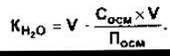
where V is the urine flow rate; Сосм and Посм are the osmolalities of urine and plasma, respectively. Thus, Косм is the volume of water required to excrete the solute load in urine that is isosmotic with plasma. The value of KH2О is negative when the urine is hypertonic and positive when it is hypotonic. For instance, Table 38-8 provides the following data: during maximal antidiuresis, the KН2О value is -1.3 mL/min (-1.9 L/day), whereas in the absence of vasopressin it reaches 14.5 mL/min (20.9 L/day).
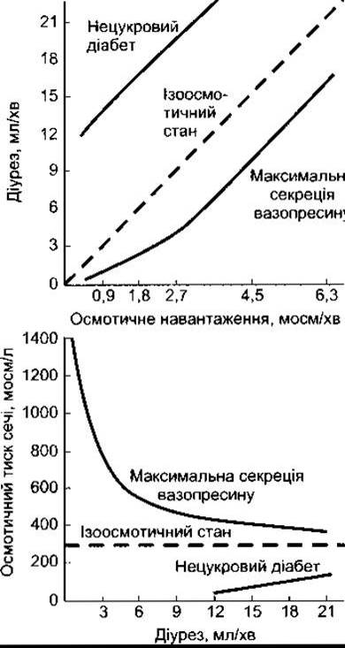
Fig. 38-19. Approximate relationship between urine concentration and urine flow rate during osmotic diuresis in humans. The dashed line in the lower graph indicates the concentration at which the osmotic pressures of urine and blood plasma are equal (reproduced with permission from Berliner RW, Giebisch G: Best and Taylor’s Physiological Basis of Medical Practice, 9th ed. Williams & Wilkins, 1979).
Last update: 10/08/2026
Editorial and Educational Adaptation: This material has been compiled based on the primary/original source text. The project team performed an editorial review, corrected technical inaccuracies, structured sections, and adapted the content for an educational format.
What was processed:
- elimination of formatting defects (OCR errors, structural breaks, corrupted characters);
- editorial organization of content;
- standardization of terminology in accordance with academic sources;
- verification of factual statements against the original source text.
All mentions of the author, publication year, and origin of the primary text have been preserved in accordance with the source.