Biological Membranes - A. N. Ogurtsov 2012
Electrogenesis of Biomembranes
Secondary Transport by Cotransporters
Aquaporins
A pure lipid bilayer does not provide a physiologically sufficient rate of Water diffusion across the membrane. Most Cell membranes contain specialized protein channels, aquaporins (Figure 106), which facilitate the required rate of water molecule diffusion. For example, such osmosis-driven diffusion through the epithelium of the renal tubules leads to urine concentration. Otherwise, The Human Body would produce several times the volume of urine, which would result in severe dehydration.
Class="center">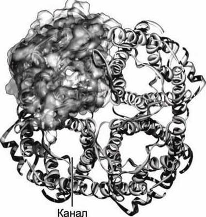
Figure 106 - Three-dimensional model of an aquaporin tetramer
In higher plants, water and dissolved mineral salts are absorbed by the roots and transport to the leaves through xylem vessels. Water evaporation from the leaf surface is the driving force behind this movement in plants.
Osmosis-driven Movement of water across The Plasma Membrane is the primary factor determining individual cell volume. Different Cells employ different mechanisms to control cell volume.
When increased membrane permeability to water molecules is required, cells express Membrane Proteins called aquaporins. Aquaporin is a tetramer of identical 28 kDa subunits (Figure 106).
Each subunit contains six transmembrane α-helices and two short α-Helix segments (Figure 107).
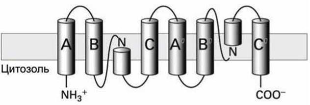
Figure 107 - Schematic of aquaporin subunit components
Three pairs of transmembrane helices (A and A', B and B', C and C'), oriented in opposite directions relative to the membrane, form a pore in the membrane through which water molecules can diffuse.
Two short loops containing asparagines (N) in the functional subunit meet in the center of the pore and, together with arginines and histidines, form a selectivity filter that allows only water molecules to pass. At the center of this filter, the pore diameter is only 0.28 nm, which is slightly larger than the diameter of a water molecule. Several water molecules move simultaneously through the channel in a single-file chain, forming Hydrogen Bonds with The amino acid residues lining the channel and pushing each other along (Figure 108).
The formation of hydrogen bonds between water molecules and amino acid side chains ensures that only water molecules can pass through the channel. Even a proton (i.e., H3O+) or other ions cannot diffuse through the aquaporin.
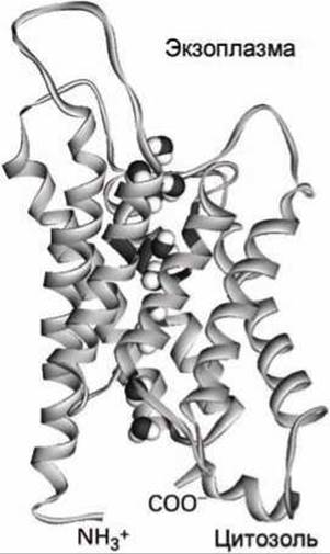
Figure 108 - Model of an aquaporin subunit and schematic of water molecule movement through the subunit channel
Just as in the case of glucose transporters (see Section 6.5), mammals express an entire family of homologous aquaporin proteins.
Aquaporin 1 is expressed in erythrocytes, and aquaporin 2 is expressed in the epithelial Cells of the renal tubules.
Other members of this family transport hydroxyl-containing molecules, such as glycerol, instead of water.
Last update: 13/08/2026
Editorial and Educational Adaptation: This material has been compiled based on the primary/original source text. The project team performed an editorial review, corrected technical inaccuracies, structured sections, and adapted the content for an educational format.
What was processed:
- elimination of formatting defects (OCR errors, structural breaks, corrupted characters);
- editorial organization of content;
- standardization of terminology in accordance with academic sources;
- verification of factual statements against the original source text.
All mentions of the author, publication year, and origin of the primary text have been preserved in accordance with the source.