Review of Medical Physiology - William F. Ganong 2002
Urine Formation and Excretion
Renal Function and Micturition
Urinary Acidification and Bicarbonate Excretion
H+ Secretion
Cells of the proximal and distal renal tubules, much like the cells of gastric glands, secrete H+ ions (see Chapter 26). Acidification also takes place in the collecting ducts. The primary mechanism for H+ secretion in the proximal tubules is Na+-H+ exchange (Fig. 38-20). This is an example of secondary Active Transport: the Na+-K+-ATPase pumps Na+ ions out of the cells into the interstitial fluid, which lowers the intracellular Na+ concentration. As a result, Na+ begins to enter the cells from the tubular fluid, driving the counter-transport of a single H+ ion. The H+ ions are generated by intracellular dissociation of H2CO3, and the resulting HCO3- diffuses into the interstitial fluid. Thus, for every H+ ion secreted, one Na+ ion and one HCO3- ion enter the interstitial fluid.
Class="center">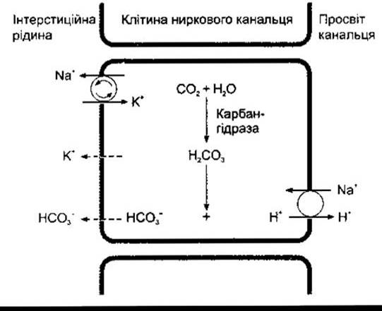
Fig. 38-20. Acid secretion by proximal renal tubule cells; H+ is transferred into the tubular lumen via an antiporter in exchange for a Na+ ion. Active transport by the Na+-K+-ATPase is indicated by circled arrows. Diffusion is represented by dashed lines. Compare with Fig. 26-10.
The reaction forming H2CO3 is catalyzed by the enzyme Carbonic anhydrase. Pharmacological agents that inhibit this enzyme simultaneously suppress H+ secretion in the proximal renal tubules along with all associated biochemical reactions.
It is believed that other pumping systems may also be involved in H+ secretion within the proximal tubule, though their existence has not been definitively proven. In any case, they play a minor role compared to the Na+-H+ exchange mechanism. In the distal tubule and collecting ducts, the situation is quite the opposite, as H+ secretion there is practically independent of the Na+ concentration in the tubular fluid. In these segments, the majority of H+ is secreted by an ATP-dependent proton pump. Aldosterone acts on this pump to enhance H+ secretion in the distal segment. Intercalated cells (I-cells) in this region of the renal tubule secrete H+ and, similar to gastric parietal cells, contain substantial amounts of carbonic anhydrase and numerous tubulovesicular structures. Literature data indicate that these vesicles and the inner membrane of tubular epithelial cells contain the so-called H+-translocating ATPase, which is actually responsible for producing H+. During acidosis, the number of proton pumps increases due to the insertion of these tubulovesicles into the luminal Cell membrane. A small amount of H+ is also secreted by H+-K+-ATPase. The basolateral membrane of I-cells contains the so-called Band 3 anion exchanger protein, which presumably Functions as a Cl--HCO3- exchanger in The transport of HCO3- into the interstitial fluid.
H+ METABOLISM in Urine
The rate of acid secretion depends on the processes described below. The Kidneys of every biological species are characterized by a maximum H+ concentration gradient against which transport mechanisms are still capable of active transport. In humans, this gradient corresponds to a urine pH of about 4.5, which means an H+ ion concentration 1,000 times greater than that in Blood Plasma. Therefore, pH 4.5 is the limiting value for urine pH. Normally, this is precisely the pH of the fluid in the collecting ducts. Were it not for buffer systems that "bind" H+ in the urine, this limiting pH would be reached very quickly, and H+ secretion would cease. However, three important Chemical Reactions allow excess free H+ ions to be readily removed from the tubular fluid, thereby sustaining further acid secretion (Fig. 38-21). These are reactions with HCO3- to form CO2 and H2O, with HPO42- to form H2PO4-, and with NH3 to form NH4+.
Reactions with Buffer Systems
The mechanics of buffer systems are discussed in Chapters 1 and 39. The pK' value is 6.1 for the bicarbonate system, 6.8 for the phosphate system, and 9.0 for the ammonia system. The concentration of HCO3- in plasma, and consequently in the glomerular filtrate, is normally 24 mEq/L, whereas the phosphate concentration is only 1.5 mEq/L. Therefore, in the proximal tubule, the bulk of H+ reacts with HCO3- to form H2CO3 (see Fig. 38-21). Subsequently, H2CO3 breaks down into CO2 and H2O. The brush border of proximal (but not distal) tubule cells contains carbonic anhydrase, which facilitates The formation of CO2 and H2O in the tubular fluid. At the same time, CO2, which easily diffuses across all Introduction/36.html">Biological Membranes, enters the tubular cells, replenishing the CO2 pool utilized for the synthesis of H2CO3. Because the vast majority of H+ is removed from the tubule, the pH of the tubular fluid does not change appreciably. This very process serves as the mechanism by which HCO3- is reabsorbed: for every mole of HCO3- removed from the tubular fluid, 1 mole of HCO3- diffuses from the tubular cells into the blood, although this is not the exact same mole that left the tubular fluid.
The secreted H+ interacts with divalent hydrogen phosphate (HPO42-), yielding monovalent dihydrogen phosphate (H2PO4-). This process is most prominent in the distal tubule and collecting ducts because Water reabsorption concentrates the phosphate ions that were not reabsorbed in the proximal tubule. The reaction with NH3 occurs in both the distal and proximal tubules; H+ ions also bind with other buffer anions, though to a much lesser extent.
Every H+ ion that reacts with buffer systems contributes to the so-called titratable acidity of urine, which is defined as The amount of alkali that must be added to urine to bring its pH to 7.4, corresponding to the pH of the glomerular filtrate. However, it is evident that titratable acidity reflects only a fraction of the secreted H+, since it does not account for the H2CO3 converted into H2O and CO2. Furthermore, the pK' of the ammonium system is 9.0, whereas it is only titrated from urine pH down to 7.4; hence, its contribution to the titratable acidity of urine is very negligible.
Ammonia Secretion
Chemical reactions within the renal tubular cells generate NH4+ and HCO3-. The NH4+ ion exists in equilibrium with the NH3 + H+ system inside the cells. Since the pK' of this reaction is 9.0, The ratio of NH3 to NH4+ at pH 7.0 is 1:100 (Fig. 38-22). Moreover, NH3 is a lipid-soluble compound and therefore readily crosses cell membranes down concentration gradients into the interstitial fluid and tubular filtrate. In the urine, it reacts with H+ to form NH4+, which remains trapped in the urine.
The primary reaction responsible for generating intracellular NH4+ is The conversion of glutamine to glutamate. This reaction is catalyzed by the enzyme glutaminase, which is abundant in renal tubular cells (see Fig. 38-22). Glutamate dehydrogenase catalyzes the conversion of glutamate to alpha-ketoglutarate, producing additional NH4+. The subsequent metabolism of alpha-ketoglutarate consumes 2 H+, thereby releasing 2 HCO3-.
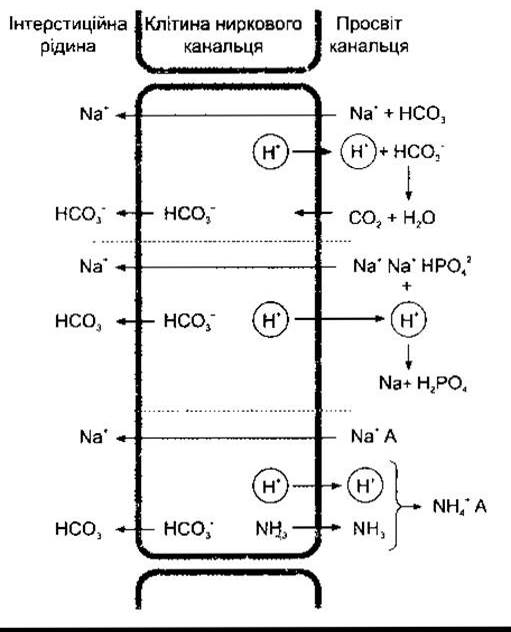
Fig. 38-21. Metabolism of H+ secreted into the tubular fluid in exchange for Na+. Top: Bicarbonate reabsorption via CO2. Center: Formation of monovalent phosphate. Bottom: Ammonia Production. Note that for every secreted H+ ion, one Na+ and one HCO3- are released; A- represents an anion.
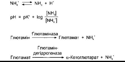
Fig. 38-22. Main reactions of ammonia production in the kidneys. See also Chapter 17.
During chronic acidosis, NH4+ excretion is enhanced. This is observed at any given pH value because a larger amount of NH3 enters the tubular fluid. This phenomenon is termed ammonia secretion adaptation, the exact cause of which is not yet fully understood. Ammonia secretion adaptation leads to an increase in H+ secretion.
The process whereby NH3 is secreted into the urine and subsequently converted into NH4+, thereby maintaining the NH3 concentration gradient, is called nonionic diffusion (see Chapter 1). The excretion of salicylates and certain other medications that are weak bases or weak acids also occurs via nonionic diffusion. Their excretion rate depends heavily on urine pH.
pH Changes Along the Nephron
In the proximal tubule, There is a slight decrease in pH; however, as noted above, H+ excretion does not significantly alter the pH of the tubular fluid due to the conversion of H2CO3 into CO2 and H2O. In contrast, the cells of the distal tubule have a much lower capacity for H+ secretion, yet secretion in this specific segment exerts the greatest influence on final urine pH.
Factors Affecting H+ Secretion
Acid secretion into the urine is modulated by intracellular PCO2, K+ concentration, carbonic anhydrase levels, and Adrenal Cortex Hormones. When PCO2 is high (respiratory acidosis), more H2CO3 is available to buffer hydroxyl ions, and acid secretion is augmented; conversely, low PCO2 produces the opposite effect. Potassium deficiency leads to increased acid excretion, presumably because K+ loss induces intracellular acidosis even when plasma pH is elevated. Conversely, intracellular K+ excess suppresses acid secretion. During carbonic anhydrase inhibition, acid secretion is also diminished due to reduced H2CO3 formation. Aldosterone, along with other Adrenocortical Hormones that promote tubular Na+ reabsorption, concurrently enhances H+ and K+ secretion.
Bicarbonate Excretion
Although the reabsorption of HCO3 does not involve the direct transport of this ion into the tubular cells, HCO3 reabsorption remains proportional to its filtration. There is no well-defined Tm; however, HCO3 reabsorption is inhibited by an unknown mechanism when the ECF volume increases (Fig. 38-23). When plasma HCO3 concentration is low, HCO3 is reabsorbed completely. However, when the concentration of HCO3 exceeds 26–28 mEq/L (the renal threshold for HCO3-), HCO3 appears in the urine, making the urine more alkaline. Conversely, when the plasma HCO3 concentration is below 26 mEq/L (the level at which all secreted H+ ions are utilized for HCO3- reabsorption), more H+ is released to bind with anions of other buffer systems. Consequently, the lower the plasma HCO3 concentration, the more acidic the urine reaction becomes and the higher its NH4+ content.
Physiological Significance of Urine pH Variations
Depending on the closely interrelated processes of H+ secretion, NH4+ production, and HCO3 excretion, human urine pH ranges widely from 4.5 to 8.0. The excretion of urine with a pH significantly different from that of Body Fluids is crucial for maintaining electrolyte and acid-base balance, as discussed in detail in Chapter 39. Acids are buffered in plasma and cells According to the chemical reaction: HA + NaHCO3 —> NaA + H2CO3. Subsequently, H2CO3 dissociates into CO2 and H2O; CO2 is absorbed into the blood and eliminated via exhalation, while NaA appears in the glomerular filtrate. The amount of Na+ replaced by H+ in the urine is returned to the body. For each H+ ion excreted with phosphate or as NH4+, the body gains one HCO3- ion, thereby replenishing the reserves of this essential buffer anion. On the other hand, when alkali enters body fluids, OH- ions bind to form additional HCO3-. If the plasma HCO3 concentration exceeds 28 mEq/L, the urine becomes alkaline, and the excess HCO3 is excreted. Because the maximum rate of tubular H+ secretion is directly dependent on arterial PCO2, HCO3 reabsorption is strongly influenced by PCO2. These relationships are described in greater detail in Chapter 39.
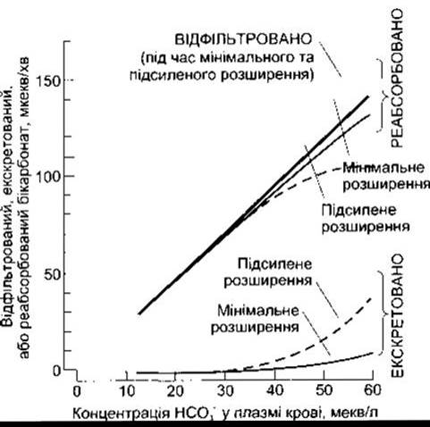
Fig. 38-23. Effect of ECF volume on filtration, reabsorption, and excretion of HCO3- in experiments on rats. In humans, HCO3- excretion follows a similar pattern. Under normal conditions, plasma HCO3- concentration is approximately 24 mEq/L (reprinted with permission from Valtin H: Renal Function, 2nd ed. Little, Brown and Co., 1983)
Last update: 10/08/2026
Editorial and Educational Adaptation: This material has been compiled based on the primary/original source text. The project team performed an editorial review, corrected technical inaccuracies, structured sections, and adapted the content for an educational format.
What was processed:
- elimination of formatting defects (OCR errors, structural breaks, corrupted characters);
- editorial organization of content;
- standardization of terminology in accordance with academic sources;
- verification of factual statements against the original source text.
All mentions of the author, publication year, and origin of the primary text have been preserved in accordance with the source.