Human Biochemistry, Volume 2 - Murray R. 1993
Biochemistry of Intracellular and Intercellular Communication
Adrenal Cortex Hormones
Classification and Mechanism of Action of Steroid Hormones
Glucocorticoid Hormones
A. Classes of glucocorticoid hormones. The initial stage of glucocorticoid hormone action is the interaction with a specific receptor. As a result of this interaction, the receptor is "activated," which is presumed to be necessary for DNA binding. In general, There is a high degree of correlation between steroid binding to the receptor and the magnitude of a specific biological response. This correlation holds true across a wide range of activities; thus, if one steroid has a 10-fold lower affinity for the receptor compared to another, its biological effect will be proportionally lower (at equivalent concentrations). "Spare receptors" do not play a role in the action of Steroid Hormones.
The biological effect of a steroid is determined by both its ability to bind to the receptor and the concentration of the free hormone in the Blood. Consider, for example, cortisol, corticosterone, and aldosterone; all three hormones exhibit high affinity for the glucocorticoid receptor, yet under physiological conditions, the action of cortisol predominates because it is present in the Blood Plasma at a relatively high concentration. Corticosterone may play an important role under certain pathological conditions (17a-hydroxylase deficiency), but aldosterone never reaches such concentrations in blood plasma for its glucocorticoid effect to be manifested.
Comparing various Steroids by their ability to mediate a well-known glucocorticoid effect—the Induction of the enzyme Tyrosine aminotransferase in the Liver—reveals that these hormones can be divided into four classes: agonists, partial agonists, antagonists, and inactive steroids (Table 48.4).
B. The glucocorticoid receptor. A series of biochemical, immunological, and genetic studies has helped shape our understanding of the glucocorticoid receptor (Fig. 48.7). Its N-terminal half contains most of the antigenic regions as well as a domain that modulates promoter function. The C-terminal region contains the DNA-binding and hormone-binding sites. The DNA-binding domain is located closer to the middle of the molecule, whereas the hormone-binding domain is closer to the C-terminus. Certain regions in the C-terminal half of the receptor are homologous to the v-erb-A oncogene protein, as well as to the DNA-binding region in TFIIIA and the corresponding region in estrogen and progesterone receptors. The Amino Acid Sequence of the receptor was established through cDNA analysis. This analysis revealed two regions rich in Cys-Lys-Arg residues within the DNA-binding domain. Comparison of these regions with other known DNA-binding Proteins, such as TFIIIA, indicated that a zinc-finger-like fold can be formed here; it is hypothesized that this "finger" Structure inserts into the DNA groove. The human glucocorticoid receptor exists in two forms, a and ß, consisting of 777 and 742 amino acid residues, respectively.
Class="center">Table 48.4. Classification of steroids by their glucocorticoid effect
|
Agonists Dexamethasone Cortisol Corticosterone Aldosterone Partial agonists 11 ß-Hydroxyprogesterone 21 -Deoxycortisol 17a-Hydroxyprogesterone Progesterone Antagonists Testosterone 17ß-Estradiol 19-Nortestosterone Cortisone Inactive steroids 11a-Hydroxyprogesterone Androstenedione 11a, 17a-Methyltestosterone Tetrahydrocortisol |
C. General characteristics of the MECHANISM OF ACTION.
The schema of The Mechanism of action of glucocorticoid hormones is described in Chapter 44 and illustrated in Fig. 44.1. Numerous Examples support the concept that these hormones affect specific intracellular processes by altering the cellular content of critically important proteins, typically Enzymes. This is governed by the ability of glucocorticoids to regulate the METABOLISM/31.html">Transcription rates of specific genes in target Cells. This process requires the steroid-receptor complex to bind to specific DNA regions near the Transcription initiation site, after which these regions determine the Specificity of the response. Exactly how this binding stimulates or inhibits transcription, how tissue specificity is ensured, and why the same Gene can be activated in one tissue and inhibited in another—these and many other fundamental questions remain open.
An illustration of current views on the mechanism of action of steroid hormones is provided by a brief description of how glucocorticoids affect the transcription of mouse mammary tumor virus DNA. This oncogenic virus system is advantageous because the steroid effect is rapid and pronounced, and the molecular biology of the virus has been studied in detail. The glucocorticoid hormone-receptor complex binds with high selectivity and specificity to a region of the viral DNA—the glucocorticoid response element—located several hundred Base Pairs upstream of the transcription initiation site. The glucocorticoid response element contains sequences highly similar to the consensus sequence AGAАТ CAGАТ found in the regulatory elements of a whole range of glucocorticoid-regulated genes. The receptor-bound glucocorticoid response element stimulates mouse mammary tumor virus transcription initiation and also activates heterologous promoters. This cis-acting element Functions when moved from one region to another in either the forward or reverse direction of transcription; furthermore, it operates independently of its forward or reverse orientation. These properties allow the glucocorticoid response element to be classified as a transcription enhancer. A number of glucocorticoid-regulated genes have been shown to share these characteristics.
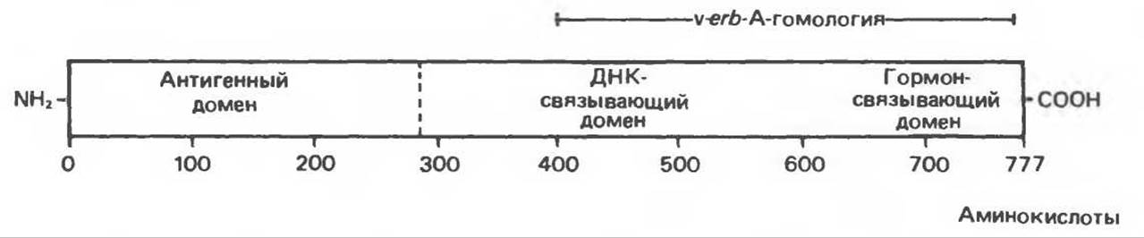
Fig. 48.7. Schematic diagram of the human glucocorticoid receptor. This receptor exists in two forms consisting of 742 or 777 amino acid residues, respectively, which differ at their C-termini. The second form is shown in the figure. The receptor can be divided into functionally distinct domains: an antigenic domain, a DNA-binding domain, and a hormone-binding domain. The region exhibiting a high degree of Homology with the v-erb-A oncogene is indicated.
The Regulation of transcription rate appears to be the most crucial element in the mechanism of action of glucocorticoid hormones, though it is not the only one. It has been found that these hormones also regulate the Processing and transport of nuclear transcripts (e.g., a1-acid Glycoproteins), the degradation rate of specific mRNAs (e.g., Growth Hormone and phosphoenolpyruvate carboxykinase), and, finally, post-translational processing (various mouse mammary tumor virus proteins). It appears that this and other classes of steroid hormones can act at any level of Genetic information transfer from DNA to protein, with the relative importance of the effect at each level varying from system to system.
Mineralocorticoid Hormones
A. General CHARACTERISTICS OF THE Mechanism of Action.
The mechanism of action of aldosterone is broadly similar to that of other steroid hormones (Fig. 48.8). Target cells contain specific receptors that bind aldosterone. The resulting hormone-receptor complex binds to Chromatin and regulates the transcription rate of specific genes. Although specific gene products have not been isolated, it is known that RNA and Protein Synthesis are required for the expression of aldosterone's effects. It is hypothesized that The Influence of aldosterone on ion transport is mediated by specific proteins.

Fig. 48.8. Mechanism of aldosterone action. The hormone induces The formation of one or more proteins, which in turn increase the permeability of the apical (luminal) membrane to Na+, enhance Active Transport of Na+ from The Cell across the basolateral membranes into the interstitial space, or improve the energy supply for the Na+ pump. (Modified from Edelman S. Candidate mediators in the action of aldosterone on Na+-transport. In: Membrane Transport Processes, vol. 1. Hoffman J.F. [editor]. Raven Press, 1978.)
B. Binding of Aldosterone to Receptors. Receptors that bind aldosterone with high affinity (Kd ~ 1 nmol/L) have been identified in the Cytoplasm and Nucleus of target cells. Total binding (receptor capacity) in the cytoplasm is 80–100 times higher than in The Nucleus; however, in terms of specificity and affinity, nuclear binding significantly exceeds the overall binding activity of the Cytosol. In vitro experiments have revealed Three types of binding proteins in the cytosol. Type I and II proteins bind aldosterone with high affinity, whereas type III proteins exhibit low affinity. Type I is the mineralocorticoid receptor, while type II is apparently the glucocorticoid receptor, which concurrently binds aldosterone. The type I receptor binds aldosterone avidly, but also exhibits a high affinity for DOC and corticosterone. Given that the plasma levels of each of these two steroids are much higher than that of aldosterone, one might assume that they would preferentially bind to the type I receptor, thereby rendering The Effect of aldosterone weak. However, recall that in blood plasma, DOC and corticosterone are bound to the steroid-transporting protein transcortin, whereas aldosterone lacks a specific transport protein. Consequently, the effective "free" concentration of aldosterone in plasma is higher than that of corticosterone or DOC. This enables aldosterone to readily enter cells, giving it a competitive advantage in binding to the type I receptor in vivo.
C. Effect of Aldosterone on Ion Transport. The Molecular Mechanism of aldosterone's action on Na+ transport remains elusive, but a substantial body of evidence Supports the model shown in Fig. 48.8. According to this scheme, Na+ from the tubular fluid bathing the apical surface of renal cells passively enters the cells via Na+ channels. This ion is subsequently transferred into the interstitial fluid, with Transport Across the serosal membrane of the cell being mediated by a Na+/K+-dependent ATPase. Thus, this active process consumes ATP energy.
Aldosterone increases the number of Na+ channels on the apical membrane of cells, which presumably leads to an elevation in intracellular Na+ levels. Additionally, aldosterone increases The activity of several mitochondrial enzymes, which should promote The production of ATP required for the operation of the Na+/K+ pump at the serosal membrane of the cell. As a result of aldosterone action, both the NADH:NAD ratio and the activity of certain mitochondrial enzymes, including citrate synthase, increase. The elevation in citrate synthase activity is due to true enzyme induction (likely mediated through effects on gene transcription), and the transient increase in The amount of this protein closely correlates with the hormone's effect on Na+ transport. Given that no direct effect of aldosterone on the Na+ pump has been detected, it appears likely that the hormone acts by increasing the intracellular Na+ concentration and generating the energy source required for the extrusion of this ion. The effects of aldosterone on K+ and H+ transport may be mediated by different mechanisms involving various hormone-regulated proteins.
Last update: 06/08/2026
Editorial and Educational Adaptation: This material has been compiled based on the primary/original source text. The project team performed an editorial review, corrected technical inaccuracies, structured sections, and adapted the content for an educational format.
What was processed:
- elimination of formatting defects (OCR errors, structural breaks, corrupted characters);
- editorial organization of content;
- standardization of terminology in accordance with academic sources;
- verification of factual statements against the original source text.
All mentions of the author, publication year, and origin of the primary text have been preserved in accordance with the source.