Biochemistry - The Chemical Reactions of Living Cells, Volume 2 - D. Metzler 1980
Biosynthesis: How New Molecules Are Formed
Steroid Compounds
Steroid Hormones
In animal organisms, Cholesterol serves as the precursor for three Major Groups of Hormones: progestins, Sex Hormones, and Adrenal Cortical Hormones (corticosteroids). The principal pathways for The Biosynthesis of these hormones are outlined in Fig. 12-17. The shortening of the side chain to two carbon atoms occurs via its hydroxylation followed by oxidative Cleavage. This yields the two-carbon side chain characteristic of pregnenolone, the central intermediate in the biosynthesis of Steroids and corticosteroids. Oxidation of the 3-OH group of pregnenolone to a C = O is accompanied by a shift in the double bond; the product of this Ketosteroid isomerase reaction is an a,β-unsaturated ketone, progesterone [equation (7-56), reaction b].
a. Progestins
Progesterone is the principal hormone of the corpus luteum, an endocrine gland that develops in the Ovary from the follicle after the release of an egg (ovulation). Progesterone is also synthesized in the Adrenal Glands, Testes, and Placenta. It undergoes extremely rapid transformation in the body, primarily via reduction to the corresponding alcohol, which is subsequently converted into conjugated derivatives and excreted as glucuronides [equation (12-12)]. Reduction of the double bond in ring A of progesterone results in a complete loss of biological activity, emphasizing the critical role of the a,β-unsaturated ketone in mediating hormonal action. Progesterone Functions as a hormone essential for the maintenance of Pregnancy and, in concert with estrogens, participates in the Regulation of the Menstrual cycle.
b. Corticosteroids
In the adrenal cortex (i.e., the outer zone of the adrenal glands), progesterone is converted into two classes of hormones, the most prominent representatives being cortisol and aldosterone. Adrenal secretion of cortisol is regulated by the pituitary hormone corticotropin (ACTH); in adults, cortisol secretion reaches 15–30 mg per day. In the Blood, cortisol circulates primarily in a protein-bound form; the cortisol-transporting plasma protein is known as transcortin. As noted in Chap. 11, Section E, 2, cortisol is a glucocorticoid that stimulates Gluconeogenesis and Glycogen storage in the Liver. In Muscle and other Tissues, cortisol inhibits Protein Synthesis, whereas in adipose tissue it enhances lipolysis, releasing Fatty acids.
Class="center">
FIG. 12-17. Biosynthetic pathways of certain steroid hormones.
The anti-inflammatory effects of cortisol and its structural analog cortisone are widely recognized. These effects stem from the ability of these hormones to stabilize lysosomal membranes and accelerate The breakdown of lymphocytes.
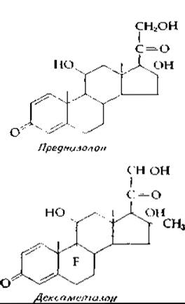
Cortisone and its synthetic derivatives, such as prednisolone and dexamethasone, rank among modern "miracle drugs." They are widely used in acute Arthritis, severe ocular inflammation, and other inflammatory conditions. However, prolonged use of these agents can induce severe adverse effects, notably muscle atrophy and bone resorption. The latter results from the specific inhibition of calcium Absorption in the gastrointestinal tract caused by these agents. Thus, in this regard, glucocorticoids act as vitamin D antagonists (Supplement 12-G).
The synthesis of aldosterone, a mineralocorticoid, is regulated by the Renin-Angiotensin System. This hormonal system is activated by imbalances in ionic Homeostasis, which are detected by sodium ion receptors in the Kidneys. Aldosterone enhances the reabsorption of sodium ions in the renal tubules, thereby regulating Water and Electrolyte balance. In healthy adults consuming a normal dietary sodium intake, aldosterone secretion is approximately 0.1–0.2 mg/day. Glucocorticoids also exhibit modest mineralocorticoid activity; for instance, patients with adrenocortical insufficiency (Addison's disease) can be maintained on glucocorticoids alone (without mineralocorticoids) provided their dietary salt intake is adequate.
Supplement 12-G
Vitamin D
Vitamin D deficiency leads to Rickets, a disorder in humans and animals characterized by bone softening, skeletal deformation, and impaired mineralizationa. The Etiology of rickets as a consequence of a dietary deficiency was recognized over a century ago, and cod liver oil began to be used for its Prevention around 1870. The Link Between rickets and insufficient exposure to sunlight was noted in 1890. However, it was not until 1924, when Steenbock and Hess demonstrated that UV irradiation of certain foods enhances their antirachitic protective activity, that vitamin D (calciferol) was recognized as the second fat-soluble vitamin. Vitamin D comprises a family of compounds generated by the photolysis of ∆5,7-unsaturated sterols, such as ergosterol and 7-dehydrocholesterolb. Ergocalciferol (vitamin D2) is derived from the former, and cholecalciferol (vitamin D3) from the latter.

At low temperatures, the intermediate product precalciferol can be isolated because a steady-state concentration of precalciferol is established during the photochemical reaction. At higher temperatures, precalciferol is converted into calciferol. Side photochemical reactions, which yield Other Compounds including toxic by-products, proceed somewhat more slowly. Consequently, the irradiation of ergosterol for dietary supplement production must be performed with caution.
Under normal conditions, humans synthesize adequate amounts of cholecalciferol from 7-dehydrocholesterol in the Skin upon sun exposure, obviating The Need for dietary vitamin D supplementation. This is particularly true for adults. For children, a daily intake of ~20 µg (400 IU) of ergocalciferol is generally recommended. Excessive amounts of vitamin D are undesirable, and a tenfold increase over the recommended dose produces significant toxicity.
The primary function of vitamin D is the Regulation of Calcium METABOLISM. Recent findings indicate that the actual active regulators are polar hydroxylated metabolites of vitamin D. Hydroxylation occurs at three positions, and the most polar metabolite currently identified is 1,24,25-trihydroxycholecalciferolc,d.
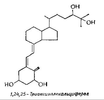
These metabolites are more appropriately classified as steroid hormones, and vitamin D itself functions as a prohormone synthesized in the skin. The investigated pathways of vitamin D hydroxylation are summarized in the scheme below. Notably, the initial hydroxylation yielding 25-hydroxycholecalciferol takes place in the liver, whereas the subsequent two hydroxylations occur in the kidneys. Because di- and trihydroxy derivatives of vitamin D serve as the true regulators of calcium ion homeostasis, patients with renal disease frequently exhibit marked bone demineralization (Renal osteodystrophy). Current research focuses on synthesizing polyhydroxylated vitamin D derivatives for therapeutic administration to patients with renal impairmente.
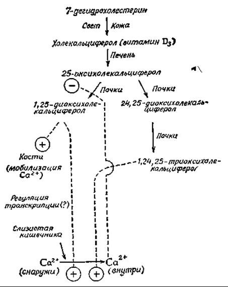
Both 1,25-dihydroxycholecalciferol and the trihydroxy derivative enhance the intestinal absorption of Calcium Ions. Like other steroid hormones, these compounds presumably exert their effects at the level of Transcriptional Regulatione. For instance, administration of vitamin D has been shown to increase the levels of calcium-binding Proteins, which likely participate in intestinal calcium absorptionc,g,h.
The relative concentrations of 1,25-dihydroxycalciferol and the 24,25-derivative synthesized in the body depend on various physiological conditions. Specifically, calcium ions exert an inhibitory effect on 1-hydroxylase, as illustrated in the provided scheme11. This calcium effect may be direct or mediated through parathyroid hormone actionk. While 1,24,25-trihydroxyvitamin D exerts a specific effect on the intestinal mucosa, the 1,25-dihydroxy derivative also acts on bone Cells, mobilizing calcium ions. This effect is partly mediated by The stimulation of Ca-activated ATPase in the Plasma Membranes of bone cellsi. 1,25-dihydroxyvitamin D also increases renal reabsorption of inorganic phosphatel. Furthermore, vitamin D metabolites influence muscle and other tissues.
a A analogous condition in cattle is known as "milk fever."
b Dyke S. F., The Chemistry of the Vitamins, pp. 271–317, Wiley (Interscience), New York, 1965.
в DeLuca H. F., Schnoes H. K., Annu. Rev. Biochem., 45, 631—666 (1976).
г Holick M. F., Kleiner-Bossaller A., Schnoes H. K., Kasten P. M., Boyle I. T., DeLuca H. F., JBC, 248, 6691—6696 (1973).
д Holick M. F., DeLuca H. F., Kasten P. M., Korycka M. B., Science, 180, 964—966 (1973).
е Procsal D. A., Okamura W. H., Norman A. W., JBC, 250, 8382—8388 (1975).
ж DeLuca H. F., Fed. Proc. Fed. Am. Soc. Exp. Biol., 33, 2211—2219 (1974).
з Emtage J. S., Lawson D. E. M., Kodicek E., Nature (London), 246, 100—101 (1973).
и Galante L., Colston К. W., Evans I. M. A., Byfield P. G. H., Matthews E. W., MacIntyre L, Nature (London), 244, 438—440 (1973).
к Russel R. G. G., Monod A., Bonjour J.-P., Fleisch H., Nature (London), New Biol., 240, 126—127 (1972).
л Tanaka Y., DeLuca H. F., PNAS, 71, 1040—1044 (1972).
b. Androgens
The primary androgen, or male sex hormone, is testosterone, which is synthesized from pregnenolone via the Cleavage of the side chain at C-17. Testosterone production amounts to 6—10 mg per day in men; it is also synthesized in women, albeit in small amounts (~0.4 mg). Interestingly, testosterone serves as the precursor for Female Sex Hormones. In the bloodstream, testosterone circulates bound to ß-globulin and exerts effects on A number of target Organs, including the Reproductive System. The most prominent effect of testosterone is the stimulation of facial Hair growth. At the same time, testosterone triggers the premature regression of scalp hair follicles in genetically predisposed individuals. Consequently, bald men typically grow magnificent beards, and when beard hair follicles are transplanted to the scalp, they remain resistant to the action of androgens. The underlying reasons for these contrasting regulatory effects remain completely unknown. Baldness can be treated using appropriate androgen antagonists, but this comes at the cost of beard loss and the cessation of sexual activity. One of the goals for medicinal chemists is to develop a compound capable of blocking the action of androgens selectively on scalp hair follicles.
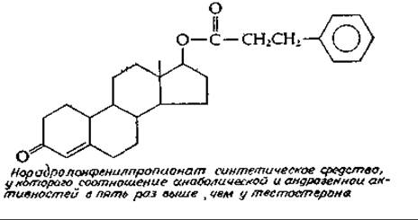
Androgens also possess general "anabolic" activity, which manifests as the stimulation of protein synthesis, particularly in Muscle tissue. They promote bone growth; the rapid growth spurt in boys and girls during Puberty is evidently driven by androgens. The greater average height of men compared to women is likewise associated with higher androgen production. Attempts to create "anabolic hormones" devoid of androgenic activity have led to The Emergence of numerous synthetic steroids. Some of these agents possess the desired properties, at least partially, and anabolic hormones are currently widely used by athletes, although the appropriateness of this practice remains highly controversial.
c. Estrogens
17ß-Estradiol is the principal estrogenic, or female, sex hormone. It is formed from testosterone via the oxidative cleavage of the C-19 atom followed by the aromatization of ring A[100]. The presence of this aromatic ring is characteristic of all estrogenic hormones. Estrogens are synthesized primarily in the Ovaries, and during pregnancy, in the placenta. Certain amounts are also synthesized in the testes. For instance, stallion testes contain more estrogens than any other endocrine gland. Target tissues for estrogens include primarily the Mammary Glands and the Uterus, as well as many other tissues throughout the body. Estrogens inhibit bone growth. The smoother skin of women and their greater proportion of adipose tissue are also attributable to the effects of estrogens.
Estradiol and progesterone are two Hormones Involved in The regulation of the menstrual cycle. At the onset of the cycle, The production of both hormones is low. This is followed by the upregulation of estrogen synthesis under the Influence of the follicle-stimulating hormone (FSH), which is secreted by the anterior Pituitary Gland and promotes the growth of ovarian Graafian follicles, where estrogens are synthesized. Around the middle of the cycle, under The Influence of the luteinizing hormone (LH), also produced by the pituitary gland, ovulation (the release of the egg) occurs, after which progesterone production begins. This hormone is essential for the maintenance of pregnancy. If blastocyst implantation does not take place, progesterone production declines, and the cycle ends with menstruation.
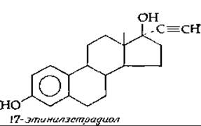
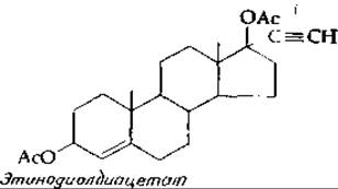
The administration of estrogens and progestins suppresses the pituitary secretion of FSH and LH via a negative feedback mechanism, thereby halting the ovulation process. This effect forms The basis of contraceptive medications. The regimen may involve taking small daily doses of the synthetic estrogen 17-ethinylestradiol for the first 10—15 days, followed by the same estrogen combined with a progestin, such as ethynodiol diacetate, for the subsequent 10—15 days. According to an alternative regimen, only progestin is administered throughout the entire cycle.
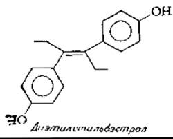
One of the synthetic compounds with estrogenic activity is diethylstilbestrol. For some time, it was widely used in livestock fattening to increase weight gain. Subsequently, it was discovered that feeding large amounts of diethylstilbestrol to rats exhibited carcinogenic effects, leading to the discontinuation of its use. Furthermore, The Use of diethylstilbestrol in tablet form as a "morning-after" pill to prevent the implantation of a fertilized egg appears highly questionable.
Mention should also be made of the existence of plant Flavonoids that exhibit estrogenic activity.
Last update: 06/08/2026
Editorial and Educational Adaptation: This material has been compiled based on the primary/original source text. The project team performed an editorial review, corrected technical inaccuracies, structured sections, and adapted the content for an educational format.
What was processed:
- elimination of formatting defects (OCR errors, structural breaks, corrupted characters);
- editorial organization of content;
- standardization of terminology in accordance with academic sources;
- verification of factual statements against the original source text.
All mentions of the author, publication year, and origin of the primary text have been preserved in accordance with the source.