Review of Medical Physiology - William F. Ganong 2002
Endocrine System, Metabolism, and Reproduction
Endocrine Function of the Kidneys, Heart, and Pineal Gland
Renin-Angiotensin System
Renin
It is well established that parenteral administration of renal tissue extracts leads to an increase in Blood pressure. This effect is mediated by renin, an enzyme belonging to the acidic protease group, which is secreted directly into the blood by the Kidneys. Renin acts in concert with the angiotensin-converting enzyme to generate angiotensin II (Fig. 24-1). Angiotensin II is a glycoprotein with a Molecular Weight of 37,326 in humans. Its molecule consists of two lobes, or domains, with the Active Site located in a deep cleft between them. The active site features two paired aspartate residues: one at position 104 and the other at position 292 (with amino acid residue numbering corresponding to human preprorenin). These residues play a crucial role in the enzymatic activity. Consequently, renin is classified as an aspartyl protease.
Like Other Hormones, renin is synthesized as a large preprohormone. Human preprorenin comprises 406 amino acid residues. Prorenin is formed upon the Cleavage of an initial 23-amino-acid signal sequence from preprorenin, yielding a 383-amino-acid residue chain. Subsequent removal of the so-called prosegment from prorenin yields the active renin, which consists of 340 amino acid residues. Prorenin is virtually devoid of biological activity; a fraction of it is converted into renin within the kidneys, while the remainder is secreted. Prorenin is also secreted by other Organs, notably the Ovaries. Following nephrectomy, circulating prorenin levels drop only slightly and may even rise in practice, whereas active renin levels fall practically to zero. Thus, only a negligible amount of circulating prorenin is converted into renin, and active renin is produced predominantly, if not exclusively, by the kidneys. While prorenin is secreted across numerous body Tissues, active renin is generated primarily within the secretory granules of juxtaglomerular Cells (see below).
The half-life of active renin in the circulating blood is up to 80 minutes. Its sole recognized function to date is the cleavage of angiotensin I from the amino terminus of the decapeptide angiotensinogen (the substrate for renin).
Angiotensinogen
Circulating angiotensinogen has been identified within the a2-globulin fraction of plasma (see Fig. 24-1). It contains approximately 13% CARBOHYDRATES and consists of 453 amino acid residues; it is synthesized in the Liver with a 32-amino-acid signal sequence that is cleaved within The Endoplasmic reticulum. Glucocorticoids, THYROID HORMONES, estrogens, certain cytokines, and angiotensin II elevate its levels in the circulating blood.
Angiotensin-Converting Enzyme and Angiotensin II
Angiotensin-converting enzyme (ACE) is a dipeptidyl carboxypeptidase that cleaves Histidine-leucine from physiologically inactive angiotensin I to form the octapeptide angiotensin II (Fig. 24-2). This same enzyme is responsible for the inactivation of bradykinin (see Chapter 31). The majority of the converting enzyme responsible for generating angiotensin II in the blood is localized within endothelial cells. This conversion occurs primarily as blood passes through the Lungs, though it also takes place in many other Regions of the body.
ACE is an ectoenzyme that exists in two forms: a somatic form, distributed throughout the body, and a testicular form, found exclusively in postmeiotic spermatogenic tissues and spermatozoa (see Chapter 23). Both ACE variants share a single transmembrane domain and a short cytoplasmic "tail." However, somatic ACE is a 170 kDa protein containing two homologous extracellular domains, each harboring an active catalytic site (Fig. 24-3). Testicular ACE has a molecular weight of 90 kDa, possessing only a single extracellular domain and one active site. Both Enzymes are transcribed from the same Gene, which utilizes two distinct promoters to generate two different mRNAs. In male mice with a targeted disruption of the ACE gene, ARTERIAL BLOOD PRESSURE is lower than normal, whereas female knockout mice exhibit normal blood pressure. Additionally, male mice display reduced fertility, a deficit not observed in females.
Class="center">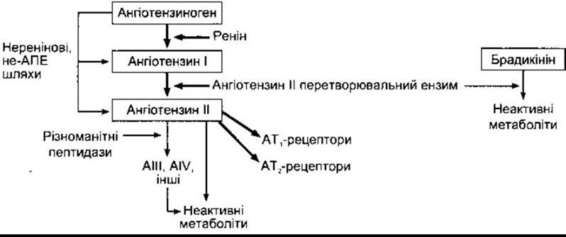
Fig. 24-1. Formation and METABOLISM of circulating angiotensins.
Metabolism of Angiotensin II
Angiotensin II is rapidly metabolized, with a half-life in human blood of 1–2 minutes. It is degraded by a variety of peptidases. Aminopeptidase cleaves the asparagine residue from the amino terminus of the peptide. The resulting heptapeptide retains physiological activity and is occasionally referred to as angiotensin III (see below). Further removal of the second terminal amino acid from angiotensin III yields the hexapeptide angiotensin IV, which likely possesses some residual activity as well. Most other peptide fragments are inactive. Furthermore, aminopeptidase can act on angiotensin I to produce (des-Asp1)-angiotensin I, which can be converted directly into angiotensin III by ACE. Both erythrocytes and other body tissues possess the capacity to metabolize angiotensins. The clearance of angiotensin II from the Circulation is presumably mediated by a uptake mechanism operating within the capillary beds of various tissues (excluding the lungs).
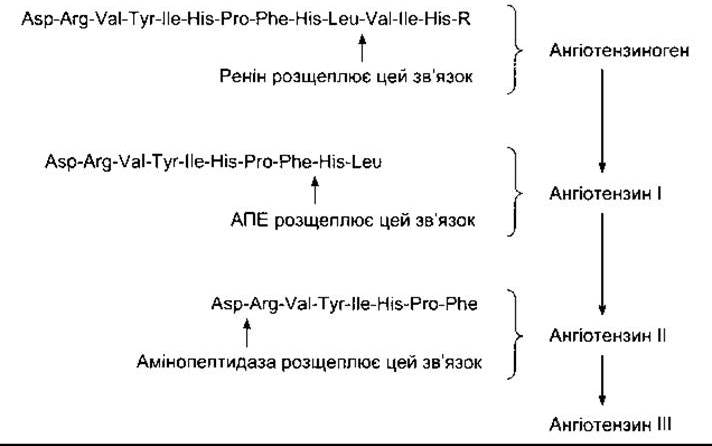
Fig. 24-2. Structure OF THE amino terminus of human angiotensinogen and angiotensins I, II, and III; R represents the protein residue. Following the Cleavage of the 24-amino-acid leader sequence, angiotensinogen consists of 453 amino acid residues. The structure of angiotensin II in dogs, rats, and many other mammals is identical to that of humans. In cows and sheep, angiotensin II contains a valine residue in position 5 instead of isoleucine.
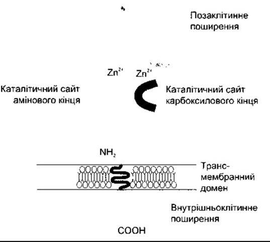
Fig. 24-3. Schematic representation of the structure of the somatic form of angiotensin-converting enzyme. Note the short endoplasmic tail of the molecule and the two extracellular catalytic sites, each binding a zinc ion (Zn2+). (Reprinted with permission from Johnston CI: Tissue angiotensin-converting enzyme in cardiac and vascular hypertrophy, repair and remodeling. Hypertension 1994;23:258)
Renin levels are typically measured by incubating the test sample and quantifying The amount of generated angiotensin I using immunoassay. This determines the plasma renin activity (PRA) of the sample. PRA can be diminished not only by a decrease in renin quantity but also by angiotensinogen deficiency, which may lead to misinterpretation of assay results. To circumvent this, exogenous angiotensinogen is frequently added to the sample, allowing the determination of plasma renin concentration (PRC) rather than PRA. In individuals on a normal dietary sodium intake, the normal supine PRA value is approximately 1 ng of angiotensin I per 1 mL of sample per hour. The plasma concentration of angiotensin II in such individuals is approximately 25 pg/mL (about 25 pmol/L).
Actions of Angiotensins
Angiotensin I Functions solely as a precursor to angiotensin II and has no other known physiological role. Angiotensin II, formerly known as hypertensin or angiotonin, causes constriction of arterial vessels and an elevation in both systolic and diastolic blood pressure. It is one of the most potent known vasoconstrictors, exhibiting 4 to 8 times The activity of norepinephrine in normal subjects. However, its pressor activity is attenuated in individuals with severe sodium depletion, cirrhosis of the liver, and certain other pathological conditions. Under these states, circulating angiotensin II levels are elevated, leading to a negative feedback downregulation of angiotensin receptors within the vascular smooth Muscle wall. This mechanism also explains why exogenous administration of angiotensin II elicits a less pronounced effect than endogenous peptide.
Angiotensin II acts directly on the adrenal cortex to stimulate aldosterone secretion. Consequently, the renin-angiotensin system serves as the primary regulatory pathway for aldosterone release (see Chapter 20). Additional effects of angiotensin II include the facilitation of norepinephrine release via direct action on postganglionic sympathetic Neurons, contraction of mesangial cells accompanied by a marked reduction in the Glomerular Filtration rate (see Chapter 38), and direct action on the renal tubular system to enhance sodium reabsorption.
Furthermore, angiotensin II influences the Central Nervous system by lowering the baroreflex threshold (see Chapter 31), which potentiates its pressor effect. It also acts on the Brain to stimulate thirst (see Chapter 14) and increase the secretion of vasopressin and ACTH. Although angiotensin II does not cross the blood-brain barrier, it exerts its effects by acting on circumventricular organs—four small structures located outside the blood-brain barrier (see Chapter 32). One of these structures, the area postrema, is primarily responsible for potentiating the pressor response, whereas the other two (the subfornical organ (SFO) and the organum vasculosum of the lamina terminalis (OVLT)) mediate the dipsogenic (thirst-stimulating) effect. However, the specific circumventricular organs responsible for the enhanced secretion of vasopressin and ACTH remain undetermined.
Angiotensin III [(des-Asp 1)-angiotensin II] retains only about 40% of the pressor activity of angiotensin II, yet possesses 100% of its aldosterone-stimulating potency. Various researchers have hypothesized that angiotensin III functions as the physiological aldosterone-stimulating peptide, whereas angiotensin II regulates blood pressure. In reality, however, angiotensin III is merely a degradation product endowed with intrinsic biological activity. The same applies to angiotensin IV, although some investigators suggest it exerts unique central nervous system effects.
Tissue Renin-Angiotensin Systems
Aside from the Endocrine System that generates circulating blood angiotensin II, numerous tissues harbor independent local renin-angiotensin systems that generate angiotensin II presumably for autocrine or paracrine actions. Components of the renin-angiotensin system are found within blood vessel walls, the Uterus, Placenta, and fetal membranes. High concentrations of prorenin have been documented in Amniotic Fluid. Moreover, the renin-angiotensin system—or at least several of its components—is present in the eyes, Exocrine Pancreas, Heart, adipose tissue, adrenal cortex, Testes, ovaries, anterior and intermediate lobes of the Pituitary Gland, Pineal Gland, and brain. The physiological roles of these tissue renin-angiotensin systems remain to be fully elucidated, although mounting evidence indicates that angiotensin II acts as a critical growth factor for cardiac and Vascular Tissues. Because tissue renin enters the general circulation only in minute amounts, bilateral nephrectomy reduces plasma renin activity to levels undetectable by current assay Methods.
Angiotensin II Receptors
There are at least two classes of angiotensin II receptors (see Fig. 24-1). AT1 receptors are the predominant angiotensin receptors in mammals, coupling via a G protein (specifically Gq) to phospholipase C, which in turn leads to an elevation of cytosolic free Ca2+ levels upon angiotensin II binding. They also activate numerous Tyrosine kinase enzymes. In vascular smooth muscle, AT1 receptors associate with caveolae (see Chapter 1), where angiotensin II increases The formation of caveolin-1, one of three isoforms of the protein specific to these Cell-surface invaginations. Rodents possess two distinct yet closely related AT1 subtypes, AT1A and AT1B, encoded by separate genes. The AT1A subtype is found in blood vessel walls, the brain, and many other organs, mediating the majority of the known Physiological effects of angiotensin II. The AT1B subtype is located in the anterior pituitary and the adrenal cortex. In humans, the gene for the AT1 receptor is situated on chromosome 3. Evidence also suggests the existence of a second class of AT1-like receptors, though it remains undetermined whether humans possess distinct angiotensin receptor subtypes homologous to the rodent AT1A and AT1B.
AT2 receptors are also present and are encoded by a gene located on the X chromosome in humans. Like AT1 receptors, they contain seven transmembrane domains but subserve entirely different functions. They signal via a G protein to activate various Phosphatases that function as growth factor antagonists and open K+ channels. Furthermore, stimulation of AT2 receptors increases NO production and consequently elevates intracellular cGMP content. The broader physiological consequences of these second-messenger effects remain to be fully elucidated. AT2 receptors are significantly more abundant in fetuses and neonates, though they are also detectable in adults within the brain and certain other organs.
The regulation of AT1 receptors in arterioles differs markedly from that in the adrenal cortex: an excess of angiotensin II exerts a negative feedback effect on vascular receptors, whereas it exerts a positive feedback effect on receptors in the adrenal cortex, thereby heightening the sensitivity of the gland to the aldosterone-stimulating actions of the peptide.
Juxtaglomerular Apparatus
Renin, found in Kidney extracts and the vascular bed, is produced by juxtaglomerular cells (JG cells). These specialized epithelial cells are located along the afferent arteriole near its entrance into the glomerulus (Fig. 24-4). They contain secretory granules that serve as the storage pool for renin. Renin is also detectable in agranular cells occupying the space between the afferent and efferent arterioles, although its function at this site remains unknown.
At the point where the afferent arteriole enters and the efferent arteriole leaves the glomerulus, the nephron tubule comes into contact with the vascular pole of its parent glomerulus. At this site, marking the beginning of the Distal convoluted tubule, There is a specialized plaque of modified tubular epithelium known as the macula densa (see Fig. 24-4). The macula densa lies in close physical proximity to the JG cells. Mesangial cells, JG cells, and the macula densa collectively constitute the juxtaglomerular apparatus.

Fig. 24-4. Left: Schematic diagram of a glomerulus and the juxtaglomerular apparatus. Right: Phase-contrast micrograph of an afferent arteriole in an unstained, frozen section of mouse kidney. Note the erythrocyte within the arteriolar lumen and the granulated juxtaglomerular cells in the vessel wall (courtesy of S. Peil).
Regulation of Renin Secretion
Renin secretion is governed by a multitude of diverse factors (Table 24-1), and at any given moment, The rate of secretion reflects the net sum of these influences. An important intrarenal mechanism is the vascular baroreceptor, which suppresses renin secretion when arteriolar pressure at the level of the JG cells rises and stimulates it when pressure falls. Another key sensory mechanism regulating renin release is the macula densa: the rate of renin secretion is inversely proportional to the rate of Na+ and Cl- transport across this tubular segment. This transport rate depends not only on macula densa activity but also on the actual load of electrolytes delivered to it. Consequently, diminished delivery of Na+ and Cl- to the distal tubules is associated with an increase in renin secretion. Prostaglandins, particularly prostacyclin (see Chapter 17), stimulate renin release, presumably by acting directly on the JG cells. There is also evidence suggesting that the signaling transmitter utilized by macula densa cells to influence renin secretion is NO (see Chapter 31). Plasma K+ concentration is inversely correlated with renin release, though this effect is likely mediated by alterations in Na+ and Cl- delivery to the macula densa cells.
Angiotensin II exerts a negative feedback inhibition on renin secretion via a direct action on the JG cells. Vasopressin also inhibits renin release both in vitro and in vivo, although it remains unclear whether its in vivo effect is direct or indirect.
Finally, enhanced activity of the sympathetic nervous system stimulates renin secretion. This response is driven by elevated circulating catecholamine levels as well as by norepinephrine released from renal postganglionic sympathetic nerve terminals. Catecholamines act predominantly on the ß1-adrenergic receptors of the JG cells. An increase in intracellular cAMP concentration stimulates renin release.
The primary clinical conditions associated with increased renin secretion in humans are summarized in Table 24-2. In most of these states, central venous pressure falls, triggering an increase in sympathetic activity that can potentially lower pressure within the renal arterioles. Stenosis of the renal artery or of the aorta proximal to the renal Arteries leads to a direct drop in renal arteriolar pressure. Psychological stressors also elevate renal sympathetic nerve activity.
Table 24-1. Factors Influencing Renin Secretion

Pharmacological Modulation of the Renin-Angiotensin System
Renin activity can now be pharmacologically suppressed through various mechanisms. Prostaglandin synthesis inhibitors, such as indomethacin, and ß-blockers (e.g., propranolol) can reduce renin secretion.
The peptide pepstatin and newly developed direct renin inhibitors, such as enalkiren, prevent renin from cleaving angiotensinogen into angiotensin I. Angiotensin-converting enzyme (ACE) inhibitors, such as captopril and enalapril, block The conversion of angiotensin I to angiotensin II. Saralasin and several other angiotensin II analogs act as competitive antagonists of angiotensin II at both AT1 and AT2 receptors. Losartan (DuP-753) selectively blocks AT1 receptors, whereas PD-123177 and other agents selectively block AT2 receptors.
Role of Renin in the Pathogenesis of Arterial Hypertension
Unilateral renal artery stenosis triggers a sharp surge in renin secretion followed by The Development of sustained hypertension (renal hypertension or Goldblatt hypertension). Removal of the ischemic kidney or correction of the vascular obstruction reverses the hypertension, provided it has not been present for too long. In general, hypertension caused by obstruction of a single renal artery in the presence of a normal contralateral kidney (see Table 33-5) is accompanied by elevated circulating plasma renin levels. The clinical counterpart of this experimental model is renovascular hypertension resulting from atherosclerotic renal artery disease or other renal vascular anomalies. However, baseline plasma renin activity in cases of unilateral occlusion often falls within the normal range.
The precise mechanisms underlying hypertension in these circumstances remain incompletely understood. In many patients with Essential Hypertension, ACE inhibitors and losartan produce a marked blood-lowering effect even when renal blood flow is normal and plasma renin activity is normal or even suppressed.
The Role of renin in the feedback loop that helps maintain extracellular fluid volume constancy through the regulation of aldosterone secretion was discussed in Chapter 20. Elevated renin secretion accounts for the hyperaldosteronism (secondary hyperaldosteronism) observed in certain normotensive patients with cirrhosis or nephrosis.
Table 24-2. Conditions Associated with Increased Renin Secretion

Last update: 10/08/2026
Editorial and Educational Adaptation: This material has been compiled based on the primary/original source text. The project team performed an editorial review, corrected technical inaccuracies, structured sections, and adapted the content for an educational format.
What was processed:
- elimination of formatting defects (OCR errors, structural breaks, corrupted characters);
- editorial organization of content;
- standardization of terminology in accordance with academic sources;
- verification of factual statements against the original source text.
All mentions of the author, publication year, and origin of the primary text have been preserved in accordance with the source.