Review of Medical Physiology - William F. Ganong 2002
Formation and Excretion of Urine
Renal Function and Micturition
Glomerular Filtration
Measurement of Glomerular Filtration Rate
The glomerular filtration rate (GFR) in humans and animals can be measured by determining the excretion and plasma concentration of a specific substance that is freely filtered through the glomeruli, and is neither secreted nor reabsorbed by the renal tubules. Per unit of time, such a substance must enter the urine exclusively through the filtration of a specific volume of Blood Plasma containing it. Therefore, if this substance is designated as X, the GFR will be equal to the concentration of X in the urine (Cx), multiplied by the urinary flow rate per unit of time V, and divided by the concentration of X in the blood plasma Px:
Class="center">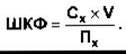
This value is also referred to as the clearance of substance X. The value of Px is, of course, identical in all PARTS OF THE arterial Circulation, and if substance X is not metabolized in the Tissues, its concentration in arterial plasma can be replaced by its concentration in peripheral venous plasma.
Substances Used to Measure GFR
The primary requirements for such substances are that they must be freely filtered in the glomeruli, neither reabsorbed nor secreted in the tubules, non-toxic, and not metabolized in the body. Inulin, a fructose polymer with a Molecular Weight of 5,200 Da found in extracts of certain dahlia varieties, fully meets these criteria and is widely used to determine GFR in both humans and laboratory animals. Various radioisotopes, such as 51Cr-EDTA, are also used, but inulin remains the gold standard for such studies. In clinical practice, a loading dose of inulin is initially administered as an intravenous bolus, followed by a continuous intravenous infusion to maintain a constant plasma level. Once the inulin has distributed evenly throughout the Body Fluids, urine is collected over a precisely measured time interval, and a blood sample is taken. The concentrations of inulin in the urine and plasma are determined, and the clearance value (CIN) is calculated.
Example. For CIN = 35 mg/mL; V = 0.9 mg/mL; PIN = 0.25 mg/mL, we obtain

In dogs, cats, rabbits, and many other mammals, creatinine clearance can also be used to determine GFR; however, in primates, particularly humans, a certain amount of creatinine is secreted by the tubules and may undergo reabsorption. Furthermore, the laboratory METHOD FOR DETERMINING plasma creatinine at low concentrations yields significant errors because, under these conditions, small amounts of other plasma constituents are measured alongside creatinine itself. Despite this, endogenous creatinine clearance measurements are widely used in clinical practice. These values are quite close to the GFR values obtained using inulin because, although the CIE × V values are overestimated due to tubular secretion, the PIN values are also overestimated due to non-specific chromogens, resulting in mutually cancelling errors. Endogenous creatinine clearance is easy to determine and serves as an important indicator of renal function; however, in cases where precise GFR determination is required, relying on a method with mutually compensating errors is inadvisable.
Normal GFR Values
The GFR in a normal human of average body mass is about 125 mL/min. This value correlates quite closely with body surface area, although in women it is 10% lower than in men, even after adjusting for body surface area. A value of 125 mL/min corresponds to 7.5 L/h, or 180 L/day, whereas normal urine output is about 1 L/day. Thus, under normal conditions, over 99% of the filtrate is reabsorbed. At a glomerular filtration rate of 125 mL/min, the Kidneys filter a volume of fluid daily that is four times the total body Water, 15 times the extracellular fluid volume, and 60 times the blood plasma volume.
Regulation of GFR
Filtration across the glomerular capillaries is determined by the same factors that influence filtration across any other capillaries (see Chapter 30)—namely, the capillary bed area, capillary permeability, and the difference between hydrostatic and osmotic pressures across the Capillary Wall. The following law holds true for each nephron:

where KF is the glomerular filtration coefficient (the product of the capillary wall's hydraulic conductivity [i.e., its permeability] and the effective filtration surface area); PGC is the mean hydrostatic pressure in the glomerular capillaries; PT is the mean hydrostatic pressure in the tubule; πGC is the plasma osmotic pressure in the glomerular capillaries; and πT is the Osmotic Pressure of the plasma in the tubular filtrate.
Permeability
The permeability of glomerular capillaries is approximately 50 times greater than that of Skeletal Muscle capillaries. Uncharged molecules with a molecular diameter of up to 4 nm pass freely through the capillary membranes, whereas the filtration rate of uncharged molecules larger than 8 nm is close to zero (Fig. 38-5). For molecules with intermediate diameters, the filtration rate is inversely proportional to molecular diameter. However, sialoproteins in the glomerular capillary wall carry a negative charge, and studies with negatively and positively charged dextrans indicate that this negative charge repels negatively charged molecules in the blood. Consequently, the filtration of negatively charged molecules up to 4 nm in diameter is more than twice as slow as that of neutral molecules of the same size. This likely explains why the concentration of albumin in the glomerular filtrate (effective molecular diameter of about 7 nm) is only 0.2% of its plasma concentration, even though consideration of molecular size alone would suggest a much higher value (circulating albumin carries a net negative charge). The filtration of positively charged substances is somewhat faster than that of neutral ones. The normal urinary protein content is up to 1000 mg/L, and its primary source is not the glomerular filtrate, but rather tubular Cells shed into the lumen. The presence of significant amounts of albumin in the urine is termed albuminuria. In nephritis of various etiologies, multiple negative charges in the glomerular walls are lost, resulting in albuminuria even without A change in membrane pore size.

Fig. 38-5. Effect of electrical charge on the clearance of dextrans of varying molecular size. The negative charge of the glomerular membrane slows the passage of negatively charged molecules (anionic dextrans) and facilitates the passage of positively charged ones (cationic dextrans) (reprinted with permission from Brenner BM, Beeuwkes R: The renal circulations. Hosp Pract [July] 1978;13:35).
The inability of Plasma Proteins to diffuse across capillary membranes gives rise to the Gibbs-Donnan effect (see Chapter 1), which involves the distribution of polyvalent cations: the concentration of anions in the glomerular filtrate is 5% higher than in blood plasma, while the concentration of polyvalent cations is correspondingly 5% lower. In most cases, however, this effect can be neglected, and the COMPOSITION OF THE filtrate can be considered identical to blood plasma.
Regulation of Capillary Lumen
It is now understood that GFR can be influenced by mesangial cells, whose contraction leads to a reduction in GFR, largely driven by a decrease in the filtration surface area. The contraction of capillary loops at their bifurcation points redistributes blood flow among the loops. The capillary lumen widens and narrows through the contraction of mesangial cells. Substances that affect mesangial cells are listed in Table 38-3. Angiotensin II is an important factor regulating mesangial Cell contraction, and renal glomeruli contain receptors for it. There is also evidence that mesangial cells synthesize renin.
Hydrostatic and Osmotic Pressures
Pressure in the glomerular capillaries is higher than in other capillaries. This is because the afferent arterioles are short, direct Branches of the interlobular Arteries. Furthermore, the efferent arterioles offer relatively high resistance. Capillary hydrostatic pressure is opposed by the hydrostatic pressure within Bowman's capsule. This is also counteracted by the osmotic pressure gradient across the glomerular capillaries (πGC - πT). Because the value of πT is normally negligibly small, this gradient is essentially equal to the colloid osmotic pressure of the plasma proteins.
Table 38-3. Factors affecting the contraction or relaxation of mesangial cells
Constriction |
Relaxation |
Endothelins Angiotensin II Vasopressin Norepinephrine Platelet-activating factor Platelet-derived growth factor Thromboxane A2 PGF2 Leukotrienes C4 and D4 Histamine |
ANP Dopamine PGE2 |
The values of these pressures, obtained in experiments on rats, are shown in Fig. 38-6. The mean filtration pressure (MFP) at the afferent end of the glomerular capillary is 15 mm Hg, but decreases to zero near the efferent end, meaning that filtration equilibrium is reached in this region. This occurs because fluid is removed from the blood plasma, and as blood flows through the glomerular capillaries, the oncotic pressure increases. Changes in Δп, calculated for the model of an "ideal" glomerular capillary, are illustrated in Fig. 38-6. It is evident that certain segments of the glomerular capillaries do not participate in The formation of the glomerular filtrate; in other words, the Transport of substances across the glomerular capillary membrane depends more on blood flow than on diffusion (see Chapter 30). It is also apparent that a decrease in The rate of rise of the Δп curve due to an increased renal plasma flow enhances filtration without altering Δп, because in this case the length of the capillary over which filtration occurs is increased.
Regarding the achievement of filtration equilibrium, significant differences are observed among various species, and furthermore, the methodology for measuring GFR has certain limitations. It remains unknown whether a state of filtration equilibrium is practically attainable in humans.
Changes in GFR
A vast array of factors discussed in previous chapters directly influence the glomerular filtration rate (Table 38-4). Changes in renal vascular resistance due to autoregulatory mechanisms serve to stabilize filtration pressure; however, when central arterial pressure falls below 90 mm Hg, There is a sharp decline in the glomerular filtration rate. GFR is maintained at a constant level as long as constriction in the efferent arterioles predominates over constriction in the afferent arterioles, though it should be borne in mind that constriction of either type reduces blood flow to the tubules.
Filtration Fraction
The ratio of GFR to renal plasma flow (RPF), termed the filtration fraction, is normally 0.16–0.20. GFR is subject to much less variation than RPF. With a drop in central arterial pressure, GFR decreases less than RPF As a result of efferent arteriolar constriction, and consequently, the filtration fraction increases.
Last update: 10/08/2026
Editorial and Educational Adaptation: This material has been compiled based on the primary/original source text. The project team performed an editorial review, corrected technical inaccuracies, structured sections, and adapted the content for an educational format.
What was processed:
- elimination of formatting defects (OCR errors, structural breaks, corrupted characters);
- editorial organization of content;
- standardization of terminology in accordance with academic sources;
- verification of factual statements against the original source text.
All mentions of the author, publication year, and origin of the primary text have been preserved in accordance with the source.