Review of Medical Physiology - William F. Ganong 2002
Urine Formation and Excretion
Renal Function and Micturition
Renal Circulation
Renal Blood Flow
In a resting adult, the Kidneys receive approximately 1.2–1.3 L of blood per minute, which accounts for nearly 25% of the Cardiac Output (see Table 32-1). Renal blood flow can be measured using electromagnetic flowmeters or by applying the Fick principle to the Kidney (see Chapter 29). This principle involves dividing The amount of a given substance delivered per unit time by the transrenal arteriovenous concentration gradient of that substance. Because the kidney filters Blood Plasma, the renal plasma flow equals the amount of the substance excreted per unit time divided by its arteriovenous concentration gradient, given that the number of erythrocytes remains virtually unchanged as blood passes through the kidneys. Any substance can serve as a reference as long as it is technically feasible to measure its concentration in arterial and renal venous plasma, it is neither metabolized, accumulated, nor synthesized by the kidney, and it does not directly affect renal blood flow itself.
Renal plasma flow can be determined by intravenous administration of $p$-aminohippuric acid (PAH) and measuring its concentration in urine and blood plasma. PAH is filtered through the glomeruli and secreted by tubular Cells such that its extraction ratio (the difference between arterial and renal venous concentrations divided by the arterial concentration) is high. For instance, when PAH is administered in small doses, 90% of the PAH dissolved in arterial plasma is cleared during a single pass of blood through the kidney. Consequently, a widely used method calculates renal plasma flow simply by dividing the urine concentration of PAH by its plasma concentration, disregarding its renal venous level. Arterial plasma can be substituted with venous plasma, which is technically much easier to obtain, since the PAH concentration in venous plasma is virtually identical to that of the arterial plasma entering the kidney. The value obtained in this manner is termed the effective renal plasma flow (ERPF), emphasizing that the concentration of the test substance in renal venous plasma was not measured. In humans, the ERPF averages 625 mL/min:
Class="center">
where $C_{\text{PAH}}$ is the urine concentration of PAH; $P_{\text{PAH}}$ is the plasma concentration of PAH; $V$ is the urine flow rate, mL/min; ERPF is the effective renal plasma flow (PAH clearance). For example,
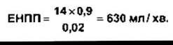
Note that the ERPF determined in this manner is essentially the clearance of PAH. METABOLISM/2.html">THE CONCEPT OF clearance is discussed in detail below.
ERPF can be converted to true renal plasma flow:

where RPF is the renal plasma flow; ERPF is the effective renal plasma flow; ER is the extraction ratio (averaging 0.9 for PAH).
Once the renal plasma flow is known, renal blood flow can be easily calculated using the following formula:

where RBF is the renal blood flow; RPF is the renal plasma flow; Hct is the hematocrit (45%).
Pressure in the Renal Vasculature
Glomerular capillary pressure has been measured directly in rat experiments and proved to be significantly lower than predicted from indirect measurements. Mean systemic arterial pressure is 100 mm Hg, whereas glomerular capillary pressure is about 45 mm Hg. The pressure drop across the glomerulus is only 1–3 mm Hg; however, a further pressure drop occurs in the efferent arteriole, resulting in a peritubular capillary pressure of about 8 mm Hg. Renal venous pressure is approximately 4 mm Hg. The pressure gradients in humans and primates are virtually identical; in both species, glomerular capillary pressure constitutes about 40% of the central arterial pressure.
Regulation of Renal Blood Flow
Norepinephrine constricts renal vessels, exerting its most pronounced effect on interlobular Arteries and afferent arterioles when administered intravenously. Dopamine is synthesized in the kidneys and induces renal vasodilation along with enhanced renal sodium excretion. Angiotensin II acts as a vasoconstrictor, with a more potent effect on efferent than on afferent arterioles. Prostaglandins increase cortical blood flow while decreasing medullary blood flow. Acetylcholine also possesses renal vasodilatory properties. A high-protein diet similarly promotes an increase in glomerular capillary pressure, leading to augmented renal blood flow.
Functions of Renal Nerves
Stimulation of renal nerves enhances renin production through a direct action of norepinephrine on $\beta_1$-adrenergic receptors of juxtaglomerular cells (see Chapter 24) and increases sodium reabsorption, likely via a direct effect of norepinephrine on renal tubular cells. The proximal and distal tubules, as well as the thick ascending limb of the Loop of Henle, are richly innervated. In animal experiments involving intense electrical stimulation of renal nerves, the initial response is an increased sensitivity of juxtaglomerular cells (Table 38-2), followed by elevated renin secretion, increased $\text{Na}^+$ reabsorption, and ultimately renal vasoconstriction, which leads to a decrease in Glomerular Filtration rate and renal blood flow. It remains unresolved whether the effect on sodium reabsorption is mediated via $\alpha$- or $\beta$-adrenergic receptors, or perhaps both receptor types. The Physiological Role of renal nerves in $\text{Na}^+$ metabolism is not yet fully understood, although most renal functions in kidney transplant recipients remain normal despite the fact that transplanted kidneys acquire functional innervation only after a considerable period of time.
Enhanced stimulation of renal sympathetic noradrenergic nerves leads to a marked reduction in renal blood flow. This effect is mediated primarily via $\alpha_1$-adrenergic receptors and to a lesser extent through postsynaptic $\alpha_2$-adrenergic receptors. Under resting conditions, a baseline tonic level of impulse activity is maintained in the renal nerves of humans and laboratory animals. When central arterial pressure drops, baroreceptor reflex activity triggers vasoconstriction, including within the kidneys. Renal blood flow also decreases during physical exertion and, to a lesser extent, upon transitioning from a supine to an upright position.
Table 38-2. Renal responses to graded electrical stimulation of renal nerves1

1 Reproduced with permission from DiBona GF: Neural Control of renal function: Cardiovascular implications. Hypertension 1989; 13:539)
Autoregulation of Renal Blood Flow
Over a moderate range of arterial pressures (90–220 mm Hg in dogs), renal vascular resistance changes such that renal blood flow remains relatively constant (see Fig. 38-4). This type of autoregulation is also observed in other Organs and is driven by multiple factors (see Chapter 31). Renal autoregulation is preserved in denervated and isolated artificially perfused kidneys, but is abolished by agents that paralyze vascular smooth Muscle. This phenomenon is undoubtedly caused, in part, by the myogenic contractile response of stretched smooth muscle cells in the afferent arteriolar walls. Nitric oxide ($\text{NO}$) likely plays a contributory role as well. At low perfusion pressures, angiotensin II becomes increasingly important by constricting efferent arterioles, thereby helping to maintain a constant glomerular filtration rate. This mechanism is believed to account for the ACUTE RENAL FAILURE occasionally seen in patients with compromised renal perfusion who are treated with angiotensin-converting Enzyme Inhibitors.
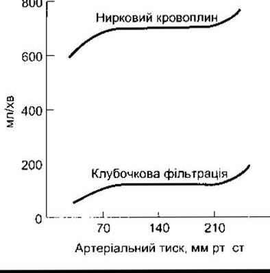
Fig. 38-4. Renal autoregulation
Renal Blood Flow and Oxygen Consumption
The primary function of the renal cortex is to filter large volumes of blood through the glomeruli; consequently, cortical blood flow is exceptionally high while oxygen extraction from the blood remains low. Renal blood flow is approximately 5 mL/min per gram of renal tissue (compared to 0.5 mL/min per gram of Brain tissue), and the overall arteriovenous oxygen concentration gradient for the kidney is only 14 mL/L of blood, compared with 62 mL/L for the brain and 114 mL/L for The Heart (see Table 32-1). The PO2 in the renal cortex is about 50 mmHg. On the other hand, maintaining the osmotic gradient in the renal medulla requires a relatively low blood flow. Therefore, it is not surprising that blood flow is approximately 2.5 mL/g/min in the outer medulla and 0.6 mL/g/min in the inner medulla.
Nevertheless, complex metabolic processes occur continuously here—most notably, sodium reabsorption in the thick ascending limb of the loop of Henle (see below)—resulting in a relatively high rate of oxygen extraction from the blood within the renal medulla. The PO2 in the medulla is approximately 15 mmHg. This renders the medullary tissue particularly vulnerable to Hypoxia if blood flow declines. Regulation mediated by nitric oxide (NO), prostaglandins, and a variety of cardiovascular Peptides via paracrine signaling helps maintain a fine balance between reduced perfusion and the metabolic demands of specific tissue regions.
Last update: 10/08/2026
Editorial and Educational Adaptation: This material has been compiled based on the primary/original source text. The project team performed an editorial review, corrected technical inaccuracies, structured sections, and adapted the content for an educational format.
What was processed:
- elimination of formatting defects (OCR errors, structural breaks, corrupted characters);
- editorial organization of content;
- standardization of terminology in accordance with academic sources;
- verification of factual statements against the original source text.
All mentions of the author, publication year, and origin of the primary text have been preserved in accordance with the source.