Review of Medical Physiology - William F. Ganong 2002
Respiration
Regulation of Respiration
Neural Control of Respiration
Regulatory Systems
Neural control of breathing is provided by two separate systems of mechanisms. One is responsible for voluntary regulation, and the other for involuntary (automatic) regulation. The voluntary system encompasses structures located in the Cerebral Cortex AND sends impulses to respiratory motoneurons via the corticospinal pathways. The automatic system is located in the Pons and Medulla Oblongata, with efferent output impulses traveling from it to the respiratory motoneurons through the White matter situated between the lateral and ventral corticospinal tracts. Nerve fibers that initiate inspiration converge on the phrenic motoneurons located in the ventral horns from C3 to C5, while the external intercostal motoneurons converge in the ventral horns of the thoracic Spinal Cord. Nerve fibers associated with expiration converge primarily on the intercostal motoneurons in the thoracic spinal cord.
Motoneurons supplying expiratory Respiratory Muscles are inhibited when those supplying inspiratory muscles are active, and vice versa. Although spinal Reflexes contribute to reciprocal innervation (see Chapter 6), it generally results from activity in descending pathways. Impulses in the descending pathways excite agonists and inhibit antagonists. An exception to reciprocal innervation is the brief, minor activity in the phrenic axons following inspiration. The Significance of this post-inspiratory output impulse lies in interrupting the elastic recoil of the Lungs and ensuring smooth breathing.
The Medullary System
Rhythmic discharge of Neurons in the medulla oblongata and pons ensures the automaticity of breathing. Transection of the Brainstem below the medulla oblongata (cut D in Fig. 36-1) stops breathing, whereas rostral transection of the pons (cut A in Fig. 36-1) allows automatic breathing to remain normal. Respiratory neurons in the brainstem are of two types: some exhibit activity during inspiration (inspiratory or I-neurons), and others during expiration (expiratory or E-neurons). Most active discharges increase in frequency during inspiration in the case of I-neurons, or during expiration in the case of E-neurons. Some discharges decrease in frequency, while others remain at a constant high level during either inspiration or expiration. However, expiration is passive during quiet breathing, and E-neurons are quiescent at this time; they become active only when pulmonary ventilation increases.
The region of the medulla oblongata associated with breathing is called the respiratory center, which contains two distinct groups of neurons (see Fig. 36-1). The dorsal group is located in or near The Nucleus tractus solitarius. The ventral group is a long Column of neurons extending through the nucleus ambiguus and retroambiguus nucleus in the ventrolateral part of the medulla oblongata. The dorsal group is formed mainly by I-neurons, some of which project and synaptically connect with phrenic motoneurons. Afferent pathways from the Airways, carotid bodies, and aortic bodies, which terminate in the nucleus tractus solitarius, likely project to these neurons. The ventral group contains E-neurons in the caudal part of the respiratory center, I-neurons in its middle part, and E-neurons at its rostral end. Some of these neurons project to respiratory motoneurons; those located at the rostral end of the group are responsible for inhibiting I-neurons during expiration.
The Main Components of the generator complex regulating Respiration, which is responsible for breathing automaticity, are located in the medulla oblongata; therefore, spontaneous breathing persists—although sometimes irregularly and with difficulty—after transection of the brainstem at the lower border of the pons (cut C in Fig. 36-1).
Neither the dorsal nor the ventral group of respiratory neurons is an obligatory part of the generator complex, since selective lesioning of either one reduces the amplitude of breathing but does not abolish it. Following such a lesion, breathing remains rhythmic, initiated by a small group of synaptically interconnected pacemaker Cells in the pre-Bötzinger complex on one or both sides of the medulla oblongata between the nucleus ambiguus and the lateral reticular nucleus (Fig. 36-2). These neurons fire rhythmically and generate rhythmic impulse activity in the phrenic motoneurons, which disappears if transection is performed between the pre-Bötzinger complex and these motoneurons.
Class="center">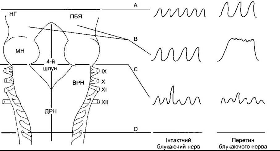
Fig. 36-1. Respiratory neurons in the brainstem. DORSAL VIEW OF the brainstem; the Cerebellum has been removed. The effects of various lesions and brainstem transections are also shown. Spirometric tracings on the right illustrate the depth and frequency of breathing. DRG - dorsal respiratory group; VRG - ventral respiratory group; PB - parabrachial nucleus (pneumotaxic center); 4th vent. - Fourth ventricle; IC - inferior colliculus; SCP - superior cerebellar peduncle. Roman numerals indicate Cranial Nerves (modified and reproduced with permission from Mitchell RA, Berger A: State of the art: Review of neural Regulation of Respiration. Am Rev Respir Dis 1975; 111:206).
Effects of the Pons and Vagus Nerves
The rhythmic discharge of medullary neurons underlies the spontaneity of breathing, but it is modulated by pontine neurons and vagal afferent fibers from receptors in the airways and lungs. In the region known as the pneumotaxic center—located in the medial and parabrachial nuclei and the Kölliker-Fuse nucleus in the dorsolateral pons—there are I-neurons, E-neurons, and neurons active during both phases of respiration. Destruction of this region (cut B in Fig. 36-1) results in slower breathing and increased tidal volume; transection of the vagus nerves in anesthetized animals leads to a prolonged inspiratory spasm resembling breath-holding. Breathing stops in the inspiratory phase (apneusis). The normal function of the pneumotaxic center is not yet fully understood, but it may act as a switch between inspiration and expiration, regulating the optimal ratio of the duration of inspiration, expiration, and the respiratory pause. Lung distension during inspiration initiates impulses in afferent pulmonary vagal fibers. These impulses inhibit inspiratory discharge. Consequently, the depth of inspiration increases following vagotomy (see Fig. 36-1); apneusis occurs when the vagus nerves are cut after destruction of the pneumotaxic center. As shown in Fig. 36-3, vagal feedback activity does not alter The rate of rise of neuronal activity in respiratory motoneurons, but without it, the activity is prolonged.
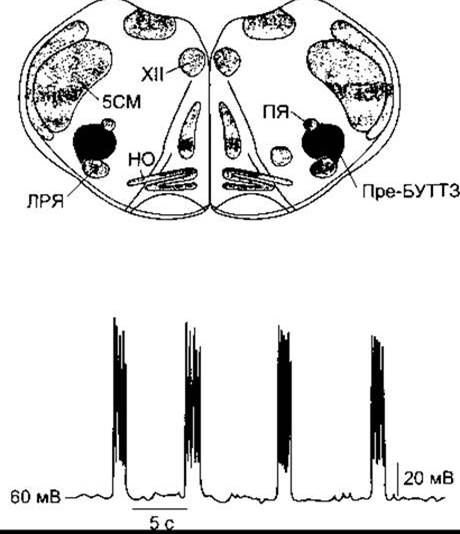
Fig. 36-2. Rhythmic discharge (bottom) of neurons in the pre-Bötzinger complex (shaded area, pre-BOTZ) in a neonatal rat brainstem preparation; IO - inferior olives; LRN - lateral reticular nucleus; NA - nucleus ambiguus; XII - hypoglossal nucleus; Sp5 - spinal trigeminal nucleus (modified from Smith JC et al: Pre-Bötzinger complex: A brainstem region that may generate respiratory rhythm in mammals. Science 1991;254:726).
In intact animals, as The activity of inspiratory neurons increases, the rate and depth of breathing increase. Breathing depth increases because the lungs are stretched to a greater extent before the strength of the inhibitory activity from the vagal and pneumotaxic centers becomes sufficient to overcome the more intense discharge of inspiratory neurons. Breathing frequency increases because the after-discharge in the vagus nerves and, presumably, in the pneumotaxic afferent fibers to the medulla oblongata rapidly predominates.
Last update: 10/08/2026
Editorial and Educational Adaptation: This material has been compiled based on the primary/original source text. The project team performed an editorial review, corrected technical inaccuracies, structured sections, and adapted the content for an educational format.
What was processed:
- elimination of formatting defects (OCR errors, structural breaks, corrupted characters);
- editorial organization of content;
- standardization of terminology in accordance with academic sources;
- verification of factual statements against the original source text.
All mentions of the author, publication year, and origin of the primary text have been preserved in accordance with the source.