Review of Medical Physiology - William F. Ganong 2002
Urine Formation and Excretion
Renal Function and Micturition
Functional Anatomy
Nephron
Each structural unit of the Kidney (the nephron) consists of an individual renal tubule and its corresponding glomerulus. The size of The Kidneys in various biological species is determined by the number of nephrons they contain. Each human kidney contains approximately 1.3 million nephrons. The Components of the nephron are schematically illustrated in Fig. 38-1. The right side of Fig. 38-2 depicts the elongated and slender Structure of nephrons.
Class="center">Table 38-1. Concentration of Certain Physiologically Important Substances in Urine and Blood Plasma
Substance |
Concentration in |
U/P Ratio |
|
Urine (U) |
Plasma (P) |
||
Glucose, mg/dL |
0 |
100 |
0 |
Na+ mEq/L |
90 |
150 |
0,6 |
Urea, mg/dL |
900 |
15 |
60 |
Creatinine, mg/dL |
150 |
1 |
150 |
The glomerulus, which is about 200 µm in diameter, is formed by the invagination of capillary loops into the expanded blind end of the nephron (Bowman's capsule). The capillaries are Branches of the afferent arteriole; blood from them collects into the efferent arteriole, which has a slightly smaller diameter (see Figs. 38-1 and 38-2). Within Bowman's capsule, blood is separated from the glomerular filtrate by two Cell layers: the capillary endothelium and the specialized capsule epithelium consisting of podocytes (see below), which envelop the glomeruli. These layers are separated from each other by a basement membrane. Star-shaped Cells known as mesangial cells lie between the basement membrane and the endothelium. They are similar to pericytes, cells found in capillary walls throughout the body. Mesangial cells are particularly common in the spaces between two adjacent capillaries; in these areas, the basement membrane forms a sheath shared by both capillaries (Fig. 38-3). Mesangial cells are contractile and participate in The regulation of Glomerular Filtration (see below). They also secrete various substances and bind circulating immune complexes, thereby contributing to glomerular diseases.
The endothelium of the glomerular capillaries is fenestrated, with pores 70–90 nm in diameter.
The epithelial cells (podocytes) have numerous interdigitating pseudopodia (see Fig. 38-3) that form filtration slits along the Capillary Wall. These slits are about 25 nm wide, and each is covered by a thin membrane. The basement membrane contains no visible slits or pores.
Normally, the glomerular membrane readily allows the passage of uncharged molecules up to 4 nm in diameter, whereas the penetration of molecules larger than 8 nm is practically impossible. However, the ability of molecules to enter Bowman's capsule is influenced not only by their size but also by their electrical charge (see below). The total surface area of the glomerular capillary endothelium in humans is 0.8 m2.
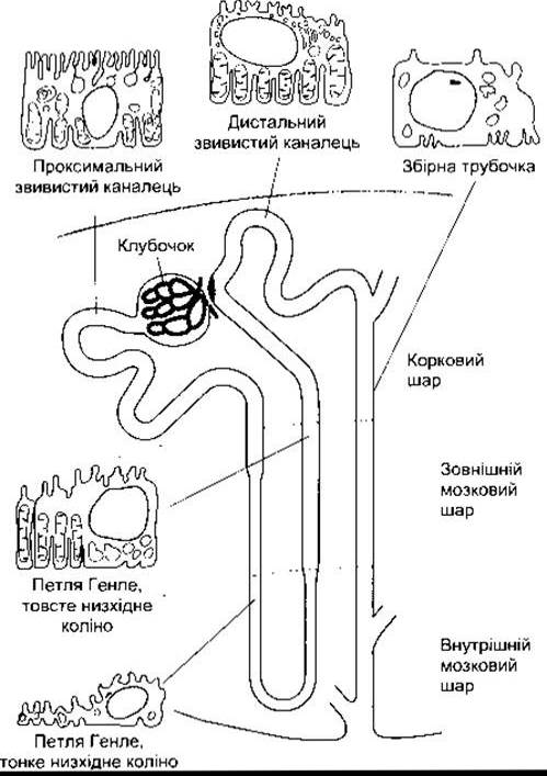
Fig. 38-1. Schematic diagram of a juxtamedullary nephron, showing the principal histological CHARACTERISTICS OF THE cells forming each part of the nephron.
The Main characteristics of the cells forming the tubular walls are illustrated in Fig. 38-1. However, specific cell subtypes are present in all tubular segments, and anatomical differences among them correlate with functional differences (see below).
The proximal convoluted tubule in humans is about 15 mm long and 55 µm in diameter. Its walls are formed by a single layer of interlocking cells connected by apical tight junctions. Between the bases of the cells are expansions of the extracellular space known as lateral intercellular spaces. The Cell surfaces facing the tubular lumen feature a brush border, so named because of numerous microvilli measuring 1.0 × 0.7 µm.
The convoluted portion of the proximal tubule (pars convoluta) transitions into the straight portion (pars recta), which forms the initial part of the Loop of Henle (see Figs. 38-1 and 38-2). The proximal tubule ends in the thin segment of the descending limb of the loop of Henle, which contains a Simple Epithelium composed of sparse squamous cells. Nephrons whose glomeruli are located in the outer part of the renal cortex (cortical nephrons) have short loops of Henle, whereas nephrons with glomeruli situated in the juxtamedullary region of the cortex (juxtamedullary nephrons) have long loops extending down into the renal pyramids. In humans, only 15% of nephrons possess long loops. The total length of the thin segment of the loop ranges from 2 to 14 mm. It terminates in the thick segment of the ascending limb, which is approximately 12 mm long. The Cells of the thick ascending limb are cuboidal in shape, rich in Mitochondria, and exhibit numerous invaginations in the basal Plasma Membrane.
The thick ascending limb of the loop of Henle reaches the nephron glomerulus from which the tubule originated and passes close to its afferent and efferent arterioles. The walls of the afferent arterioles contain juxtamedullary (juxtaglomerular) cells that produce renin. In this region, the glomerular epithelium is histologically modified, forming the macula densa. The juxtaglomerular cells, the macula densa, and the adjacent mesangial cells are collectively termed the juxtaglomerular apparatus (see Fig. 24-4).
The length of the Distal convoluted tubule is about 5 mm. Its epithelium is thinner than that of the proximal tubule, and although some microvilli are present, it lacks a well-defined brush border. Distal tubules merge to form collecting ducts, which are about 20 mm long, pass through the renal cortex and medulla, and empty into the renal pelvis via the apices of the renal pyramids. The epithelium of the collecting ducts consists of principal cells and intercalated cells. The principal cells, which are predominant in number, are relatively tall and contain few Organelles. They participate in the reabsorption of Na+ and Water under METABOLISM/18.html">The Influence of vasopressin. Intercalated cells, found in small numbers in the collecting ducts and distal tubules, contain more microvilli, cytoplasmic vesicles, and mitochondria. They are responsible for the secretion of H+ ions and The transport of HCO3-. The total length of nephrons including the collecting ducts ranges from 45 to 65 mm.
Renal cells with a secretory function include not only the juxtaglomerular cells but also certain cells within the renal medullary interstitium. These cells are called type I medullary interstitial cells. They contain lipid inclusions and presumably secrete Prostaglandins, predominantly PGE2 (see Chapter 17). In addition, PGE2 is secreted by collecting duct cells, while prostacyclin (PGI2) and other prostaglandins are produced by arterioles and glomeruli.
Renal Circulation is schematically illustrated in Fig. 38-2. Afferent arterioles are short, direct branches of the interlobular Arteries. Each of them divides into multiple capillary branches that form a tuft of glomerular vessels. The capillaries coalesce to form the efferent arteriole, which subsequently branches into capillaries supplying blood to the tubules (peritubular capillaries). These capillaries ultimately form interlobular Veins. Thus, the arterial segments between the glomeruli and tubules constitute a portal system. Glomerular capillaries are the only capillaries in The Human Body that drain into arterioles.
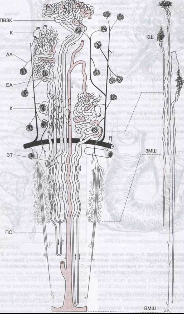
Fig. 38-2. Renal circulation in the dog. In general outlines, it is identical to that in the human kidney. The vascular bed structure is shown in a simplified form. Details of nephron structure are shown enlarged on the left; on the right, the components of the nephron are shown while preserving their geometric proportions; AA – afferent arterioles; Ctx – cortex; CD – collecting duct; EA – efferent arteriole; G – glomerulus; IM – inner medulla; OM – outer medulla; PCT – proximal convoluted tubule; VR – vasa recta. Distal convoluted tubules and collecting ducts are highlighted in color (reproduced with permission from Beeuwkes R III: The vascular Organization OF THE kidney. Annu Rev Physiol 1980;42:531).
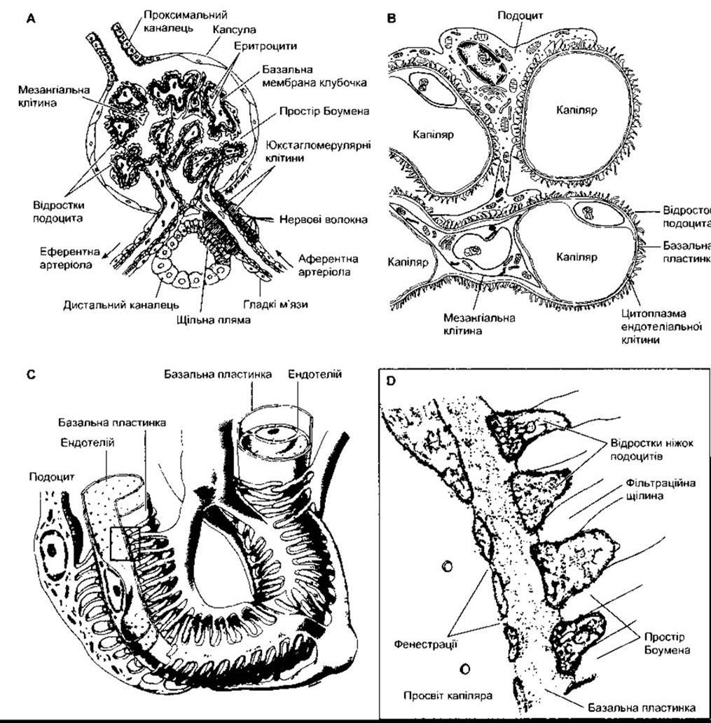
Fig. 38-3. Structural details of the glomerulus: A – section through the vascular pole showing capillary loops; B – relationship between mesangial cells and podocytes in glomerular capillaries; C – filtration slits in the basement membrane formed by podocytes, and the relationship between the membrane and capillary endothelium; D – enlarged view of C, showing podocyte FOOT processes. The blurred substance on their surface is the glomerular polyanion.
Furthermore, efferent arterioles contain few smooth Muscle fibers.
Capillaries collecting blood from cortical nephrons form the peritubular network, whereas efferent arterioles originating from juxtamedullary glomeruli empty not only into the peritubular network but also into vessels forming hairpin loops (vasa recta). These loops penetrate deep into the renal pyramids alongside the loops of Henle (see Fig. 38-2).
Descending vasa recta have a non-fenestrated endothelium containing urea transport systems, whereas ascending vasa recta feature a fenestrated endothelium consistent with their function of solute conservation (see below).
The efferent arteriole of each glomerulus divides into capillaries that supply blood to numerous different nephrons. Consequently, the tubule of each nephron receives blood not solely from its own efferent arteriole. In humans, the total surface area of the renal capillaries roughly corresponds to the total surface area of the tubules, amounting to about 12 m2. The volume of blood contained within the renal capillaries at any given time is 30–40 ml.
The kidneys feature a well-developed lymphatic network. Lymph drainage occurs via the thoracic lymphatic duct into the thoracic veins.
Capsule
The renal capsule is thin yet dense. When the kidney swells, the capsule restricts the expansion of the volumetric process, resulting in increased tissue pressure (renal interstitial pressure). This leads to a reduction in the glomerular filtration rate and is also believed to contribute to the severity and prolonged duration of anuria in ACUTE RENAL FAILURE (see Chapter 33).
Innervation of Renal Vessels
The renal nerves accompany the renal vessels until they enter the kidney. They contain a high density of postganglionic sympathetic nerve fibers and numerous afferent fibers. Cholinergic innervation mediated by the Vagus nerve also plays some role, although its specific function remains to be fully elucidated. Preganglionic sympathetic innervation is primarily supplied by the lower thoracic and upper lumbar segments of the Spinal Cord. The cell bodies of postganglionic Neurons are located in the sympathetic chain ganglia, the superior mesenteric ganglion, and along the renal artery. Sympathetic fibers project mainly to the afferent and efferent arterioles, the proximal and distal tubules, and the juxtaglomerular cells (see Chapter 24). In addition, a prominent noradrenergic Innervation of the thick ascending limb of the loop of Henle is observed.
Pain receptor fibers responsible for renal pain run parallel to the sympathetic efferent fibers and enter the spinal cord via the thoracic and upper lumbar dorsal roots. Other afferent renal nerves are thought to mediate the so-called renorenal reflex, which involves a decrease in efferent nerve activity in one kidney in response to an elevation of ureteral pressure in the contralateral kidney. This results in enhanced excretion of Na+ and water.
Last update: 10/08/2026
Editorial and Educational Adaptation: This material has been compiled based on the primary/original source text. The project team performed an editorial review, corrected technical inaccuracies, structured sections, and adapted the content for an educational format.
What was processed:
- elimination of formatting defects (OCR errors, structural breaks, corrupted characters);
- editorial organization of content;
- standardization of terminology in accordance with academic sources;
- verification of factual statements against the original source text.
All mentions of the author, publication year, and origin of the primary text have been preserved in accordance with the source.