Cytology, General Histology and Embryology - V. K. Naphanyuk 2002
Tissues
Epithelial Tissues
Simple Epithelium
Simple squamous epithelium (epithelium simplex squamosum) in the body is represented by endothelium and mesothelium.
Endothelium (endothelium) lines Blood and Lymphatic vessels and consists of a single layer of flat Cells called endothelial cells (endotheliocytes). These are polygonal cells with irregular, wavy borders; their Cytoplasm is poor in Organelles and contains pinocytotic vesicles. Some cells contain 2–3 nuclei.
Mesothelium (mesothelium) covers the serous membranes (pleural folds, visceral and parietal Peritoneum, and Pericardium). It is formed by flat, polygonal cells (mesotheliocytes) with uneven edges. The cells are slightly thicker in the region of the nuclei, and some contain two or even three nuclei. The free surface of mesotheliocytes bears sparse microvilli.
Simple cuboidal epithelium (epithelium simplex cuboideum) lines PARTS OF THE renal tubules (proximal and distal), the excretory ducts of many glands, and the small Bronchi of the Lungs. The cells of this epithelial type are equal in height and width. Cells of the proximal renal tubules feature a brush border and basal striation, which is caused by the accumulation of Cell/35.html">Mitochondria in the basal regions along with deep infoldings of the Plasmalemma.
Simple columnar epithelium
(epithelium simplex columnare) lines the internal surface of The Stomach, small and large intestines, Gallbladder, and certain ducts of The Liver and Pancreas.
Depending on Cell Structure, the following types of this epithelium are distinguished:
— striated / brush-bordered (located in the intestine, gallbladder);
— ciliated (Uterus, uterine tubes);
— glandular (stomach).
Pseudostratified ciliated columnar epithelium (epithelium pseudostratificatum) lines the respiratory tract, Nasal cavity, Trachea, bronchi, and several other Organs.
This epithelium comprises four cell types: ciliated, short and long intercalated (intercalary), mucous (goblet), and basal-granulated (endocrine) cells.
Slides for study
Slide 8. Simple squamous epithelium (mesothelium) (Fig. 32, see Fig. 35).
Low power magnification. Examine the slide. Under this magnification, identify the area where The Cell borders are most distinct.
High power magnification. Note that the mesothelial cells are flat and polygonal with irregular edges. Some cells contain 2–3 nuclei.
Draw the slide. Label the following on your drawing: 1) cell borders; 2) cell nuclei; 3) blood vessel beneath the epithelium.
Slide 9. Simple cuboidal and columnar epithelium of the renal tubules (Fig. 33).
Low power magnification. Examine the slide. Under this Microscope magnification, locate the renal medulla and identify the cross-sectioned tubules within it.
High power magnification. Observe the cell shape. It is evident that the epithelial cells have approximately equal height and width, which is a characteristic feature of cuboidal epithelium. Cell borders are very distinct, appearing as thin lines in this preparation; intercellular spaces are not visible here. The cell nuclei are round and located roughly in the center. The Cell Cytoplasm is somewhat granular.
Class="center">
Fig. 32. Simple squamous epithelium (mesothelium). Silver impregnation; hematoxylin staining. ×400:
1 — cell borders; 2 — cell nuclei
Draw the slide. Label the following on your drawing: 1) simple columnar epithelium; 2) simple cuboidal epithelium; 3) Connective Tissue; 4) Blood Vessels.
Slide 10. Pseudostratified ciliated epithelium of the trachea (Fig. 34, see Fig. 36).
Low magnification. Examine the slide. Determine the arrangement of the epithelium relative to other Tissues. It can be seen that the epithelium lines the inner surface of the trachea.
High magnification. Locate the ciliated cells, goblet cells, short intercalated (interstitial) cells, long intercalated cells, and the basement membrane. In ciliated cells, the nuclei are located in the upper row. Goblet cells feature a light cytoplasm. The nuclei of both short and long intercalated cells lie closer to the basement membrane.
Draw a diagram of the slide. Label the following in the drawing: 1) epithelium; a) cilia; b) rows of nuclei; 2) goblet cell; 3) connective tissue; 4) glands; 5) hyaline Cartilage.
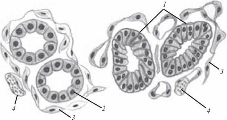
Fig. 33. Simple cuboidal and simple columnar epithelium of the renal tubules. Hematoxylin and eosin staining. x 400:
1 — simple columnar epithelium; 2 — simple cuboidal epithelium; 3 — connective tissue; 4 — blood vessels

Fig. 34. Pseudostratified ciliated epithelium of the trachea. Hematoxylin and eosin staining. x 600:
1 — epithelium (a — cilia; b — rows of nuclei); 2 — goblet cell; 3 — connective tissue; 4 — glands; 5 — hyaline cartilage

Fig. 35. Mesothelial cells. Electron micrograph of the rat oviduct mesothelium. Magnified to the scale indicated on the micrograph:
1 — nuclei of mesothelial cells; 2 — microvilli; 3 — borders between two mesothelial cells; 4 — basement membrane (after Odor)

Fig. 36. Cilia of the ciliated epithelium. Electron micrograph of the distal region of a ciliated epithelial cell from the rat trachea (a — cross-section of cellular cilia, x 96,000; b — Cytology/practical/54.html">Longitudinal section of cellular cilia, x 68,000):
1 — cell membrane (plasmalemma); 2 — cytoplasm of the cilium; 3 — peripheral doublet microfibrils; 4 — central doublet microfibril; 5 — basal body (kinetosome); 6 — cytoplasm of the distal region of the cell (after Sjöstrand)
Control Questions
1. Definition of the tissue concept. General characteristics of tissues.
2. Classification of tissues.
3. Sources of tissue development.
4. Epithelial Tissues. General characteristics.
5. Morphological classification of epithelial tissues.
6. Phylogenetic classification of epithelial tissues.
7. CHARACTERISTICS OF THE endothelium.
8. Characteristics of the mesothelium.
9. Characteristics of the simple cuboidal epithelium.
10. Characteristics of simple columnar epithelium.
11. Characteristics of pseudostratified epithelium.
Case studies
1. The specimen demonstrates tissues with the following structures: 1) a layer of closely apposed cells; 2) cells separated by Extracellular matrix. Which of the specified tissues is epithelial and why?
2. The specimen shows an epithelial layer formed by cells whose nuclei are located at different levels relative to the basement membrane. What type of epithelium is this?
Sample examination questions
1. Tissues. Histogenesis of tissues. Classification of tissues.
2. Epithelial tissues. Classification of epithelial tissues.
3. Structure and histophysiology of endothelium and mesothelium.
4. Structure and histophysiology of simple cuboidal and columnar epithelium.
5. Structure and histophysiology of pseudostratified epithelium.
Last update: 10/08/2026
Editorial and Educational Adaptation: This material has been compiled based on the primary/original source text. The project team performed an editorial review, corrected technical inaccuracies, structured sections, and adapted the content for an educational format.
What was processed:
- elimination of formatting defects (OCR errors, structural breaks, corrupted characters);
- editorial organization of content;
- standardization of terminology in accordance with academic sources;
- verification of factual statements against the original source text.
All mentions of the author, publication year, and origin of the primary text have been preserved in accordance with the source.