Cytology, General Histology and Embryology - V. K. Napkhanyuk 2002
Tissues
Epithelial Tissues
Glandular Epithelium
Glandular epithelium (epethelium glandulare) is formed by glandular or secretory Cells known as glandulocytes, which synthesize and release specific products—secretions—onto the Skin surface, mucous membranes, and into the cavities of certain Internal Organs (external or exocrine secretion), or directly into the Blood AND Lymph (internal or endocrine secretion).
Glandulocytes (glandulocyti) are highly specialized cells capable of synthesizing, accumulating, storing, and discharging secretions.
Glandulocytes rest on the basal lamina and exhibit a variety of shapes that change depending on the phase of secretion.
The nuclei of glandulocytes are large, with an irregular surface.
The Cytoplasm of glandulocytes producing proteinaceous secretions contains a well-developed rough Endoplasmic reticulum. In the cytoplasm of cells synthesizing non-protein secretions, the smooth endoplasmic reticulum is prominent. Additionally, glandulocytes possess a well-developed Golgi apparatus, the shape and Location of which may vary depending on the secretory phase. A large number of Cell/35.html">Mitochondria and secretory granules are also present. The Size and Structure of secretory granules depend on The chemical composition of the secretion, whereas their number depends on the secretory phase. The cytoplasm of certain glandulocytes (such as the gastric glandulocytes that produce Hydrochloric acid) contains intracellular secretory canaliculi, which represent deep invaginations of the Plasmalemma whose walls are covered with microvilli.
The structure of the glandulocyte plasmalemma differs across various cell surfaces.
On the lateral surfaces, it forms desmosomes and tight junctions (terminal bars).
The apical surface is covered with microvilli.
The basal surface of the plasmalemma forms a small number of narrow folds that penetrate into the cytoplasm.
Glandulocytes display a well-defined polar differentiation driven by the directionality of secretory processes.
Glands
Glands (glandulae) are organs whose primary functional tissue is epithelial (with the exception of The adrenal medulla, the posterior pituitary, and the Pineal Gland), capable of synthesizing and releasing specific secretions.
Glands can be unicellular or multicellular. Each type of gland possesses characteristic features. The entire diversity of specific differentiations among secretory cells represents a Modification of the metabolic apparatus common to all cytoplasm; therefore, the differentiation of glandular cells is associated with the hypertrophy and specialization of general systems for the synthesis, transport, and accumulation of substances within cells. The ability to release secretions emerged and evolved from the fundamental cellular capacity to expel Metabolic waste products into the external environment.
Unicellular glands can vary in shape, residing either within the thickness of the epithelial layer (endoepithelial glands) or outside it (exoepithelial glands), and are capable of synthesizing protein, mucopolysaccharide, mucoprotein, lipoprotein, and other secretions (Figs. 40, 41).
Class="center">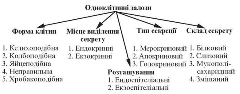
Fig. 40. Classification of unicellular glands
Multicellular glands contain cells of two main types: secretory (acinar, glandular) cells, which produce the secretion specific to the given gland; and duct cells, through which the secretion is transported or temporarily stored, releasing excess Water and salts or being supplemented with mucous, protein, mineral, and other components.
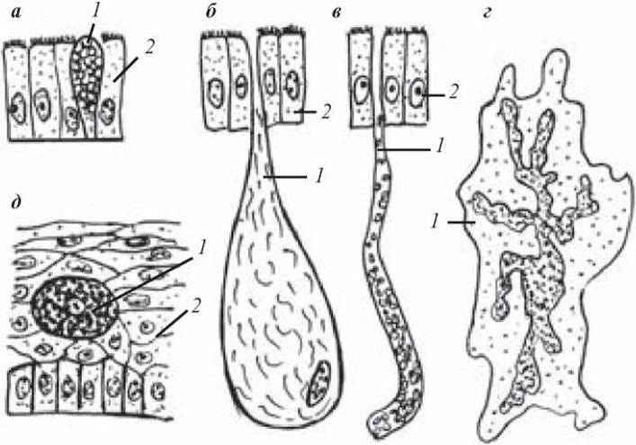
Fig. 41. Diagram of the structure of unicellular glands:
a — goblet endoepithelial gland of the intestine; b — flask-shaped exoepithelial gland of the skin; c — tubular exoepithelial gland; d — unicellular endocrine gland with a branched Nucleus; e — oval endoepithelial gland of the skin (Leydig cell); 1 — unicellular gland; 2 — epithelial layer proper
Although the STRUCTURE AND Functions of these cells vary, duct cells frequently perform a secretory function In addition to filtration and mechanical roles.
Multicellular glands may feature branched or unbranched excretory ducts. Glands with a single excretory duct are termed simple, whereas those with branched ducts are termed compound. Secretory portions, in turn, can be branched or unbranched. In the former case, several secretory units empty into a single duct (branched glands). If the terminal units are unbranched, each duct terminates in a single secretory endpiece; such glands are designated as "unbranched." Based on the shape of their terminal portions, glands are classified as tubular, alveolar, or tubulo-alveolar. Multicellular glands can be either exo- or endoepithelial, although the majority belong to the exoepithelial type. According to the direction of secretion release, multicellular glands are divided into exocrine and endocrine. The former discharge their secretion onto a free surface or into internal Body Cavities (exocrine glands), while the latter release their secretions directly into the blood (Endocrine glands). Multicellular glands can also be categorized by the Chemical composition of their secretion and by their mode of secretion (Fig. 42).
Secretory Cycle
Secretion is a complex process involving the synthesis, accumulation, and expulsion of cell-synthesized substances beyond its boundaries.
The secretory cycle consists of four phases:
— absorption is the process by which glandular cells extract various substances from blood and lymph, including Inorganic Compounds (water, mineral salts, ions) and Organic compounds (Amino Acids, Monosaccharides, Fatty acids);
— synthesis and accumulation is the process during which a secretion is formed from these compounds (proteinaceous in the granular endoplasmic reticulum; non-proteinaceous in the agranular reticulum). The synthesized product is transported to the Golgi apparatus zone, where it gradually accumulates, undergoes chemical modification, and is packaged into granules;
— discharge — secretory granules are released from The Cell via one of the pathways (holocrine, apocrine, merocrine);
— restoration — the process by which the initial state of the glandular cells is recovered.
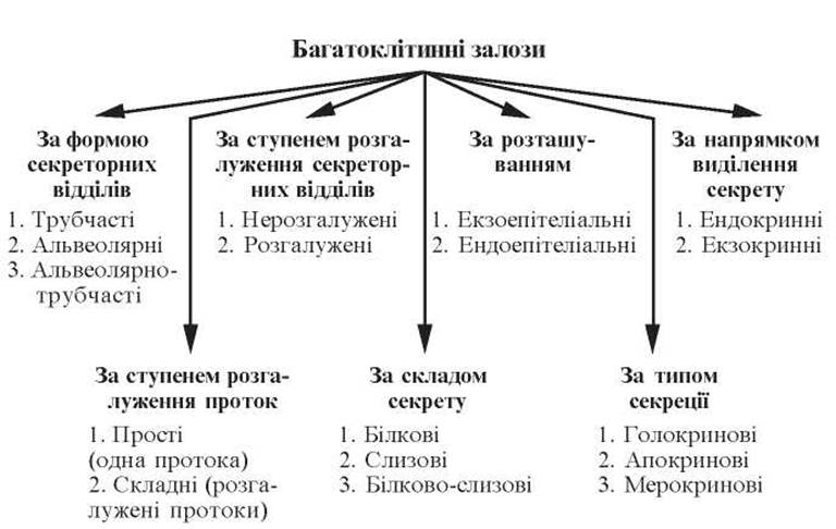
Fig. 42. Classification of multicellular glands
These phases may occur sequentially, cyclically, as part of a secretory cycle. Sometimes they occur simultaneously, which is characteristic of diffuse or spontaneous secretion.
Control Questions
1. Classification of Stratified Epithelium.
2. Characteristics of stratified squamous non-keratinizing epithelium.
3. Characteristics of stratified squamous keratinizing epithelium.
4. Characteristics of transitional epithelium.
5. Glandular epithelium. Characteristics of glandulocytes.
6. Unicellular glands. Structure. Classification.
7. Multicellular glands. Structure. Classification.
8. Secretion. Secretory cycle.
Situational Problems
1. Two glandular preparations are provided. One gland has an unbranched excretory duct and a branched secretory portion. The other gland has both a branched excretory duct and a branched secretory portion. To which gland types do they belong?
2. A gland has a single layer of secretory cells (exocrinocytes), whose cytoplasm contains a well-developed Golgi apparatus and secretory granules. The Cell Nucleus is of normal structure (neither condensed nor fragmented), and cytoplasmic Organelles are preserved. No signs of apical cytoplasm detachment are detected under light or Electron Microscopy. By which mechanism does this gland release its secretion?
Sample Examination Questions
1. Structure of various types of stratified epithelium.
2. Glandular epithelium. Structural Features of glandulocytes.
3. Glands. Classification of glands. Structure. Secretory cycle.
Last update: 10/08/2026
Editorial and Educational Adaptation: This material has been compiled based on the primary/original source text. The project team performed an editorial review, corrected technical inaccuracies, structured sections, and adapted the content for an educational format.
What was processed:
- elimination of formatting defects (OCR errors, structural breaks, corrupted characters);
- editorial organization of content;
- standardization of terminology in accordance with academic sources;
- verification of factual statements against the original source text.
All mentions of the author, publication year, and origin of the primary text have been preserved in accordance with the source.