Review of Medical Physiology - William F. Ganong 2002
Functions of the Digestive System
Digestion and Absorption
Water and Electrolyte Absorption
Water, sodium, potassium, chloride
The overall water balance in the gastrointestinal tract is summarized in Table 25-3. Each day, the intestinal lumen receives about 2000 mL of ingested fluid and 7000 mL of various secretions produced by the gastrointestinal mucosa and associated glands. Of this fluid, 98% is reabsorbed, and daily losses in stool amount to 200 mL. Only a small amount of water passes directly across the gastric mucosa, whereas in the small and large intestines, water moves in both directions depending on the osmotic gradient. A fraction of Na+ diffuses into or out of the Small Intestine along its concentration gradient. Because the luminal membranes of all enterocytes in the small and large intestines are permeable to Na+, and the basolateral membranes contain Na+-K+-ATPase, Na+ is also actively absorbed in both the small and large intestines. In the small intestine, secondary Active Transport of Na+ plays a crucial role in supplying energy during the absorption of glucose, Certain Amino Acids (see above), and other substances, while the presence of glucose in the intestinal lumen facilitates Na+ reabsorption. This physiological principle forms the basis for treating Na+ and water losses in diarrhea *per os* using various solutions containing NaCl and glucose. Carbohydrate-containing cereal flakes are also used to treat diarrhea. This approach is equally valuable in the management of cholera—a disease characterized by severe and, if left untreated, frequently fatal diarrhea.
Class="center">
Fig. 25-5. Fat absorption. Triglycerides are synthesized within mucosal Cells from monoglycerides and Fatty acids. Some glycerides are also formed from glucose via phosphatidic acid. Subsequently, triglycerides are incorporated into chylomicrons and released via exocytosis. From the extracellular space, they enter the lymphatics. Bold lines indicate major pathways; * denotes a reaction inhibited by monoglycerides; MGAT = monoacylglycerol acyltransferase; DGAT = diacylglycerol acyltransferase.
Under normal conditions, Cl- enters enterocytes from the interstitial fluid via basolateral Na+-K+-2Cl- cotransporters (Fig. 25-7). It is then secreted into the intestinal lumen through channels regulated by numerous protein Kinases. One of these is activated by protein kinase A and subsequently by cAMP. In cholera, the intracellular concentration of cAMP increases significantly. The cholera bacterium resides in the intestinal lumen, where it produces a toxin that binds to the GM1 ganglioside receptor, allowing a portion of the toxin's A-subunit (the A1 peptide) to enter The Cell. The A1 peptide catalyzes the ADP-ribosylation of the Gs *alpha*-subunit, which inhibits its GTPase activity (see Chapter 1). Consequently, the constitutively activated G protein causes prolonged stimulation of adenylate cyclase and a marked elevation in intracellular cAMP concentration. Alongside increased Cl- secretion, the mucosal carrier function for Na+ is diminished, leading to reduced NaCl absorption. The net result is an increase in the fluid volume and electrolyte content of the intestine, culminating in diarrhea. Furthermore, neither Na+-K+-ATPase nor the Na+/glucose cotransporter is fully activated, rendering the coupled reabsorption of glucose and Na+ defective.
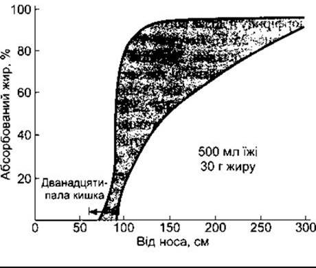
Fig. 25-6. Dynamics of fat absorption in humans, based on measurements following a fatty meal. The arrow indicates the duodenum (reprinted with permission from Davenport HW: PHYSIOLOGY OF THE Digestive Tract, 2nd ed. Year Book, 1966).
Water moves into or out of the intestine only when the Osmotic Pressure of the intestinal contents matches that of plasma. Depending on the ingested food, the osmolality of duodenal contents may be hypertonic or hypotonic; however, by the time the chyme reaches the jejunum, its osmolality approaches that of plasma, a level maintained throughout the interdigestive period in the intestine. As osmotically active particles generated during Digestion are absorbed, water moves passively out of the intestine along the newly established osmotic gradient. In the colon, active extrusion of Na+ occurs, driving the passive Movement of water along with it, again down an osmotic gradient. Mineral laxatives, such as magnesium sulfate—poorly absorbable osmotically active salts—retain water in the gut to restore osmotic equilibrium. The resulting increase in intestinal volume produces the laxative effect.
Table 25-3. Daily Water Balance in the Gastrointestinal Tract1, mL
Ingested |
2000 |
|
Endogenous secretions |
7000 |
|
1500 |
||
2500 |
||
500 |
||
1500 |
||
Intestine |
1000 |
|
7000 |
||
Total amount |
9000 |
|
Reabsorbed |
8800 |
|
Jejunum |
5500 |
|
Ileum |
2000 |
|
Colon |
1300 |
|
8800 |
||
Fecal content |
200 |
1 Data from Moore EW: Physiology of Intestinal Water and Electrolyte Absorption, American Gastroenterological Society, 1976.

Fig. 25-7. Movement of ions across small intestinal enterocytes: Cl- enters the cell via the basolateral Na+-K+-2Cl- cotransporter and is secreted into the intestinal lumen through Cl- channels, some of which are activated by cAMP; K+ is recycled into the interstitial fluid (IF) via basolateral K+ channels (reprinted with permission from Field M, Rao MC, Chang EB: Intestinal electrolyte transport and diarrheal disease. N Engl J Med 1989;321:800).
Only a small amount of K+ is secreted into the intestinal lumen specifically as a component of mucus, whereas the majority arrives via passive diffusion of K+ across the gastrointestinal mucosa. Additionally, K+ is secreted into the colon because K+ channels are present in both the luminal and basolateral membranes of colonic enterocytes. This movement of K+ occurs passively along the electrochemical gradient.
The accumulation of K+ in the colon is partially counterbalanced by H+-K+-ATPase located in the luminal membrane of distal colonic cells, which mediates the net active uptake of K+ into the cells. Despite this compensatory mechanism, fluid losses during chronic diarrhea can lead to profound hypokalemia.
During prolonged excessive Dietary intake of K+, aldosterone secretion increases, leading to enhanced delivery of K+ to the colon. This is partly mediated by an upregulation of Na+-K+-ATPase pumps in the basolateral membranes of the cells, resulting in an elevated intracellular K+ content and increased K+ diffusion across the luminal membranes.
Last update: 10/08/2026
Editorial and Educational Adaptation: This material has been compiled based on the primary/original source text. The project team performed an editorial review, corrected technical inaccuracies, structured sections, and adapted the content for an educational format.
What was processed:
- elimination of formatting defects (OCR errors, structural breaks, corrupted characters);
- editorial organization of content;
- standardization of terminology in accordance with academic sources;
- verification of factual statements against the original source text.
All mentions of the author, publication year, and origin of the primary text have been preserved in accordance with the source.