Human Biochemistry, Volume 2 - Murray R. 1993
Biochemistry of Intracellular and Intercellular Communication
Hormones Regulating Calcium Metabolism
Hormones Involved in Calcium Homeostasis - Parathyroid Hormone (PTH)
PTH is a single-chain peptide consisting of 84 amino acid residues (mol. wt. 9500) and containing no CARBOHYDRATES or other covalently linked components (Fig. 47.1). The entire biological activity resides in the N-terminal third of the molecule: $ ext{PTH}_{1-34}$ is fully active. The 25–34 region is primarily responsible for receptor binding.
PTH is synthesized as a precursor molecule consisting of 115 amino acid residues (Fig. 47.1). The immediate precursor of PTH is proPTH, which differs from the active hormone by having an additional basic hexapeptide at the N-terminus of uncertain function. The primary Gene product and the direct precursor of proPTH is preproPTH; it differs from proPTH by possessing an additional N-terminal sequence of 25 amino acid residues that exhibits hydrophobic properties (typical of leader or signal sequences characteristic of secretory Proteins). The complete structure of preproPTH as well as the sequences of proPTH and PTH are shown in Fig. 47.1.
Class="center">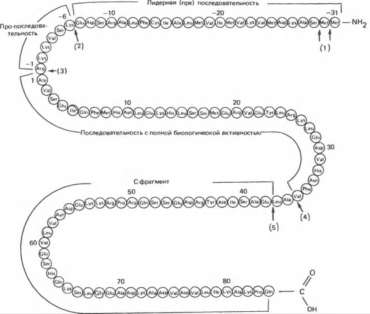
Fig. 47.1. Structure of bovine preproparathyroid hormone. Arrows indicate sites of enzymatic Cleavage during hormone Processing in the parathyroid gland (1–5) and after hormone secretion in the Liver (4, 5). Attached to the biologically active region of the molecule is a sequence not involved in receptor-mediated activity. (Slightly modified and reproduced, with permission, from Habener J. F., Recent advances in parathyroid hormone research. Clin. Biochem 1981; 14:223.)
PreproPTH was the first preprohormone to be identified. The Sequential Stages of its conversion into PTH are shown in Fig. 47.2. As preproPTH molecules are synthesized on Ribosomes, they are translocated into the cisternae of The Endoplasmic reticulum. During translocation, the 25-amino-acid prepeptide (signal or leader peptide) is cleaved off, yielding proPTH. Next, proPTH is transported to the Golgi apparatus, where Enzymatic cleavage of the propeptide takes place, producing the final product, PTH. From the Golgi apparatus, PTH enters secretory vesicles, where the hormone may 1) be stored, 2) undergo degradation, or 3) be secreted immediately.
Involvement of PTH in Mineral Homeostasis
A. Calcium Homeostasis. The Central Role of PTH in calcium METABOLISM is highlighted by the observation that in the course of evolution, this hormone first appears in animals attempting to adapt to terrestrial life. The physiological mechanism maintaining calcium balance relies on the long-term effects of PTH, which regulates intestinal calcium absorption by stimulating calcitriol production. In cases of chronic dietary $ ext{Ca}^{2+}$ deficiency, intestinal absorption fails to meet physiological demands, thereby engaging a complex regulatory system in which PTH also participates. Here, PTH restores normal extracellular fluid calcium levels through direct actions on bone and Kidneys and an indirect action (via stimulation of calcitriol synthesis) on the intestinal mucosa. PTH 1) increases The rate of bone dissolution (leaching out both organic and Inorganic Components), which drives $ ext{Ca}^{2+}$ into the ECF; 2) decreases renal clearance, i.e., calcium excretion, thereby promoting an increase in the concentration of this cation in the ECF; 3) increases the efficiency of intestinal $ ext{Ca}^{2+}$ absorption by stimulating calcitriol production. The renal effects of PTH manifest most rapidly, whereas its actions on bone are the most robust. Thus, PTH prevents The Development of hypocalcemia during dietary calcium deficiency, albeit at the expense of bone substance.

Fig. 47.2. Precursors and cleavage products of PTH, and the localization of individual degradation steps in the Parathyroid glands and liver. Numbers in parentheses indicate the number of Amino Acids in the pre (31) and pro (6) fragments.
B. Phosphate Homeostasis. Phosphate is the physiological ion paired with calcium; hydroxyapatite crystals in bones consist of calcium phosphate. When PTH stimulates the dissolution of the bone mineral matrix, phosphate is released alongside calcium. PTH also enhances the renal clearance of phosphate. Consequently, the net effect of PTH on bone and kidneys results in an increased calcium concentration and a decreased phosphate concentration in the ECF. Crucially, this prevents the supersaturation of Blood Plasma with calcium and phosphate.
Biochemistry
A. Regulation of Synthesis. The concentration of $ ext{Ca}^{2+}$ in the medium does not affect the rate of proPTH synthesis, but the rates of PTH formation and secretion increase markedly when $ ext{Ca}^{2+}$ concentrations drop. It has been shown that 80–90% of synthesized proPTH cannot be recovered as intact PTH accumulated in Cells or in the incubation medium during in vitro experiments. Hence, it was concluded that the bulk of synthesized proPTH undergoes rapid degradation. Later, it was discovered that the rate of this degradation decreases at low $ ext{Ca}^{2+}$ concentrations and increases at high ones. Thus, calcium regulates PTH production through the modulation of degradation rather than synthesis. Total proPTH synthesis can be gauged by The amount of PTH mRNA; notably, this level remains unchanged even during substantial fluctuations in extracellular $ ext{Ca}^{2+}$ concentrations. Apparently, an increase in systemic PTH synthesis can only occur As a result of an increase in the number and size of the chief Cells of the parathyroid glands that produce PTH.
B. REGULATION OF METABOLISM. The degradation of PTH begins approximately 20 min after proPTH synthesis and initially proceeds independently of $ ext{Ca}^{2+}$ concentration; the molecules undergoing degradation are those residing within secretory vesicles. Newly formed PTH is either secreted immediately or stored in vesicles for subsequent secretion. Degradative processes are initiated after secretory vesicles enter the storage compartment.
Proteolytic cleavage of PTH yields highly specific fragments (Figs. 47.1 and 47.2), with large amounts of C-terminal PTH fragments entering the Circulation. Their molecular weight is approximately 7000. These are primarily the $ ext{PTH}_{37-84}$ sequence, and to a lesser extent, $ ext{PTH}_{34-84}$. Most newly synthesized PTH undergoes proteolysis; overall, approximately two moles of C-terminal fragments are secreted per mole of intact PTH. Consequently, circulating PTH is represented predominantly by these molecules. The Biological Role of C-terminal PTH fragments remains elusive, but they may serve to prolong the half-life of the hormone in the bloodstream. A number of Proteolytic Enzymes, including cathepsins B and D, have been identified in parathyroid tissue. Cathepsin B cleaves PTH into two fragments—$ ext{PTH}_{1-16}$ and $ ext{PTH}_{37-84}$; the latter undergoes no further proteolysis, whereas $ ext{PTH}_{1-36}$ is rapidly and sequentially degraded down to di- and tripeptides. ProPTH does not enter the bloodstream, and $ ext{PTH}_{1-34}$ escapes the gland in minimal quantities (if at all). PreproPTH was successfully identified by decoding the coding sequence of the PTH gene. PTH proteolysis occurs mainly within the parathyroid gland, although several studies indicate that secreted PTH also undergoes proteolysis in other Tissues. However, THE CONTRIBUTION OF this extra-endocrine process to the overall proteolytic degradation of PTH has not been quantified, nor is it known which proteases are involved or how closely their cleavage sequences and products match.
The peripheral metabolism of secreted PTH involves both The Liver and the kidneys. Following hepatectomy, fragments 34–84 virtually vanish from the blood, implying that the liver serves as the principal organ for their generation. The Role of the kidneys appears to be the clearance and excretion of these fragments from the blood. Peripheral proteolysis takes place primarily in Kupffer cells lining the lumen of hepatic sinusoids. The endopeptidase responsible for the initial step of proteolysis (cleavage into N- and C-terminal fragments) is localized On the surface of these macrophage-like cells, which are in direct contact with blood plasma. This enzyme, also identified as cathepsin B, cleaves PTH between residues 36 and 37; similarly to the events in the parathyroid gland, the resulting C-terminal fragment continues to circulate in the bloodstream, whereas the N-terminal fragment is rapidly degraded.
C. Regulation of Secretion. PTH secretion is inversely dependent on the concentration of calcium and magnesium ions in the medium, as well as on the circulating level of immunoreactive PTH. As shown in Fig. 47.3, a linear relationship exists between serum PTH content and serum calcium concentration (ranging from 4 to 10.5 mg%).
The presence of biologically active PTH in blood serum when calcium levels reach 10.5 mg% or higher is indicative of hyperparathyroidism.
A linear relationship also exists between PTH release and intracellular cAMP levels in parathyroid cells. This relationship is likely mediated by shifts in intracellular $ ext{Ca}^{2+}$ levels, given the inverse correlation between intracellular concentrations of $ ext{Ca}^{2+}$ and cAMP. This may stem from the well-known activating effect of calcium on phosphodiesterase (via $ ext{Ca}^{2+}$/calmodulin-dependent protein kinase) or an inhibitory effect (via a similar mechanism) on adenylate cyclase. Phosphate has no effect on PTH secretion.
The parathyroid glands contain relatively few storage granules, and their hormone content can sustain maximal secretion for only about 1.5 hours. This contrasts sharply with pancreatic islet tissue, where Insulin stores suffice for several days of secretion, and The Thyroid Gland, which holds a hormone reserve lasting several weeks. Thus, the Synthesis and Secretion of PTH must proceed continuously.
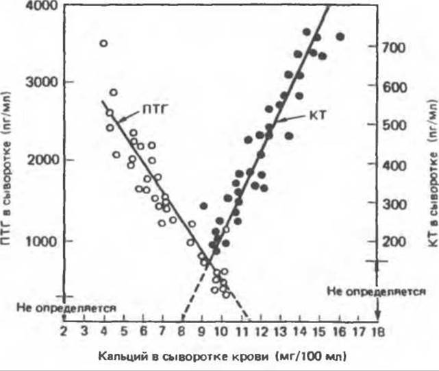
Fig. 47.3. Concentration of Calcitonin (CT) and parathyroid hormone (PTH) as a function of plasma calcium concentration. (Modified and reproduced, with permission, from Arnaud C. D., Littleluke T., Tsao H.S. Calcium homeostasis and the simultaneous measurement of calcitonin and parathyroid hormone in pig. Pages 95 — 101. In: Calcitonin: Proc. of the 2 nd intern. Symp. Taylor S. (ed.). Heinemann, 1969.)
A. The PTH Receptor. PTH binds to a membrane receptor, which is a monomeric protein with a Molecular Weight of approximately 70,000. Receptors in Kidney and bone cells appear identical; this protein is absent in cells that are not targets for PTH. The interaction of the hormone with its receptor triggers a classic cascade of events: activation of adenylate cyclase $ ightarrow$ elevation of cellular cAMP concentration $ ightarrow$ increase in intracellular calcium content $ ightarrow$ phosphorylation of specific intracellular proteins by Kinases $ ightarrow$ activation of designated intracellular enzymes or proteins that ultimately dictate the biological actions of the hormone. Like the systems for other protein and Peptide Hormones, the PTH-responsive system is subject to down-regulation of receptor number; moreover, it exhibits The phenomenon of "desensitization," The Mechanism of which is linked not to changes in cAMP content, but to downstream reactions within the cascade.
B. Effects of PTH on Bone. PTH exerts multifaceted effects on Bone tissue, apparently acting on various Cell types within it. The net effect of PTH is bone destruction accompanied by the release of calcium, phosphorus, and organic matrix elements, including Collagen breakdown products. The cells responsible for this process may be osteoclasts, which have been proven to resorb bone during chronic PTH stimulation, or osteocytes, which are also capable of bone resorption. Alternatively, PTH may stimulate the differentiation of precursor cells into bone-resorbing cells. At low concentrations, presumably corresponding to physiological levels, PTH exerts an anabolic effect and is responsible for bone remodeling. Exposure to these hormone concentrations leads to an increased number of osteoblasts, elevated alkaline phosphatase activity indicative of new Bone Formation, and enhanced incorporation of radioactive sulfur (as sulfate) into Cartilage. Calcitriol may play a permissive role in the action of PTH on bone.
The intracellular mediator of PTH appears to be Ca2+. The initial manifestation of the PTH effect is a decrease in the concentration of Ca2+ in the pericellular space alongside an increase within The Cell itself. This rise in intracellular calcium stimulates RNA Synthesis in bone cells and the release of enzymes involved in bone resorption. These processes are presumably mediated by the binding of calcium to calmodulin. In the absence of extracellular calcium, PTH still increases cAMP levels, yet it no longer stimulates bone resorption. Thus, a paradoxical influx of ionized calcium into bone-resorbing cells may be a crucial prerequisite for the stimulatory action of PTH on bone resorption.
B. Effect of PTH on the kidneys. PTH exerts a wide range of effects on the kidneys, specifically influencing The transport of certain ions and regulating the synthesis of calcitriol. Under normal conditions, over 90% of the Ca2+ contained in the glomerular filtrate undergoes reabsorption, but PTH increases this figure to 98% or more. Phosphate reabsorption normally ranges from 75–90% depending on diet and other factors; however, PTH inhibits phosphate reabsorption regardless of its baseline level. PTH also inhibits the transport of sodium, potassium, and bicarbonate ions. The Effect of PTH on calcitriol metabolism (see below) appears to be mediated through the same cellular sites as its action on Mineral Metabolism.
Infusion of PTH leads to a rapid increase in cAMP concentration within renal cells and enhanced urinary excretion of cAMP. This effect precedes the phosphaturia characteristic of PTH action and is evidently responsible for it. PTH-stimulated adenylate cyclase is located on the basolateral membrane of cells within the cortical segments of the renal tubules; it differs from renal adenylate cyclase stimulated by calcitonin, catecholamines, or ADH. Intracellular cAMP receptor proteins (commonly considered to be protein kinases) are found in the brush border of these cells on the luminal surface of the tubules. Consequently, cAMP synthesized under The Influence of PTH migrates from the basolateral region of the cell to its luminal surface, where it exerts its effect on ion transport.
Calcium appears to be involved in the mechanism of PTH action on the kidneys. Indeed, the primary Physiological Effect of PTH administration is a reduction of Ca2+ levels in the extracellular fluid and an increase within the cell. However, these shifts occur following changes in intracellular cAMP concentration; consequently, The Link Between the influx of Ca2+ into cells and the action of PTH is less distinct in the kidney than in bone.
C. Effect of PTH on the intestinal mucosa. PTH apparently does not have a direct effect on Ca2+ Transport Across the intestinal mucosa, but it serves as a critical regulatory factor in The Biosynthesis of calcitriol (see below) and exerts a vitally important indirect effect on the intestines.
PTH deficiency leads to hypoparathyroidism. The biochemical hallmarks of this condition are a decreased serum level of ionized calcium and an elevated serum phosphate level. Symptoms include heightened neuromuscular excitability, causing Muscle cramps and tetany (in moderate cases). Severe acute hypocalcemia leads to tetanic paralysis of the Respiratory Muscles, laryngospasm, severe convulsions, and death. Prolonged hypocalcemia is accompanied by Skin changes, cataract development, and calcification of the Basal Ganglia of the Brain. Hypoparathyroidism is typically caused by the accidental removal of or damage to the parathyroid glands during neck surgery (secondary hypoparathyroidism), though the disease occasionally arises from autoimmune destruction of the parathyroid glands (primary hypoparathyroidism).
Pseudohypoparathyroidism is discussed in Chapter 44. In this inherited disorder, the endocrine gland produces biologically active PTH, but the target Organs are resistant to it, rendering the hormone ineffective. As a result, the same biochemical shifts occur as in hypoparathyroidism. These are typically associated with developmental abnormalities such as short stature, shortened metacarpals and metatarsals, and mental retardation. Several types of pseudohypoparathyroidism exist; they are attributed either 1) to a partial deficiency of the regulatory Gs protein of the adenylate cyclase complex or 2) to a disruption at some step unrelated to the cAMP generation mechanism.
Hyperparathyroidism, i.e., the Excessive production of PTH, typically results from a parathyroid adenoma, but it may also be caused by parathyroid hyperplasia or ectopic PTH production by a malignant tumor. The biochemical criteria for hyperparathyroidism include elevated levels of ionized calcium and PTH alongside a decreased serum phosphate level. In advanced cases of hyperparathyroidism, pronounced skeletal bone resorption and various renal pathologies can be observed, including Nephrolithiasis, nephrocalcinosis, frequent Urinary Tract infections, and (in some instances) impaired renal function. Secondary hypoparathyroidism, characterized by parathyroid hyperplasia and hypersecretion of PTH, is observed in patients with renal failure. It is believed that the development of hyperparathyroidism in these patients stems from impaired synthesis of 1,25-(OH)2-D3 from 25-OH-D3 within the pathologically altered renal parenchyma, leading to impaired intestinal calcium absorption; this impairment, in turn, triggers a secondary release of PTH as a compensatory mechanism to maintain normal ECF calcium levels.
Last update: 06/08/2026
Editorial and Educational Adaptation: This material has been compiled based on the primary/original source text. The project team performed an editorial review, corrected technical inaccuracies, structured sections, and adapted the content for an educational format.
What was processed:
- elimination of formatting defects (OCR errors, structural breaks, corrupted characters);
- editorial organization of content;
- standardization of terminology in accordance with academic sources;
- verification of factual statements against the original source text.
All mentions of the author, publication year, and origin of the primary text have been preserved in accordance with the source.