BIOCHEMISTRY - V. V. Emelyanov - 2016
SECTION 6. METABOLIC REGULATION. BIOCHEMISTRY OF BLOOD
1.1. Metabolic Regulation
METABOLISM is a unified, integrated process. The separate consideration of carbohydrate, protein, and Lipid Metabolism is strictly a pedagogical approach. Various aspects of metabolism are integrated by regulatory systems: the nervous, endocrine, and immune systems. The Endocrine System and BIOLOGICALLY ACTIVE SUBSTANCES known as Hormones play the leading role in metabolic regulation.
The science that studies the hormonal Regulation of Metabolism and the physiological Functions of the Organism is called endocrinology. It encompasses both fundamental biological and clinical disciplines. Modern endocrinology emerged as an independent discipline about 100 years ago. Today, it is a high-tech and rapidly developing branch of biology equipped with a full range of morphological, chemical, biochemical, and molecular-genetic Research Methods. These studies have deepened our understanding of the physiology and pathology of Endocrine glands, revealed the chemical structures and Mechanisms of action of hundreds of biologically active substances, and enabled the synthesis of pharmaceutical drugs based on them.
Throughout our study of biochemistry, we have repeatedly encountered various molecules with regulatory properties that serve to transmit information in living systems. Such molecules are referred to as informones or signaling molecules. Among the latter, a specific group is traditionally classified as hormones based on A number of shared properties.
6.1.1. Hormones: Definition and Properties
Hormones are biologically active substances produced by specialized endocrine Cells, secreted into the Blood or Lymph, and capable of exerting effects on the Structure AND FUNCTIONS of the organism at a site distant from their production. A prohormone is a biologically inactive precursor of a hormone that is converted into the active hormone through specific metabolic reactions.
KEY FEATURES OF hormone action:
1. Synthesis and Secretion by specialized cells. It is now evident that the endocrine system comprises not only well-known endocrine glands but also individual cells that secrete signaling molecules into the bloodstream. Such cells are found within the gastrointestinal tract, Kidneys, Liver, Lungs, Heart, adipose tissue, and The Immune System. All these cells are collectively referred to as the Cytology/cytology/65.html">Diffuse endocrine system.
2. High biological activity. Hormones exhibit extremely high biological activity and are present in biological fluids at micro-, nano-, and picomolar concentrations. Consequently, the Quantitative determination of hormones in biological fluids is performed using highly sensitive immunochemical methods (such as radioimmunoassay, enzyme-linked immunosorbent assay [ELISA], immunochemiluminescence, etc.).
3. Specificity of action. Each hormone elicits a unique spectrum of biological effects exclusively in cells that possess specific receptors for it, known as target cells. Cells lacking these receptors are insensitive to the hormone and are described as hormone-resistant. Typically, a hormone has a primary (main) and secondary effects. For instance, glucocorticoids primarily stimulate Gluconeogenesis, while their secondary effect is anti-inflammatory action. In some cases, several hormones share a unidirectional effect; for example, cortisol, adrenaline, and Glucagon all increase blood glucose levels, yet they differ in their other physiological effects, which provides the unique profile of each hormone.
4. Distant action. A defining criterion for a hormone is that it exerts its biological effect far from its site of origin and is transported via the blood or lymph (hemocrine action). If a substance secreted by an endocrine Cell exerts its effect not only via blood transport but also through tissue fluid, it is referred to as isocrine action. For example, evidence has been obtained regarding the mutual influence of various hormones on neighboring cells within the same endocrine gland, such as the interactions among cells in the islets of Langerhans, adenohypophysis, and adrenal cortex. A specific case of isocrine action is the neurocrine action of hypothalamic releasing and inhibiting factors (liberins and statins).
Substances that do not meet this criterion may be considered hormone-like substances (hormonoids, autacoids):
a) "cellular hormones" act on the same cell in which they are produced (autocrine action);
b) "tissue hormones" act on neighboring cells (paracrine action); if the hormone does not enter the intercellular fluid but is transmitted directly through The Plasma Membrane of an adjacent cell, it is termed juxtacrine action.
5. Regulability of action. Hormonal regulation is designed to ensure the optimal adaptation of the organism to environmental conditions; therefore, the quantity and spectrum of hormones present in the blood and acting on target cells at any given moment are subject to diverse influences and exhibit biorhythms of various periodicities. The regulation of hormonal activity can occur at all stages of their metabolism.
6.1.2. Classification of hormones
In biochemistry, the most convenient classification of hormones is based on their chemical nature:
1. Protein-Peptide Hormones: oligopeptides (liberins and statins, ACTH, ADH, oxytocin, glucagon, etc.), simple Proteins (Insulin, GH, PRL), and Glycoproteins (LH, FSH, TSH).
2. Amino Acid Derivatives (catecholamines, THYROID HORMONES, melatonin).
3. Steroids (glucocorticoids, mineralocorticoids, androgens, estrogens, progestogens).
Classification by chemical nature is useful not only because it allows every hormone to be definitively assigned to a specific group, but also because the chemical structure predetermines the features of its biological action (blood transport, interaction with receptors).
Additionally, hormones are classified:
- by their site of production (Hormones of the Hypothalamus, Pituitary Gland, Pineal Gland, thyroid, Parathyroid glands, Pancreas, Gonads, and Adrenal Glands).
- by physiological functions (regulators of carbohydrate, protein, lipid, Water-salt, and calcium-phosphorus metabolism, as well as reproductive functions and The activity of peripheral endocrine glands).
Based on the mechanisms of reception by target cells, hormones are classified into:
- those acting on Membrane Receptors (proteins, amino acid derivatives);
- those acting on intracellular receptors (steroids);
- those acting on both types of receptors (thyroid hormones).
Principles of Organization OF THE Neuroendocrine System
1. Hierarchy. The endocrine system operates across several levels of regulation:
a) Cerebral Cortex;
b) subcortical structures;
c) hypothalamus;
d) pituitary gland;
e) peripheral endocrine glands;
f) target cells (receptors and post-receptor signal Transduction mechanisms).
Modern endocrinology posits the existence of three interacting regulatory systems in the organism: nervous, endocrine, and immune. The interaction among the nervous, endocrine, and immune systems can be represented by a diagram (Fig. 29).
Class="center">Fig. 29. Interaction of the nervous, endocrine, and immune systems in Homeostasis regulation

According to this scheme, any target cell (effector cell) is subject to The Influence of regulatory molecules from all three systems.
2. Cascade signal Amplification and the presence of hormone action mediators: each successive level in the hormonal regulation hierarchy amplifies the transmitted signal due to an exponential increase in the PRODUCTION OF BIOLOGICALLY active substances at the next hierarchical level.
3. Self-regulation via positive feed-forward and negative feedback loops. The hypothalamus stimulates the secretion of tropic Pituitary Hormones, which, in turn, stimulate hormone secretion by peripheral endocrine glands—representing positive feed-forward loops. Hormones produced by the peripheral endocrine glands inhibit the secretion of pituitary tropic hormones and hypothalamic liberins—representing negative feedback.
6.1.3. Stages of Hormone Metabolism
1. Biosynthesis and storage.
Protein hormones are synthesized on polyribosomes initially as pre-prohormones, prohormones, and active hormones, which are then stored within the endocrine cell. Amino acid-derived hormones (from phenylalanine, Tyrosine, Tryptophan) are synthesized via specific enzyme systems. Cholesterol serves as the precursor for steroid hormone synthesis, the subsequent transformations of which involve microsomal oxidation Enzymes located in the membranes of the smooth Endoplasmic reticulum.
2. Activation and secretion.
Each endocrine cell maintains a specific reserve of hormone within Golgi vesicles (secretory granules); upon receiving regulatory stimuli, exocytosis occurs, releasing the hormones into the bloodstream. In this case, hormone entry into the blood is pulsatile, which accounts for the existence of biorhythms of hormonal secretion with varying periodicity. Less frequently, hormones
enter the bloodstream via simple diffusion. Under these conditions, their secretion rate is determined by the blood flow intensity within the gland.
3. Blood transport.
Hormones are transported in the bloodstream either in a free state (soluble proteins) or bound to proteins or formed elements (steroid and thyroid hormones). A distinction is made between specific binding proteins (thyroxine-binding globulin, transcortin, sex hormone-binding globulin) and nonspecific ones (albumin and prealbumin). The activity of the latter is significantly lower. The free hormone fraction in the blood usually does not exceed 1% of the total amount, yet it is this fraction that remains biologically active. A dynamic equilibrium is established between the free and bound hormone fractions. The Biological Significance of hormone binding in the blood lies in protecting it from proteolytic degradation, titrating the hormonal effect (the buffering function of proteins), and hormone storage (depot function).
4. Reception and biological effect.
The characteristics of receptor types and post-receptor mechanisms are detailed below.
Despite the wide diversity of physiological effects, hormones are generally classified into the following groups:
a) metabolic: influencing metabolic processes;
b) morphogenetic (formative): stimulating Cell Division, growth, and the differentiation of Tissues and Organs;
c) kinetic (initiating): triggering specific effector activities, such as adenohypophyseal trophic hormones;
d) correcting: altering the intensity of physiological processes that can also occur independently of hormones.
5. Inactivation and excretion.
No hormone acts indefinitely; systems exist for their inactivation, represented by hepatic enzyme systems (microsomal oxidation and conjugation with PAPS, UDPGA, and Glycine). The liver excretes hormone metabolites into the Bile for subsequent elimination via feces and blood, from which they are excreted by The Kidneys in the urine. Peptide hormones can be degraded in the kidneys by proteases (e.g., insulinase) into individual Amino Acids. No more than 10% of circulating hormones are eliminated from the body unchanged. The primary route of elimination for hormones and their metabolites is via urine and feces, with minor excretion through sweat, saliva, and milk.
6.1.4. Receptors and Mechanism of hormone Action
A receptor is a macromolecule capable of specifically interacting with a limited set of biologically active substances and transducing the signal of this interaction into a specific cellular response.
In terms of chemical structure, all known receptors are proteins with molecular weights in the hundreds of kDa. An essential component of the receptor is
a transmembrane spanning protein featuring 1, 4, or 7 such loops. Virtually all receptors form a quaternary structure with membrane Phospholipids, CARBOHYDRATES, and Metal Ions.
The interaction between a hormone and its receptor proceeds via THE PRINCIPLE OF structural complementarity, resembling enzyme-substrate interaction to form a hormone-receptor complex. Various Functional groups of amino acids (COOH, NH2, SH, OH, Histidine imidazole rings) play a role in this process; however, receptors exhibit a much higher affinity for hormones than enzymes do for substrates. Following hormone binding to the recognition domain of the receptor, a conformational shift occurs in its effector domain, ensuring the activation of post-receptor pathways that mediate the hormonal signal. The quantitative measure of receptor affinity is the dissociation constant of the hormone-receptor complex. The Structure and properties of receptors are genetically determined, though their cellular Abundance can vary.
Key properties of receptor systems for signal transduction and amplification:
1. High receptor sensitivity driven by signal-amplifying cascade mechanisms.
2. The presence of a receptor reserve — a maximal cellular response can be achieved even when only a fraction of receptors are hormone-bound. Under physiological conditions, the number of receptors for various hormones ranges from 500 to 30,000 per target cell, with no more than 5% of them actively interacting with the hormone at any given time.
3. Receptor adaptation to fluctuating hormone concentrations in the microenvironment through hormone-induced down-regulation and up-regulation. Adaptation to reduced hormone levels involves receptor clustering — aggregating into specific areas to increase local density per unit of membrane surface. The activity of the hormone-receptor interaction also depends on the receptor's membrane microenvironment and its microviscosity.
Often, the manifestation or enhancement of a hormone's biological effect requires its chemical modification at the target cell level (e.g., The conversion of thyroxine to triiodothyronine, or testosterone to 5-α-dihydrotestosterone).
Secondary messengers are intracellular molecules that couple receptors to intracellular effectors (enzymes, Ion Channels, The Genome).
Intracellular systems for hormonal signal transduction:
1. The cyclic nucleotide system.
2. The Inositol trisphosphate and diacylglycerol system.
3. Tyrosine kinase and tyrosine phosphatase system.
4. Calcium system.
5. Eicosanoid system.
6.2. Blood Biochemistry
The Significance of biochemical testing in modern medical practice can hardly be overstated. These tests are utilized for establishing diagnoses, monitoring Treatment efficacy, identifying adverse therapeutic effects, and tracking drug concentrations. In daily clinical practice, physicians routinely need to compare biochemical test results against established normal values. In modern medicine, the term “reference intervals” for laboratory parameters is preferred. This emphasizes that normality is a statistical concept: The values of a given parameter for 95% of healthy individuals within a population must fall within the reference interval, meaning only 5% of healthy individuals will have values outside this range. Far from every alteration in a laboratory parameter indicates disease. Laboratory parameters typically exhibit 3 types of variation (temporal Variability): biological (associated with fluctuations in a healthy individual's parameters throughout the day, seasons, etc.), analytical (caused by errors in the analytical Procedure), and pathological (associated with The impact of disease).
Stages of laboratory testing:
1) pre-analytical (includes patient preparation, sample collection, transport to the laboratory, and Sample preparation for analysis—such as plasma Separation, etc.). This is precisely the stage where the majority of errors distorting the test results occur;
2) the analysis itself;
3) interpretation of the obtained results by the physician.
Materials used for biochemical studies: Blood and Its fractions (95% of cases), urine, saliva, tears, gastric juice, duodenal contents, bile, feces, semen, effusions (transudates), and inflammatory exudates from serous cavities, etc. Certain substances can also be measured in Nails, Hair, and biopsy specimens.
It must be kept in mind that the concentration of any compound in the blood is determined by two opposing processes: entry into the blood from the body's cells or from external sources, and elimination from the blood via all possible pathways. Table 10 summarizes data on the most important blood biochemistry parameters used in medical practice.
Table 10. Selected blood biochemical parameters
Parameter |
Reference interval for Blood Plasma (serum) |
Diagnostic |
significance |
Increase |
Decrease |
||
Glucose |
3.3-6.1 mmol/L |
Observed in healthy individuals after meals. Pathologically - in Diabetes Mellitus, hyperproduction of counter-regulatory hormones |
Observed in healthy individuals during fasting or strenuous physical exertion. Pathologically - in Insulin Excess and counter-regulatory hormone deficiency, liver and Kidney dysfunction |
Cholesterol |
3.5-5.0 mmol/L |
In healthy individuals, depends on dietary habits. Elevated cholesterol concentration is a risk factor for atherosclerosis. May be observed in liver and kidney pathology, hypothyroidism |
In hyperthyroidism, severe hepatic impairment |
Triglycerides |
Up to 1.7 mmol/L |
In healthy individuals, depends on dietary habits. Hypertriglyceridemia is a risk factor for atherosclerosis |
Unknown |
Urea |
2.5-8.3 mmol/L |
With excess dietary protein or enhanced breakdown of endogenous proteins. Most commonly - an indicator of impaired renal nitrogen-excretory function (renal failure) |
Physiological during Pregnancy. Characteristic of severe Liver failure |
Creatinine |
60-100 mcmol/L |
The parameter depends on Muscle mass. Pathologically - the main laboratory parameter for assessing renal nitrogen-excretory function, increases in renal failure |
Unknown |
Bilirubin |
5-20.5 mcmol/L |
Physiological for newborns in the 1st week of life. Pathologically associated with excessive heme destruction (erythrocyte hemolysis), impaired hepatic bilirubin uptake, or obstruction of bile outflow into the intestine |
Unknown |
Total protein |
65-85 g/L in blood serum, 67-89 g/L in blood plasma |
Characteristic of hemoconcentration (blood thickening due to dehydration). Sometimes observed in inflammation or associated with The production of abnormal proteins by hematopoietic tumors (multiple myeloma) |
Normally occurs during starvation. Pathologically associated with intense Protein Catabolism (trauma, Burns), urinary protein loss (Nephrotic Syndrome), impaired Protein Digestion and amino acid absorption |
Uric acid |
220-380 mcmol/L |
Associated with excess dietary Purines. Observed in predisposed individuals with Gout |
Unknown |
Automatic biochemical analyzers are an essential component of any modern clinical biochemistry laboratory. Traditional (manual) analysis methods rely on the direct involvement of a laboratory technician at every stage: biological sample collection, reagent dispensing and mixing, incubation, recording measurement results, and calculating the concentration of the substance being determined. Even minor deviations in testing conditions, which inevitably occur when Processing large numbers of samples, can significantly distort the final analysis result.
Automating most Stages of the process standardizes the entire analytical procedure, making testing faster and more accurate,
while significantly reducing reagent and biological sample consumption. Operation of the automated biochemical analyzer is controlled by a computer with specialized software. Modern commercial reagent kits designed for measuring human body fluid parameters are fully optimized for use in automated analyzers. A major advantage of analyzers is The ability to determine up to 20-40 different parameters (substance concentrations, enzyme activities, etc.) from a single biological fluid sample. Modern automated biochemical analyzers are capable of performing up to 800 or more tests per hour, making it possible to complete a comprehensive blood chemistry panel for several hundred patients within a single workday.
The operation of biochemical analyzers is based on photoelectrocolorimetry. Photoelectrocolorimetry (from Greek photos - light, Latin color - color) is a METHOD FOR DETERMINING the concentration of colored solutions based on the Beer-Lambert-Bouguer law of Light absorption:
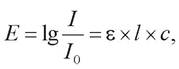
where E is the optical density (extinction) of the solution; I and I0 are the intensities of light passing through the solution and incident on the solution, respectively; ε is the extinction coefficient; l is the optical path length (solution thickness) in cm; c is the concentration of the solution in mol/L. The extinction coefficient serves as a proportionality factor indicating what fraction of the light flux is absorbed by a 1 cm thick layer of the given solution.

Thus, by measuring the optical density of a solution with a known extinction coefficient placed in a transparent cuvette of known thickness using a photoelectrocolorimeter, we can determine its concentration.
The schematic diagram of a photoelectrocolorimeter comprises several mandatory components (Fig. 30).
Fig. 30. Schematic diagram of a photoelectrocolorimeter: a - light source - incandescent lamp or ultraviolet lamp; b - optical filter - colored Glass that transmits monochromatic light; c - glass cuvette containing the test solution; d - photocell for recording the intensity of the light flux passing through the solution
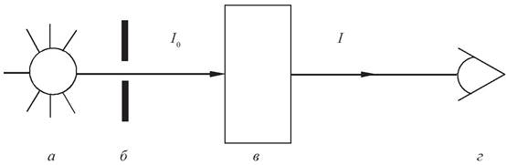
To determine I0 in this case, a reference solution is used in which the concentration of the target substance is zero. To convert optical density (extinction) units into concentration units, measurements are taken on a series of control solutions with known substance concentrations, followed by the Construction of a calibration curve.
It is by no means always that a solution of the substance of interest has a color of its own. In this case, the measurement method is based on a color reaction specific to the substance in question, where the concentration of the reaction product is directly proportional to the concentration of the test substance and is determined using a photoelectrocolorimeter.
Review Questions for Comprehension Check
1. Regulation of metabolism. Hormones: definition, properties, and biological role.
2. Stages of hormone metabolism. Hormone Receptors.
3. Significance of biochemical studies in medical practice. Reference intervals for laboratory parameters. Preparation of blood for biochemical analysis. Difference between blood plasma and serum.
4. Blood parameters characterizing Carbohydrate Metabolism: glucose, lactate, Pyruvate.
5. Blood parameters characterizing lipid metabolism: cholesterol, triglycerides, Lipoproteins, Ketone Bodies.
6. Nitrogenous substances in blood plasma: proteins and non-protein nitrogen, their composition, origin, and biological significance.
7. Mineral constituents of blood (potassium, sodium, calcium, magnesium, chloride, bicarbonate, and phosphate ions).
Written Homework Assignment
1. Draw a schematic diagram of a photoelectrocolorimeter in your notebook and describe its operating principle.
2. State the main advantages of automated biochemical testing methods over manual Procedures.
Sample Test on the Topic “Regulation of Metabolism. Blood Biochemistry”
Instructions: Unless otherwise specified in the test question, select a single correct answer.
1. How do the hormones insulin and adrenaline affect blood glucose concentration:
a) insulin increases it, adrenaline decreases it;
b) insulin decreases it, adrenaline increases it;
c) both insulin and adrenaline increase it;
d) both insulin and adrenaline decrease it.
2. Select The change in the plasma lipid profile that increases the risk of atherosclerosis:
a) decreased total cholesterol concentration;
b) decreased triglyceride concentration;
c) increased LDL cholesterol concentration;
d) increased HDL cholesterol concentration.
3. Indicate the main plasma protein fraction:
a) albumins;
b) α1-globulins;
c) α2-globulins;
d) β-globulins;
e) y-globulins.
4. Choose the correct ratio between cation concentrations in cells and blood plasma:
a) blood plasma has a higher concentration of potassium and magnesium, while cells have a higher concentration of sodium and calcium;
b) blood plasma has a higher concentration of sodium and magnesium, while cells have a higher concentration of potassium and calcium;
c) blood plasma has a higher concentration of sodium and potassium, while cells have a higher concentration of magnesium and calcium;
d) blood plasma has a higher concentration of sodium and calcium, while cells have a higher concentration of potassium and magnesium.
5. Which parameter differs between cells and macromolecules that are effectively separated by centrifugation:
a) charge;
b) density;
c) chemical nature;
d) light absorption;
e) light refraction.
6. Which method allows Blood Plasma Proteins to be separated into fractions based on the charge-to-molecular-mass ratio:
a) spectrophotometry;
b) Electrophoresis;
c) Chromatography;
d) centrifugation;
e) fluorimetry.
7. Indicate the main component of non-protein nitrogen in blood plasma:
a) urea;
b) uric acid;
c) creatine;
d) creatinine;
e) amino acids.
8. An increase in enzyme activity in blood plasma may be caused by:
a) increased enzyme synthesis within cells;
b) increased cell membrane permeability;
c) destruction of cell membranes;
d) all of the above.
9. Which blood enzymes are most commonly assayed in biochemical laboratories for diagnosing human diseases:
a) phospholipase, amylase, Pepsin;
b) citrate synthase, succinate dehydrogenase, aconitase;
c) hexokinase, Phosphofructokinase, aldolase;
d) creatine kinase, Lactate dehydrogenase, Alanine aminotransferase;
e) nucleotidase, xanthine oxidase, uricase.
10. Fill in the missing words (3 Answers) in the sentence: "Bilirubin is the end product of ... breakdown, circulates in the blood as direct and ... fractions, and when its concentration exceeds 40 µmol/L, it causes ..."
11. What pathological features are present in the given biochemical blood test (3 answers):
Parameter, unit of measurement |
Value |
Glucose, mmol/L |
12,6 |
Cholesterol, mmol/L |
4,8 |
Triglycerides, mmol/L |
3,5 |
Urea, mmol/L |
5,5 |
Creatinine, µmol/L |
78 |
Uric acid, µmol/L |
495 |
Bilirubin, µmol/L |
12 |
Potassium, mmol/L |
4,0 |
Sodium, mmol/L |
138 |
a) hyperglycemia;
b) hypocholesterolemia;
c) hypertriglyceridemia;
d) hypoazotemia;
e) hyperuricemia;
f) hypobilirubinemia;
g) hyperkalemia;
h) hyponatremia.
Last update: 06/08/2026
Editorial and Educational Adaptation: This material has been compiled based on the primary/original source text. The project team performed an editorial review, corrected technical inaccuracies, structured sections, and adapted the content for an educational format.
What was processed:
- elimination of formatting defects (OCR errors, structural breaks, corrupted characters);
- editorial organization of content;
- standardization of terminology in accordance with academic sources;
- verification of factual statements against the original source text.
All mentions of the author, publication year, and origin of the primary text have been preserved in accordance with the source.