Review of Medical Physiology - William F. Ganong 2002
Endocrine System, Metabolism, and Reproduction
Pituitary Gland
Hormones of the Intermediate Lobe
Proopiomelanocortin
Cells of the pars intermedia and corticotrophs of the anterior pituitary synthesize a large precursor protein that is cleaved to yield a group of Hormones. Following the removal of the signal peptide, a prohormone known as proopiomelanocortin (POMC) is formed. The synthesis of this molecule also occurs in the Hypothalamus and other parts of The Nervous system, Lungs, gastrointestinal tract, and Placenta (see Chapter 4). The molecular Structure is illustrated in Fig. 22-2. In corticotrophs, this prohormone is hydrolyzed to ACTH, ß-LPH, and small amounts of ß-endorphin, after which these substances are secreted. In the pars intermedia, POMC is further hydrolyzed to corticotropin-like intermediate lobe peptide (CLIP), γ-LPH, and significant amounts of ß-endorphin. The Functions of CLIP and γ-LPH remain unknown, whereas ß-endorphin is an opioid peptide (see Chapter 4) containing the five amino acid residues of met-enkephalin at its N-terminus in humans. Furthermore, α- and ß-MSH are also produced.
Class="center">Table 22-2. Hormone-Secreting Cells of the Human Anterior Pituitary
Cell Type |
Secreted Hormones |
% of Total Secretory Cells |
Staining Affinity |
Secretory Granule Diameter, nm |
Somatotrophs |
50 |
Acidophilic |
300-400 |
|
Lactotrophs |
Prolactin |
10-30 |
Acidophilic |
200 |
Corticotrophs |
ACTH, ß-LPH |
10 |
Basophilic |
400-550 |
Thyrotrophs |
TSH |
5 |
Basophilic |
120-200 |
Gonadotrophs |
FSH, LH |
20 |
Basophilic |
250-400 |
The pars intermedia is a vestigial structure, and in adults, α- or ß-MSH are sometimes not secreted.
Control of Skin Pigmentation
Fish, reptiles, and amphibians change their skin color for thermoregulation, camouflage, and behavioral responses. They achieve this through the movement of black or brown granules toward or away from the periphery of pigment cells called melanophores. These granules are composed of melanin, which is synthesized from DOPA (see Chapter 4) and DOPAquinone. Granule movement is regulated by various hormones and Neurotransmitters, including α- and ß-MSH, melanin-concentrating hormone, melatonin, and catecholamines.
Mammals lack melanophores with pigment granules that disperse and aggregate; instead, they possess melanocytes containing melanosomes, which synthesize Melanins. Melanocytes subsequently transfer these melanosomes to skin cells (keratinocytes) and Hair follicles, accounting for hair and skin pigmentation. Melanocytes express melanocortin 1 receptors, one of several cloned melanocortin receptor types. Treatment with MSH enhances melanin synthesis and causes a subtle darkening of human skin within 24 hours. As noted above, α- and ß-MSH do not circulate in healthy adults, and their physiological function remains unclear. ACTH binds to melanocortin 1 receptors.
Human Pigmentary Disorders
Alterations in pigmentation associated with certain endocrine disorders are driven by changes in circulating ACTH levels. For instance, abnormal pallor is a hallmark of Hypopituitarism. Hyperpigmentation occurs in patients with adrenocortical insufficiency due to primary adrenal disease. In fact, the presence of hyperpigmentation combined with adrenocortical insufficiency indicates that the deficiency is primary rather than secondary to pituitary or hypothalamic disease, since plasma ACTH levels are not elevated in the latter conditions (see Chapter 20).
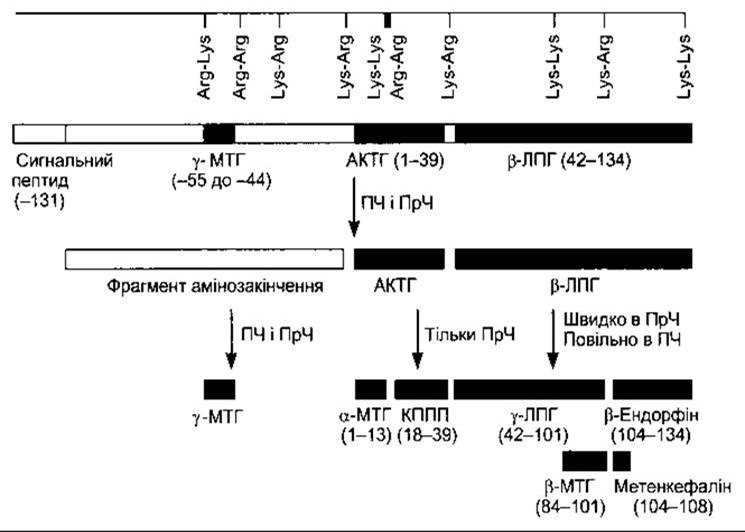
Fig. 22-2. Schematic representation of the preproopiomelanocortin molecule produced in pituitary cells, Neurons, and other Tissues. Numbers in parentheses indicate the Amino Acid Sequence positions within each polypeptide fragment. For convenience, the Amino acid sequences are numbered starting from the N-terminus of ACTH and read toward the C-terminus of the parent molecule, whereas The sequence of the remaining portion of the molecule extends to the left up to -131 toward the N-terminus of the parent molecule. The locations of Lys-Arg and other pairs of basic amino acid residues are also indicated; these represent sites of proteolytic Cleavage yielding smaller fragments of the parent molecule; AL - anterior lobe; IL - intermediate lobe.
Albinism is an inherited inability to synthesize melanin. It occurs in humans and many other mammalian species and can result from various genetic defects along the melanin biosynthetic pathway. Partial albinism manifests as skin patches associated with a localized reduction in melanin caused by congenital defects in the migration of pigment cell precursors from the neural crest during embryonic development. Not only the condition itself, but also the exact pattern of pigment loss is heritable. Vitiligo, a disorder caused by a similar loss of melanin, develops after birth and progresses throughout life.
Last update: 10/08/2026
Editorial and Educational Adaptation: This material has been compiled based on the primary/original source text. The project team performed an editorial review, corrected technical inaccuracies, structured sections, and adapted the content for an educational format.
What was processed:
- elimination of formatting defects (OCR errors, structural breaks, corrupted characters);
- editorial organization of content;
- standardization of terminology in accordance with academic sources;
- verification of factual statements against the original source text.
All mentions of the author, publication year, and origin of the primary text have been preserved in accordance with the source.