Fundamentals of Biochemistry - A. A. Anisimov 1986
Protein and Amino Acid Metabolism
Protein Catabolism
5.6.1. Protein Digestion and The Role of Proteins in Nutrition. In the Human and Animal body, Cell destruction and protein breakdown and consumption occur continuously: this includes the degradation of "aged" protein molecules across all Tissues, epithelial desquamation, growth of Nails, horns, and hooves, consumption of digestive and Other Enzymes, hormone expenditure, and antibody production. Replenishing continuously consumed proteins in all these processes is possible only through a sufficient dietary intake. The daily protein requirement for humans is 80–100 g, and up to 120–150 g for those engaged in physical labor, with at least 50–70 g of these being animal-derived proteins. Protein deficiency rapidly impairs the functioning of The Thyroid Gland, Adrenal Glands, and Gonads. The Central Nervous system, particularly the Cerebral Cortex, is especially sensitive to protein starvation. Even during total starvation, the Brain and Heart retain their mass for a long time, renewing their proteins at the expense of protein degradation in the Muscles and Liver.
Dietary proteins are classified as complete or incomplete. Complete proteins contain all 10 Essential Amino Acids, which animals and humans, unlike plants and microorganisms, are incapable of synthesizing. The capacity for their synthesis was lost during evolution because these amino acids were supplied to the Organism in sufficient quantities via plants, microorganisms, and other animals.
Essential amino acids include Lysine, Arginine, Histidine, valine, leucine, isoleucine, Threonine, Methionine, phenylalanine, and Tryptophan. This list requires a few notes. Arginine can be synthesized in The Human Body and other animals, but this occurs in very small quantities. Since Tyrosine is formed directly from phenylalanine in a single step, the requirement for phenylalanine is effectively a requirement for both amino acids, and tyrosine can be classified as an essential amino acid. For the same reason, Cysteine is a non-essential amino acid only when methionine is present in the diet. Glycine, which is a non-essential amino acid for all other animals, is essential for chicks.
Incomplete proteins lack one or several essential amino acids. Complete proteins include milk casein, egg albumin, milk albumin, and wheat gluten, whereas incomplete proteins include many plant proteins (corn zein, legume legumin), which are typically deficient in lysine, methionine, and tryptophan. Another drawback of plant proteins is their relatively low concentration in plant-based foods; consequently, on a vegetarian diet (consisting exclusively of plant products), a very large amount must be consumed to meet the body's daily protein requirements. Furthermore, some plant proteins are poorly digested.
Animal products contain large amounts of proteins, and relatively small quantities are needed to sustain the organism (it is particularly important that animal proteins are typically complete). All of this explains why more than half of the daily protein intake should be of animal origin.
However, this does not imply that plant proteins should be ignored. Vegetable proteins are relatively rich in tryptophan and phenylalanine, and potato proteins boast a favorable amino acid profile, although the protein content in tubers is low—around 2%. Moreover, different plant proteins lack different essential amino acids, meaning that mixtures of two or three plant Proteins can be nutritionally complete. For example, a mixture of roughly equal amounts of unbolted corn flour, sorghum, and cottonseed meal supplemented with 3% Yeast, as well as vitamin A and CaCO3, is nearly comparable to cow's milk in nutritional value.
The minimum dietary protein content required to establish nitrogen balance (where the body's protein expenditure is offset by dietary intake) is termed the protein minimum. Protein balance is also maintained when dietary protein is in excess. Unlike CARBOHYDRATES or fats, proteins cannot be stored as reserves; therefore, accelerated protein breakdown begins, accompanied by The excretion of their catabolic products in urine and feces until equilibrium is restored. This process generates an excess of harmful acidic protein breakdown products in the body, leading to tissue acidification (acidosis).
One such breakdown product is uric acid, which, in excess, deposits in the joints (causing Gout). These harmful acidic products are neutralized and eliminated from the body by binding with alkaline minerals, which are abundant in plant foods such as vegetables and fruits. This is the primary reason why pairing meat dishes with vegetable side dishes is beneficial—a practice humans have discovered through centuries of experience. Furthermore, an excessive, one-sided diet high in animal protein triggers intensive putrefactive processes in the intestines, generating toxic substances. A plant-and-dairy diet positively influences intestinal microflora and inhibits putrefaction.
Protein digestion. The organism utilizes not the dietary proteins themselves, but their breakdown products—Amino Acids and simple Peptides. Dietary proteins undergo hydrolytic digestion within "protected compartments." In Protozoa, digestion occurs in digestive vacuoles, whereas in humans and animals, it takes place in the intestinal lumen. Thus, the internal contents of animal Cells remain inaccessible to hydrolytic digestive enzymes. The latter are synthesized and secreted as inactive proenzymes known as zymogens.
Following synthesis on the Ribosomes of The Endoplasmic reticulum in specialized secretory cells, proenzymes are "packaged" into zymogen granules, which then migrate to The Cell surface and are secreted into the extracellular environment. Upon reaching their Site of Action, zymogens are converted into active enzymes, sometimes through the action of another enzyme molecule that cleaves a polypeptide chain fragment—the so-called inhibitor peptide—from the precursor.
Protein digestion begins in The Stomach. In an adult human, secretions from the ducts of 10 to 30 million gastric glands enter the stomach. Secretion is performed by glands comprising three cell types: chief cells, mucous cells, and parietal cells. Chief cells synthesize and secrete pepsinogen, mucous cells produce mucus, and parietal cells secrete Hydrochloric acid along with the human intrinsic factor—a mucoprotein essential for the normal intestinal absorption of dietary vitamin B12. The proton concentration in the stomach is 106 times greater than that in Blood Plasma. The exact mechanism of HCl formation remains unknown. The simplest hypothesis suggests that the secretion mechanism is analogous to proton transport driven by ATP during mitochondrial inner membrane functioning. Carbonic anhydrase plays a major role in HCl secretion.
Parietal cell secretion is stimulated by histamine and gastrin Hormones. The latter are produced in the pyloric region of the stomach. Gastrin production is inhibited by secretin (a duodenal mucosal hormone) and Somatostatin (a hypothalamic hormone also present in the Pancreas).
A cascade mechanism has been proposed for the REGULATION OF GASTRIC acid secretion. This model is based on the hypothesis that histamine acts as a mediator for gastrin. Gastrin induces The formation of histamine, which activates adenylate cyclase. The resulting cAMP activates protein Kinases; one of these kinases phosphorylates a low-activity carbonic anhydrase isoenzyme, thereby enhancing its activity, which is required for HCl secretion.
The primary proteolytic enzyme of gastric juice is Pepsin. The molecular mass of its zymogen form is 40,000. The activation of pepsinogen is autocatalytic, driven by pepsin itself and the highly acidic environment of the gastric contents. The molecular mass of pepsin is 32,700, which is lower than that of pepsinogen. During activation, 42 amino acid residues are cleaved from the N-terminal region of pepsinogen as a mixture of inhibitor peptides. In the stomach, substrates for pepsin can include both denatured proteins resulting from culinary Processing and native proteins.
Pepsin hydrolyzes peptide bonds formed by the amino groups of cyclic amino acids: Phe, Trp, and Tyr. Peptide bonds formed by dicarboxylic Amino acids are hydrolyzed very slowly by pepsin. The optimal pH for pepsin activity is 2–3. If HCl secretion fails to maintain this acidity, protein digestion is severely impaired. In cases of achylia gastrica, digestion does not occur in the stomach at all, as neither pepsin nor acid is present in the contents. Under in vitro conditions, pepsin is capable of hydrolyzing proteins down to individual amino acids, but this process requires a considerable amount of time. Since food remains in the stomach for a limited duration, protein digestion halts here at the stage of polypeptide mixture formation.
A proteolytic enzyme named gastrixin has been isolated from the human gastric mucosa. It acts on proteins similarly to pepsin, but features a lower molecular mass and a less acidic pH optimum. In humans, pepsin causes milk to curdle, whereas in ruminants, this function is performed by a specialized enzyme found in the abomasum (the fourth stomach compartment) of suckling calves, known as chymosin or rennin. Structural studies of all gastric Proteolytic Enzymes reveal significant Structure/154.html">Sequence Homology, indicating that they share a common precursor.
Proteins undergo further DIGESTION IN THE slightly alkaline environment of the Small Intestine. Here, their Hydrolysis is catalyzed by several enzymes: Trypsin, Chymotrypsin, elastases, and peptidases. Trypsin and chymotrypsin operate more efficiently following pepsin action, yet they are also capable of breaking down proteins without prior Pepsin Hydrolysis. Consequently, patients who have undergone a gastrectomy retain The ability to utilize dietary proteins.
The pancreas secretes trypsinogen, chymotrypsinogen (see section 3.3), procarboxypeptidases A and B, and proelastase. The intestine secretes the enzyme enteropeptidase (enterokinase), which specifically and rapidly activates trypsinogen into trypsin. Enteropeptidase was first discovered and studied by N. P. Shepovalnikov in I. P. Pavlov's laboratory. Trypsinogen activation can also occur autocatalytically via trypsin itself, though this process is 2,000 times slower.
The role of enteropeptidase cannot be overstated, as the resulting trypsin serves as the activator for all other proteolytic zymogens into their respective active forms. Trypsinogen consists of a single polypeptide chain. During activation, hydrolysis of a single peptide bond at the N-terminus releases a hexapeptide; a partial conformational change occurs in the molecule, enzymatic activity emerges, and trypsin is formed.
The sequence Val—(Asp)4—Lys of the cleaved hexapeptide is conserved in the trypsinogens of most vertebrates, ranging from fish to mammals. Pepsin, trypsin, and chymotrypsin Complement one another in substrate Specificity (see section 3.3). Their combined action results in the deep Hydrolysis of Proteins into small peptides.
Carboxypeptidase A is a zinc-containing enzyme that primarily cleaves C-terminal amino acid residues with aromatic side chains. It is secreted as procarboxypeptidase A and activated by trypsin.
Carboxypeptidase B is also secreted in an inactive form. Once activated, it attacks C-terminal residues containing exclusively Arg and Lys.
Proelastase is secreted by the pancreas and converted into Elastase under METABOLISM/18.html">The Influence of trypsin; it acts on peptide bonds between residues of various neutral amino acids and is particularly active against the protein Elastin.
The mucosa of the small intestine also contains proteolytic enzymes. Although they can be secreted into the intestinal juice, they function primarily intracellularly. This group of enzymes includes aminopeptidases, which sequentially release N-terminal amino acids. Leucine aminopeptidase is an exopeptidase with broad specificity; it is a zinc-containing enzyme that is also activated by Mn2+. The intestinal mucosa also contains dipeptidases, such as glycylglycine dipeptidase, which is activated by Co2+ or Mn2+.
As a result of the combined action of proteolytic enzymes secreted by the gastric wall, pancreas, and intestinal mucosa, dietary proteins undergo nearly complete hydrolysis into amino acids within the small intestine.
Unhydrolyzed small peptides account for a very small fraction of dietary proteins. They can also be absorbed from the intestine; therefore, following protein digestion, the content of peptide nitrogen in the blood increases slightly. The penetration of native proteins across the intestinal mucosa into the bloodstream is a rare exception. It may occur, for example, when an animal is fed blood serum Proteins of the same species. After the ingestion of a large amount of egg white, it appears in the urine, accompanied by signs of intoxication and poisoning. In newborns of most mammalian species, the permeability of the intestinal mucosa is elevated, allowing colostral Antibodies to enter the bloodstream. These Examples merely highlight the exceptional nature of this phenomenon, which is extremely rare in nature—the absorption of intact proteins from the intestine into the blood.
Absorption of Amino acids from the intestine. Amino acids are absorbed primarily in the small intestine. This is an active process that requires energy and depends on the concentration of Na+. There are more than five specific transport systems, each of which transports structurally related amino acids. Amino acids compete with one another for binding sites. Once absorbed in the intestine, amino acids are transported via the portal system to the liver.
Protein decomposition during putrefactive processes in the intestine. Under normal conditions, the Oral Cavity and stomach lack the environmental conditions necessary for the proliferation of putrefactive Bacteria. In the intestine, a portion of the amino acids is utilized by microbes as a nutrient source prior to absorption. Excessive consumption of animal proteins and certain pathological conditions can trigger putrefactive and fermentative processes in the intestine. Microbial Decarboxylation of amino Acids produces amines, some of which are toxic (such as putrescine and cadaverine). Deamination yields various products, including saturated and unsaturated acids, hydroxy acids, and keto acids.
One of the primary conditions for the bacterial decarboxylation of amino acids is an acidic environment (pH 3.5–5.5). Under normal physiological conditions, however, the intestinal environment is weakly alkaline. Nevertheless, infection with certain pathogenic bacteria can lead to dyspepsia (digestive disorders not associated with organic Changes in the gastrointestinal tract), during which the pH of the intestinal environment drops (typically locally) to 3–5. Fermentation processes actively develop in these acidified areas.
The bacterial degradation of cystine, cysteine, and methionine yields hydrogen sulfide (H2S), methyl mercaptan (CH3SH), and other sulfur-containing compounds. Through the gradual shortening of its side chain, tyrosine can be converted by microorganisms into toxic cresol and phenol. After absorption via the PORTAL VEIN SYSTEM, these substances are transported to the liver. They are detoxified through the formation of conjugated compounds with sulfuric or glucuronic acids. These conjugates are non-toxic and are excreted in the urine.
During protein putrefaction, The amino acid tryptophan gives rise to indole and skatole. Skatole features a partially degraded side chain, whereas indole lacks it entirely. The ring structure remains unaltered.
Class="center">
Indole and skatole are toxic substances responsible for the characteristic odor of feces; they are also detoxified in the liver by conjugation with sulfuric or glucuronic acids, following preliminary oxidation into hydroxyl-containing compounds (indoxyl and skatoxyl, respectively), and are subsequently excreted in the urine as conjugates.
5.6.2. Animal Tissue Proteinases. Proteolysis in Plants and Microorganisms. Active proteolytic enzymes—both proteinases (Endopeptidases, peptidyl-peptide Hydrolases) and peptidases—have been detected in all animal tissues. Their activity is particularly high in rapidly growing or dividing cells, as well as in glandular cells and other Organs characterized by a high rate of Protein Synthesis. This is because the synthesis of tissue proteins relies heavily on amino acids generated during the Catabolism of aged protein molecules. Alongside supplying building blocks for biosynthetic reactions, intracellular proteolysis plays a role in specific processes, such as intracellular digestion during morphogenesis in insects and vertebrates, natural Immunity reactions, and the Synthesis and degradation of physiologically active substances (enzymes, hormones). The latter is well illustrated by numerous examples of The conversion of proenzymes into active enzymes, as well as the presence in many organs and tissues of proteinases that degrade excess amounts of specific hormones.
Intracellular proteolysis is localized primarily in Lysosomes, which contain a diverse array of active hydrolases, including acidic proteinases. Intracellular proteinases that hydrolyze proteins in the mildly acidic pH range are known as cathepsins. Currently, five well-characterized types of cathepsins are recognized, designated by the letters A, B, C, D, and E. They differ in their pH optima, substrate specificity, and several other properties. In certain animals, cathepsin D accounts for approximately 2/3 of the total proteolytic activity in Spleen and Kidney homogenates. Unlike cathepsins B and C, it is not a sulfhydryl proteinase and is not inhibited by sulfhydryl group Reagents. Cathepsin E is similar to cathepsin D in its substrate specificity and other properties.
In addition to cathepsins, which function as acidic tissue proteinases, neutral and alkaline proteinases have been discovered in various organs and tissues (such as erythrocytes, Lungs, skeletal muscles, and the brain). Their biological Functions and catalytic properties remain insufficiently studied. A distinct group of intracellular proteinases with an alkaline pH optimum comprises kallikreins, which are widely distributed in tissues and Body Fluids. They are particularly active in the blood, Salivary Glands, and pancreas. Plasma kallikrein acts on substrates to produce bradykinin, whereas pancreatic and glandular kallikreins yield kallidin (see Section 2.5.1), which is converted into bradykinin in the blood by aminopeptidase.
Proteinases of higher plants predominantly belong to the sulfhydryl type and are activated by cysteine, Glutathione, and other reducing agents. Their optimal activity occurs in mildly acidic, neutral, and mildly alkaline pH ranges, depending largely on The Nature of the substrate. A typical representative is Papain, derived from the fruit juice of the papaya tree (Carica papaya). Plant proteinases that are not activated by reducing agents are significantly less common. Proteinases from carnivorous plants (such as sundew and pitcher plants) exhibit a sharply acidic pH optimum of 3–3.5.
Microorganisms predominantly produce secreted (extracellular) proteinases that supply them with low-molecular-weight products of environmental protein degradation. Intracellular enzymes are mainly peptidases, which break down peptides that have entered the cell from the external environment. Intracellular proteinases are found much less frequently. Proteases from actinomycetes possess broad substrate specificity, hydrolyzing both globular and Fibrous proteins.
Due to their broad spectrum of activity and high potency, protease preparations from Str. griseus are widely utilized in industry and biochemical research laboratories, and are marketed under the names pronase (Japan, USA) or protelin (USSR).
Alkaline proteinases from Bac. subtilis—known as subtilisins—have been thoroughly investigated and are widely used in research and industry because they hydrolyze proteins more extensively and rapidly than many other proteinases (including pepsin and trypsin). These are typical Serine proteinases, with an active center structure closely resembling that of chymotrypsin.
5.6.3. Deamination and Further Catabolism of Amino Acids in Animals, Plants, and Bacteria. Amino acids produced via protein proteolysis undergo further transformations. The initial stage of catabolism for Most amino acids involves the removal of the α-amino group, either through Transamination with a keto acid (most commonly α-ketoglutaric acid) or through various types of deamination: oxidative, reductive, hydrolytic, or intramolecular.
During transamination, the α-amino group of an amino acid is transferred to the α-carbon atom of α-ketoglutaric acid, and much less frequently to pyruvic or oxaloacetic acid. This reaction yields the α-keto analogue of the original amino acid along with a new amino acid (see Section 2.5.1 [or 5.2.3]). The overall result of the transamination of various amino acids (excluding lysine and threonine) is that all their Amino groups are "channeled" into glutamic acid, or, in some organisms, into aspartate or Alanine. Ultimately, however, these Two amino acids also react with α-ketoglutaric acid to form glutamate, which transfers the amino groups into the final series of reactions leading to the End products of Nitrogen metabolism. Subsequently, the amino groups collected from various amino acids into L-glutamic acid are released as NH+4 ions.
The Oxidative Deamination reaction is catalyzed by a specific and active L-Glutamate dehydrogenase:

Thus, the glutamic acid ⇄ α-ketoglutaric acid system plays a leading role in keto acid amination, transamination, and deamination reactions alike.
L-Glutamate dehydrogenase is particularly active in the liver Cytosol and Mitochondria and functions as an allosteric enzyme. Its activity is enhanced in the presence of ADP, GDP, and Certain amino acids, while being inhibited by effectors such as ATP, GTP, and NADH. In many organisms, oxidative deamination can be mediated by flavin dehydrogenases, specifically L-Amino Acid Oxidase and D-Amino Acid Oxidase. However, they do not make a significant contribution to Amino Group Metabolism and are localized within the Endoplasmic reticulum and Microbodies of liver and kidney cells. In higher plants, oxidative deamination—proceeding via the same pathway as in animal liver—likewise serves as the primary route of AMINO ACID DEAMINATION. This pathway is also characteristic of Fungi, Yeasts, and bacteria.
In plants, amino acid deamination may additionally occur through the action of polyphenol oxidase and polyphenols According to the following scheme:

In bacteria, and occasionally in plants, three Other types of deamination are observed:
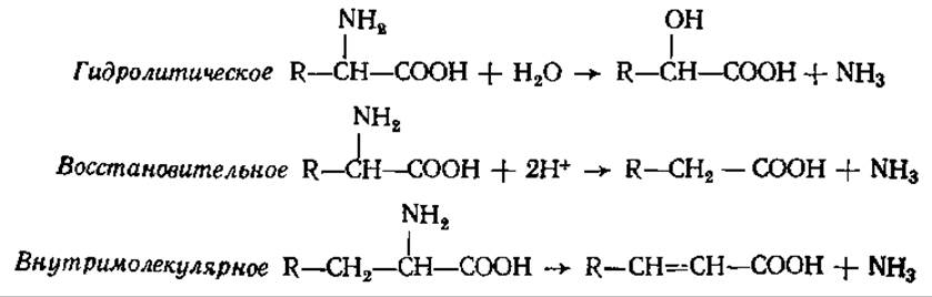
Under anaerobic conditions, in the presence of ammonifying microorganisms, amino acids undergo degradation such that one is oxidized while the other is reduced, releasing ammonia in the process:

The keto acid reacts with another starting amino acid. The keto acids and other Organic compounds resulting from Amino Acid Catabolism undergo further degradation, yielding substances that either enter The Tricarboxylic Acid Cycle to serve as an energy source or act as precursors for the synthesis of metabolites, including biologically essential compounds. The remarkable diversity of products derived from Amino acids can be seen in Fig. 5.10.

Fig. 5.10. Main Pathways of Amino acid metabolism
There are several ways amino acids enter the Krebs cycle, depending on the Nature of the amino acid's side chain. Sometimes one part of the carbon Skeleton enters the Krebs cycle through one "gateway," while another part enters through a different one. For instance, the carbon skeletons of Phenylalanine and Tyrosine enter the cycle as fumaric and acetoacetic acids.
Thus, amino acid deamination serves as the primary mechanism for converting nitrogenous substances into non-nitrogenous compounds, which can subsequently be processed into carbohydrates, fats, and Other Compounds.
5.6.4. End Products of Nitrogen Metabolism: Evolutionary and Ecological Aspects. Ammonia is produced as a result of amino acid catabolism in All living organisms. However, being toxic even at the lowest concentrations, it does not accumulate in the cell; instead, it is rapidly removed either by excretion into the external environment or by conversion into non-toxic compounds. In many organisms, the initial binding of ammonia within cells occurs through the formation of amides: glutamine and asparagine. As demonstrated by the prominent Soviet agricultural chemist N. D. Pryanishnikov, this pathway is especially characteristic of plant tissues, which store nitrogen—a constantly scarce element for plants—in the form of these amides. Nevertheless, glutamine formation (and to a lesser extent, asparagine) is also widespread in animals, having been detected in the muscles, brain, liver, and Kidneys of mammals, as well as in the fat body and hemolymph of insects.
In animals and humans, glutamine produced in various tissues and organs enters the bloodstream and is transported to The Liver and kidneys, thus acting as a specialized transport form and temporary reservoir for ammonia. In recent years, it has been discovered that aspartic and glutamic acids can undergo amidation while incorporated within a protein molecule, meaning protein amidation is also possible.
The NH3 released in tissues can be immediately utilized for the synthesis of new amino acids, most commonly via reductive amination (see Section 5.2.3).
In humans, as well as all mammals, amphibians, and several other animals, urea is the primary end product of nitrogen metabolism excreted from the body.
As early as the late 19th century, I. P. Pavlov, M. V. Nencki, I. Zalessky, and S. S. Salazkin established that urea synthesis takes place in the liver. The first theory of urea synthesis was developed by M. V. Nencki, correctly asserting that the starting compounds for urea formation are NH3 and CO2. In the 1930s, H. Krebs, while INVESTIGATING THE REACTION chain of Urea formation in detail, discovered that this pathway is cyclical and that Ornithine plays a crucial role in the process. Consequently, the entire process of urea Biosynthesis became known as the ornithine cycle.
In the first reaction of the cycle, NH3, CO2, and two ATP molecules interact to form carbamoyl phosphate (see Section 5.2.2). The enzyme carbamoyl phosphate synthetase is localized in the liver mitochondria, and NH3 serves as the exclusive nitrogen donor for this enzyme. Carbamoyl phosphate transfers its carbamoyl group to ornithine, resulting in the formation of citrulline. This reaction is catalyzed by ornithine transcarbamylase:
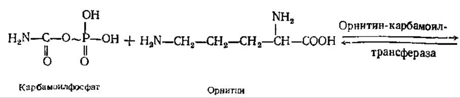

The second NH2 group of the future urea molecule enters the cycle with the help of aspartic acid, which is produced via transamination between glutamic acid and oxaloacetate. The conversion of glutamic acid into aspartic acid is catalyzed by the enzyme aspartate aminotransferase. The amino group of aspartic acid condenses with the carbamoyl group of citrulline. The reaction proceeds in the presence of ATP and is catalyzed by argininosuccinate synthetase. This yields argininosuccinate, which is then reversibly cleaved into arginine and fumarate.
Arginine is cleaved by arginase into urea and ornithine, which re-enters the cycle (Fig. 5.11). Therefore, the synthesis of a single urea molecule requires two molecules of NH3, one molecule of CO2, and three ATP molecules. The amount of urea excreted by the body depends on Dietary Protein Intake and normally ranges from 25 to 35 g per day in healthy adults.
Urea is far from being the primary end product of protein catabolism across all animal taxonomic groups. In the course of evolution, different animal species have developed distinct biochemical pathways for producing excreted nitrogenous end products depending on their habitat conditions (Table 5.2). Thus, the specific forms of nitrogenous end products are determined both by the level of evolutionary development and by environmental conditions (with Water availability playing the primary role among the latter).
Table 5.2. Main end products of nitrogen metabolism in various animal groups
Animals |
Main end product of Protein metabolism |
Animals |
Main end product of protein metabolism |
Aquatic invertebrates |
Ammonia |
Turtles |
Urea and uric acid |
Bony Fishes |
Ammonia, some urea |
Insects |
Uric acid |
Bivalves |
Urea |
Terrestrial gastropods |
» » |
Crocodiles |
Ammonia, some uric acid |
Lizards |
|
Amphibian embryos |
Ammonia |
Snakes |
» » |
Adult amphibians Mammals |
Urea |
Birds |
» » |

Fig. 5.11. The ornithine cycle
The ability to detoxify ammonia through the synthesis of organic compounds emerged in animals at a specific stage of their evolutionary development. In many invertebrates, particularly aquatic dwellers, ammonia and ammonium salts account for up to 80% of the nitrogenous substances excreted in urine. This group of animals is referred to as ammonotelic. In large volumes of water, the excreted ammonia is diluted to concentrations so low that they become non-toxic. In these animals, ammonia is produced directly in the renal tubules as a result of glutamine breakdown and immediately enters the urine; Glutamine + H2O → Glutamic acid + NH3. Invertebrates are also characterized by the excretion of certain amounts of amino acids in their urine (up to 25% of dietary proteins). Apparently, invertebrate nitrogen metabolism is insufficiently advanced, as they are unable to fully utilize dietary proteins.
Freshwater bony fishes are also largely ammonotelic, as their chemical ammonia-binding system is still poorly developed. In marine bony fishes, ammonium salts account for 40–60% of total urinary nitrogen. Furthermore, they excrete a considerable amount of trimethylamine oxide O = N ≡ (CH3)3, the formation of which is likely associated with the oxidative degradation of Choline.
Small quantities of ammonium salts (3–6% of total urinary nitrogen) are also found in the urine of humans and vertebrates. However, for them, this is an atypical end product of nitrogen metabolism. In certain pathological conditions (such as diabetes), acidic metabolic products accumulate in the body, leading to acidosis. In such cases, the amount of ammonium salts in the urine increases sharply, and NH3 is used to neutralize acids. This allows the body to spare other useful cations (K, Mg, etc.) and maintain normal electrolyte balance.
Most terrestrial vertebrates—mammals, amphibians (in their adult state), and cartilaginous fishes—form a group of ureotelic animals, in which the primary end product of nitrogen metabolism is highly water-soluble urea. Birds and terrestrial reptiles consume limited amounts of water; in flying birds, it imposes a mass overload, while terrestrial reptiles often inhabit arid environments. Their urine is a semi-fluid mass containing crystals of poorly water-soluble uric acid. Such organisms are termed uricotelic. In addition to birds and terrestrial reptiles, insects also belong to this group.
The formation of uric acid, which occurs in the liver and partly in the intestinal mucosa, is a complex process preceded by the Synthesis of purine rings. In this pathway, two nitrogen atoms of the purine are provided by the amide groups of glutamine, one nitrogen atom by aspartate, and one by glycine (see Section 4.5.2).
In humans and primates, uric acid is present in urine, but in very small quantities as the end product of purine catabolism (normally, humans excrete 0.6–0.7 g of uric acid per day). In gout, the blood concentration of uric acid increases sharply, and its sparingly soluble sodium salt deposits in Cartilage, tendons, and joint capsules. The deposition of uric acid and its salts (urate stones) also occurs in the kidneys. The onset of gout is promoted by an excess of meat-rich diet containing high amounts of purine bases.
Lungfishes from tropical water bodies provide an interesting example of the dependence of nitrogen metabolism on environmental conditions. During the hot dry season, they burrow into the mud, and their urine contains high amounts of urea; in the tropical rainy season, when rivers fill with water, lungfishes rid themselves of the excess urea accumulated during the drought and begin excreting ammonium salts, much like many other fishes. Aquatic turtles living in humid habitats excrete urea, whereas turtles from arid regions excrete uric acid. In aquatic frogs, the tadpole excretes mainly ammonium salts in its urine and can thus be classified as an ammonotelic animal. Adult frogs, upon transitioning to land, excrete urea and are ureotelic.
The conditions of embryonic development—a closed environment within an egg surrounded by water-impermeable membranes—greatly influence the formation of end products of nitrogen metabolism in birds and reptiles. The amount of water in the egg is very small, and the accumulation of toxic ammonium salts would quickly lead to the embryo's death. Urea synthesis would result in a sharp increase in osmotic concentration, which would also negatively impact embryonic development. Only the formation of poorly water-soluble uric acid does not interfere with the embryo's development; it precipitates out as a solid sediment, effectively isolated from metabolism. Interestingly, during the earliest stage of development, the avian embryo excretes ammonium salts into the environment, followed by urea, and finally uric acid—a pattern that persists throughout the postembryonic stage. Thus, this exemplifies a biochemical manifestation of the famous biogenetic law of Müller and Haeckel, wherein ontogeny recapitulates phylogeny.
In humans and mammals, the ureotelic type of nitrogen metabolism is established as early as during Embryogenesis. The urea produced by the fetus passes through the Placenta into the maternal bloodstream and is subsequently removed from the body by the kidneys.
In most mammals, uric acid, produced in small amounts as the end product of Purine base catabolism, is further oxidized to allantoin by the liver enzyme urate oxidase: In dogs, for example, almost all purine nitrogen is excreted in the urine as allantoin. Humans, primates, and birds lack urate oxidase; therefore, uric acid itself serves as the end product of purine metabolism in these species. Pigs excrete guanine in their urine in addition to allantoin due to the low activity of guanine deaminase. Consequently, cases of guanine gout have been described in pigs, where guanine crystals deposit in the joints. Guanine serves as the metabolic end product in spiders.
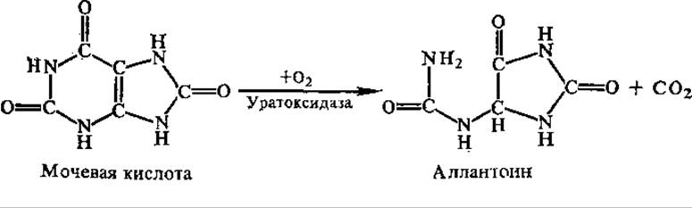
In many animals (excluding mammals), allantoin can undergo further degradation to yield allantoic acid (in some fishes), and subsequently urea and glyoxylic acid (amphibians, most fishes, freshwater bivalve Mollusks), or ammonia (crustaceans, some marine invertebrates). From an evolutionary perspective, It is interesting to note that as animal forms became more complex, individual enzymes responsible for uric acid Cleavage disappeared; in humans and primates, these enzymes are entirely absent.
A constant nitrogen-containing component of urine in humans and many vertebrates is creatinine, the anhydride of creatine (methylguanidinoacetic acid). Humans excrete 1.5–2.5 g per day. Creatine is particularly abundant in muscles, where its high-energy phosphate ester (creatine phosphate) serves as the primary energy storage form. A. V. Palladin was the first to suggest that arginine participates in the synthesis of creatine. Modern experiments using isotopic tracers have confirmed this: creatine synthesis (which takes place in the kidneys and liver) begins with The transfer of the guanidino group from arginine to glycine, after which the resulting guanidinoacetic acid is methylated. Creatinine is formed either directly from creatine via dehydration (1) or from creatine phosphate upon the cleavage of H3PO4 (2):

Free creatine is typically absent from the urine of healthy adults or is present only in trace amounts. However, There is a direct correlation between the content of creatine (and creatine phosphate) in muscles and creatinine in urine. In particular, urinary creatinine levels show a remarkably stable correlation with an individual's Muscle mass.
The excretion of free creatine in urine is quite common during childhood. Its appearance in the urine of adults (creatinuria) is a consequence of various pathologies, such as different forms of muscular dystrophy, myositis (Skeletal Muscle inflammation), diabetes, hyperthyroidism, and vitamin E deficiency. In women, creatinuria can occur during Pregnancy and the early postpartum period.
In invertebrate animals, arginine is present in the urine instead of creatinine, and creatinine is detected extremely rarely. The metabolic and energetic roles of Creatine phosphate in invertebrates are fulfilled by various other phosphorylated guanidine-containing substances, most commonly arginine phosphate.
Human urine also consistently contains a certain amount (0.1–2 g per daily volume) of hippuric acid. It is exceptionally abundant in the urine of herbivores, as it is formed from benzoic acid, a breakdown product of Aromatic Compounds found in plant tissues.

A comparison of protein catabolism pathways and mechanisms across various living organisms leads to the Conclusion that the core features of this process share a common origin. Consequently, these biochemical processes were established at fairly Cytology/cytology/16.html">Early stages of evolution, before the unified stream of living matter diverged into separate branches. During subsequent evolution, various taxonomic groups inhabiting specific ecological niches developed distinctive Features of protein metabolism that facilitated their survival under particular environmental conditions and helped them utilize those conditions more effectively.
Higher plants, with their sessile lifestyle, rapidly consume nitrogen salts within the ROOT zone; therefore, the evolution of their nitrogen metabolism proceeded toward the economical use of nitrogen under conditions of strictly limited supply. Consequently, plants do not excrete ammonia detoxification products into the external environment. Asparagine and glutamine serve as their storage forms of nitrogen. Besides ammonia detoxification and nitrogen storage, asparagine and glutamine are also crucial as reserve pools of dicarboxylic amino acids required for numerous metabolic processes. It is hardly surprising, therefore, that the amide content in plant proteins reaches very high levels: in corn and wheat seed proteins, dicarboxylic acids account for 35–45%, with the majority present in the form of amides.
In plants characterized by a high content of organic acids in their tissues (sorrel, rhubarb), ammonia detoxification occurs through the formation of ammonium salts of organic acids. Urea has also been detected in the tissues of certain higher plants, synthesized via the ornithine cycle. Large amounts of urea accumulate in some fungi (up to 13% in cultivated mushrooms). Research by the renowned GDR plant biochemist and physiologist K. Mothes (1962) demonstrated the relatively wide distribution of allantoin in plants. Certain plants (such as alder, birch, and hazel) accumulate significant amounts of citrulline, which likely serves as a form for ammonia detoxification. However, the synthesis of urea, allantoin, and citrulline is generally atypical for plants as a whole (none of these are excreted into the external environment).
There are many peculiar and still poorly understood aspects regarding the relationship between protein catabolism and biosynthesis in bacteria. For a long time, the applicability of the term "protein turnover" to bacteria was questioned, and certain proteins were assumed to be stable (within the lifespan of the cell). It was not until the 1960s that it was reliably demonstrated that protein turnover does occur in living, non-dividing cells of E. coli and yeast. It was established that in E. coli cells during the logarithmic growth phase, protein degradation proceeds at a rate of about 3% per generation. Furthermore, the degradation rate is often independent of the physiological state of the cell. Evidently, the concentration of proteins in bacterial cells is primarily a function of their biosynthesis processes.
During the Deamination of Amino acids in bacteria, along with deamination pathways common to all living organisms, bacteria-specific pathways also function: reductive, hydrolytic, and intramolecular. Another characteristic feature of several bacteria is the presence of highly active, low-specificity L- and D-amino acid oxidases. Many microorganisms (primarily anaerobes) ferment amino acids, utilizing them not only as structural building blocks but also as an energy source. Certain bacteria are known to specialize in the fermentation of purine and pyrimidine bases, as well as uric acid.
A fundamental difference between bacteria and animals lies in the Regulation of Amino acid metabolism. When bacteria encounter an environment rich in all amino acids, the synthesis of enzymes involved in Amino acid biosynthesis is repressed. Conversely, when animals consume a diet rich in amino acids, their amino acid degradation processes are activated.
Last update: 06/08/2026
Editorial and Educational Adaptation: This material has been compiled based on the primary/original source text. The project team performed an editorial review, corrected technical inaccuracies, structured sections, and adapted the content for an educational format.
What was processed:
- elimination of formatting defects (OCR errors, structural breaks, corrupted characters);
- editorial organization of content;
- standardization of terminology in accordance with academic sources;
- verification of factual statements against the original source text.
All mentions of the author, publication year, and origin of the primary text have been preserved in accordance with the source.